. 2024 Jan 3;21(1):4.
doi: 10.1186/s12984-023-01295-5.

First-in-human demonstration of floating EMG sensors and stimulators wirelessly powered and operated by volume conduction

Affiliations

First-in-human demonstration of floating EMG sensors and stimulators wirelessly powered and operated by volume conduction

Laura Becerra-Fajardo et al. J Neuroeng Rehabil. .

Abstract

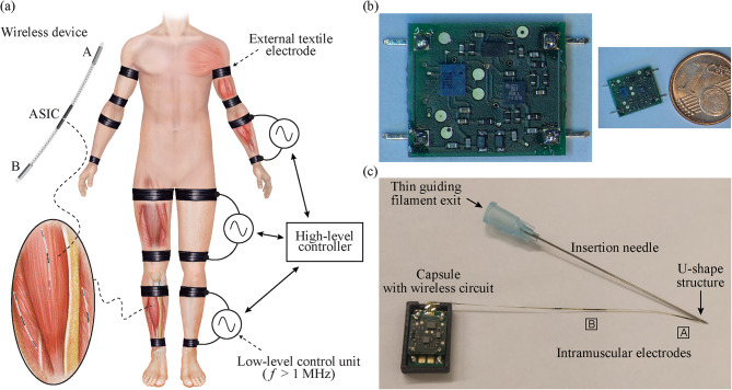
Background: Recently we reported the design and evaluation of floating semi-implantable devices that receive power from and bidirectionally communicate with an external system using coupling by volume conduction. The approach, of which the semi-implantable devices are proof-of-concept prototypes, may overcome some limitations presented by existing neuroprostheses, especially those related to implant size and deployment, as the implants avoid bulky components and can be developed as threadlike devices. Here, it is reported the first-in-human acute demonstration of these devices for electromyography (EMG) sensing and electrical stimulation.

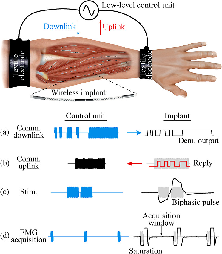
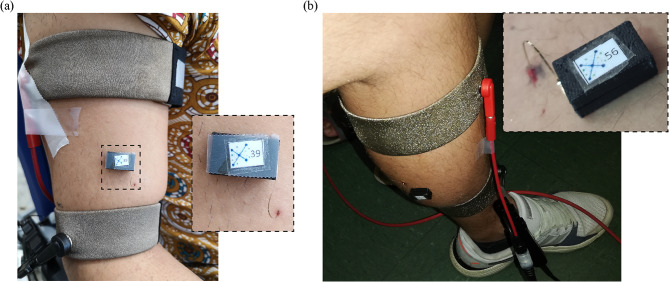
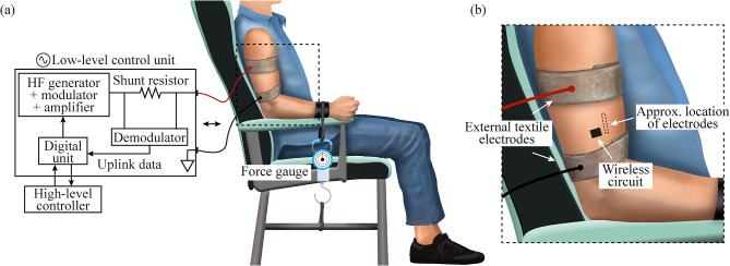
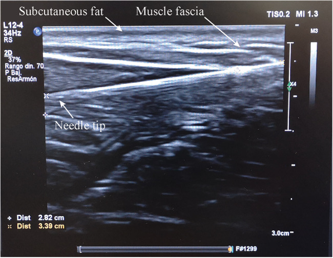
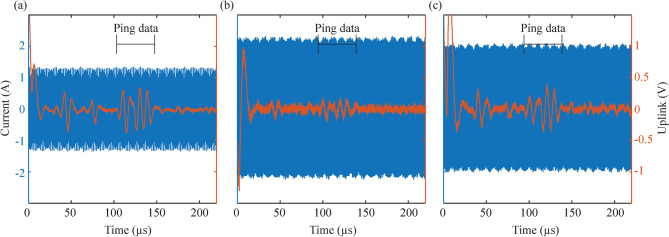
Methods: A proof-of-concept device, consisting of implantable thin-film electrodes and a nonimplantable miniature electronic circuit connected to them, was deployed in the upper or lower limb of six healthy participants. Two external electrodes were strapped around the limb and were connected to the external system which delivered high frequency current bursts. Within these bursts, 13 commands were modulated to communicate with the implant.

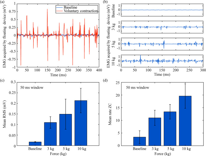
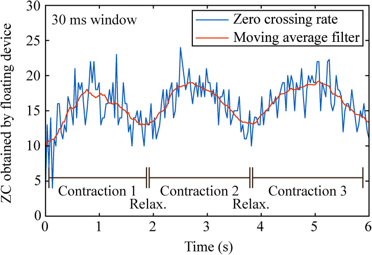
Results: Four devices were deployed in the biceps brachii and the gastrocnemius medialis muscles, and the external system was able to power and communicate with them. Limitations regarding insertion and communication speed are reported. Sensing and stimulation parameters were configured from the external system. In one participant, electrical stimulation and EMG acquisition assays were performed, demonstrating the feasibility of the approach to power and communicate with the floating device.

Conclusions: This is the first-in-human demonstration of EMG sensors and electrical stimulators powered and operated by volume conduction. These proof-of-concept devices can be miniaturized using current microelectronic technologies, enabling fully implantable networked neuroprosthetics.

Keywords: AIMDs; Bidirectional communications; Electrical stimulation; Electromyography; Human validation.; Neuroprostheses; Semi-implantable devices; Sensor; Volume conduction; Wireless power transfer.


Conflict of interest statement

The authors declare no competing interests.

Figures

Fig. 1
Fig. 1
Bidirectional Hyper-Connected Neural System (BHNS) concept and proof-of-concept semi-implantable devices. (a) Schematic representation of the envisioned BHNS. (b) Miniature electronic circuit for EMG sensing and stimulation and its comparison with a 1 cent euro coin for acute human demonstration of the BHNS concept. (c) Complete proof-of-concept semi-implantable device showing the dedicated insertion needle, the thin-film intramuscular electrodes, the thin guiding filament that exits through the needle hub, and the polymer capsule that houses the miniature circuit (opened lid) [17]
Fig. 2
Fig. 2
HF current bursts sequence. It shows the different sequences that can be obtained with the combination for the external system and its low-level control unit, and the wireless implantable devices. See main text for detailed information
Fig. 3
Fig. 3
Proof-of-concept semi-implantable devices deployed in the target muscles of two participants. (a) Device in biceps brachii of participant 2. (b) Device in gastrocnemius medialis of participant 5
Fig. 4
Fig. 4
Setup used during the study in upper limb. The participant was always sitting on a chair, with the upper arm aligned with the torso and the elbow at an angle of 120°. The strapped external textile electrodes were connected to the external system. (a) For the force measurement during isometric contractions of the biceps brachii, the wrist was strapped to the armrest of the chair, and to a force gauge. (b) Zoom of the region where the wireless circuit was located, and approximate location of thin-film electrodes
Fig. 5
Fig. 5
Ultrasounds image obtained from participant 2 immediately after the needle was inserted in the biceps brachii. The needle was injected from the top right corner (not shown in the image), passed through subcutaneous fat and the muscle fascia, and ended up in the target muscle. The tip of the needle is seen at the left of the image at a depth of approximately 1 cm
Fig. 6
Fig. 6
Waveforms obtained with a floating oscilloscope and the probes connected to the external system. These waveforms are obtained after the external system requests a “Ping” to a specific device. In blue, HF current delivered by external system for uplink; in orange, output of external system’s demodulator, showing the filtered and amplified voltage across the shunt resistor when the floating device does an ACK ping message by load modulating the HF current burst (i.e., shows changes in the current flowing through the textile electrodes). (a) Participant 2, biceps brachii. (b) Participant 5, gastrocnemius medialis. (c) Participant 6, gastrocnemius medialis
Fig. 7
Fig. 7
Examples of raw EMG obtained by the floating EMG sensor and stimulator in biceps brachii of participant 2. (A) Comparison between baseline and sustained contraction. (b) Comparison between baseline and three different target forces exerted by the participant. (c) Offline calculation of average of root mean square (RMS) value for the raw signals shown in b. d) Offline averaging of zero crossing (ZC) calculation for the raw signals shown in b. In both cases, the window used for calculation was set to 50 ms
Fig. 8
Fig. 8
Parametric acquisition using ZC rate. Participant 2 did three contractions during 6 s (progressive contraction followed by relaxation), which can be clearly seen by the parametric waveform obtained. After the samples were uploaded to the external system, a moving average filter was added to show the envelope of the contractions

References

    1. Pascual-Valdunciel A, Gonzalez-Sanchez M, Muceli S, Adan-Barrientos B, Escobar-Segura V, Perez-Sanchez JR, et al. Intramuscular stimulation of muscle afferents attains prolonged tremor reduction in essential tremor patients. IEEE Trans Biomed Eng. 2020;68(6):1768–76. doi: 10.1109/TBME.2020.3015572. - DOI - PubMed
    1. Koller JR, Jacobs DA, Ferris DP, Remy CD. Learning to walk with an adaptive gain proportional myoelectric controller for a robotic ankle exoskeleton. J Neuroeng Rehabil. 2015;12(1):1–14. doi: 10.1186/s12984-015-0086-5. - DOI - PMC - PubMed
    1. Salminger S, Sturma A, Hofer C, Evangelista M, Perrin M, Bergmeister KD, et al. Long-term implant of intramuscular sensors and nerve transfers for wireless control of robotic arms in above-elbow amputees. Sci Rob. 2019;4(32):eaaw6306. doi: 10.1126/scirobotics.aaw6306. - DOI - PubMed
    1. Fleming A, Stafford N, Huang S, Hu X, Ferris DP, Huang HH. Myoelectric control of robotic lower limb prostheses: a review of electromyography interfaces, control paradigms, challenges and future directions. J Neural Eng. 2021;18(4):41004. doi: 10.1088/1741-2552/ac1176. - DOI - PMC - PubMed
    1. Jung MK, Muceli S, Rodrigues C, Megía-García Á, Pascual-Valdunciel A, del-Ama AJ, et al. Intramuscular EMG-Driven Musculoskeletal Modelling: towards implanted muscle interfacing in spinal cord Injury patients. IEEE Trans Biomed Eng. 2022;69(1):63–74. doi: 10.1109/TBME.2021.3087137. - DOI - PubMed

Publication types

LinkOut - more resources