. 2024 Jan 3;16(1):8.
doi: 10.1186/s13148-023-01615-5.

Histone acetylation: a key determinant of acquired cisplatin resistance in cancer

Affiliations

Histone acetylation: a key determinant of acquired cisplatin resistance in cancer

Abhiram Natu et al. Clin Epigenetics. .

Abstract

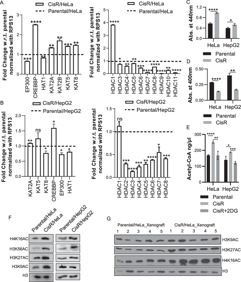
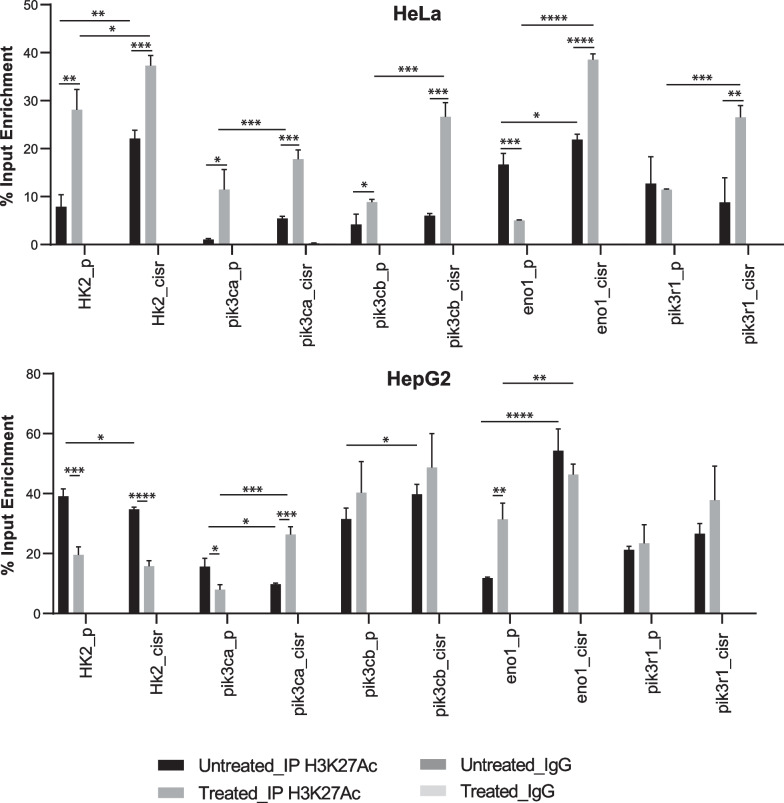
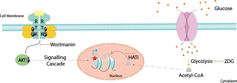
Cisplatin is an alkylating class of chemotherapeutic drugs used to treat cancer patients. However, cisplatin fails in long-term treatment, and drug resistance is the primary reason for tumor recurrence. Hence, understanding the mechanism of acquirement of chemoresistance is essential for developing novel combination therapeutic approaches. In this study, in vitro cisplatin-resistant cancer cell line models were developed. Gene ontology and GSEA of differentially expressed genes between parental and resistant cells suggest that PI3K-AKT signaling, central carbon metabolism, and epigenetic-associated phenomenon alter in cisplatin-resistant cells. Further, the data showed that increased glucose transport, alteration in the activity of histone-modifying enzymes, and acetyl-CoA levels in resistant cells paralleled an increase in global histone acetylation. Enrichment of histone acetylation on effectors of PI3K-AKT and glycolysis pathway provides evidence of epigenetic regulation of the key molecules in drug resistance. Moreover, cisplatin treatment to resistant cells showed no significant changes in histone acetylation marks since drug treatment alters cell epigenome. In continuation, targeting PI3K-AKT signaling and glycolysis leads to alteration in histone acetylation levels and re-sensitization of resistant cells to chemo-drug. The data provide evidence of histone acetylation's importance in regulating pathways and cisplatin-resistant cells' cell survival. Our study paves the way for new approaches for developing personalized therapies in affecting metabolic pathways and epigenetic changes to achieve better outcomes for targeting drug-resistant cells.

Keywords: 2DG; Cisplatin; Hyperacetylation; PI3K; Resistance.


Conflict of interest statement

The authors declare no competing interests.

Figures

Fig. 1
Fig. 1
Establishment and characterization of cisplatin resistant cell lines. A Percent cell viability for the cell lines plotted as a line chart at different concentrations of the cisplatin. B Bar graph representing colonies obtained for cisplatin-resistant model. C Effect of cisplatin treatment on cisplatin resistant xenograft model. D Estimation of intracellular platinum concentration using TXRF method. Error bar represents mean ± S.D from 3 replicates. *p < 0.05, **p < 0.01, ***p < 0.001 and ****p < 0.0001
Fig. 2
Fig. 2
Differential gene expression and pathway analysis in cisplatin resistant model. A Volcano plot representing differentially expressed genes in parental vs. cisplatin-resistant cells. B Gene ontology and pathway analysis of upregulated in the cisplatin-resistant model. C Gene set enrichment analysis plot depicting enrichment of PI3K-AKT signaling in cisplatin resistant cells. D Network analysis of upregulated pathways in cisplatin resistance
Fig. 3
Fig. 3
Alteration in histone modifying enzymes and their activity. A, B Bar graphs representing qPCR analysis for HAT & HDAC genes in HeLa and HepG2 cisplatin resistant model systems. C Estimation of HAT activity in cisplatin resistant cell lines. D Investigation of HDAC activity in resistant cells. E Assessment of acetyl-CoA levels in cisplatin-resistant cells. F Western blot analysis for histone lysine acetylation in in vitro cisplatin resistant models. G Western blot analysis for histone lysine acetylation in in vivo HeLa cisplatin resistant model. Error bar represents mean ± S.D from 3 replicates. *p < 0.05, **p < 0.01, ***p < 0.001 and ****p < 0.0001
Fig. 4
Fig. 4
Enrichment of H3K27Ac in response to cisplatin treatment. Bar graph representing percent input enrichment of H3K27Ac on the promoter of depicted genes. The comparison carried out between parental & resistance w or w/o treated with cisplatin. Error bar represents mean ± S.D from 3 replicates. *p < 0.05, **p < 0.01, ***p < 0.001 and ****p < 0.0001
Fig. 5
Fig. 5
Role of histone acetylation in survival of cisplatin resistant cells. A Western blot analysis to check the effect of cisplatin in time-dependent manner on histone PTM levels. T.P. depicts Time point. B Percent apoptotic cells determined using Annexin-FITC/PI staining in CisR/HeLa cells. C Long-term survival assay upon 2DG treatment in CisR/HepG2 cells. Data with error bar represent mean ± S.D from 3 biological replicates *p < 0.05, **p < 0.01, ***p < 0.001 and ****p < 0.0001
Fig. 6
Fig. 6
Effect of 2DG treatment on tumorigenic potential of cisplatin resistant cells. A Estimation of tumor progression of CisR/HeLa cells after Cisplatin or 2DG treatment. ↑ represents 1 round of dose. B Representative image for tumor size after 8 weeks of treatment in different groups. C Effect of 2DG on histone PTMs and Caspase3 in cisplatin resistant xenograft tissues. Bar graph representing the H-Score desired antigens in the experimental setup. The scale bar denotes 10 μm. The bar graph represents H-Score for mean ± S.E.M. from 4–5 biological replicates. *p < 0.05, and ****p < 0.0001
Fig. 7
Fig. 7
Schematic representation highlighting the molecular alterations in cisplatin resistant model. Cisplatin-resistant cells display more activation of PI3K signaling, Acetyl-CoA production and histone hyperacetylation. The occupancy of histone acetylation marks on the promoter of genes related to PI3K subunits and glycolysis in resistant cells indicates their importance. Inhibiting glycolysis using 2-DG or PI3K signaling may imporove therapeutic outcome in terms of reduces tumor growth

References

    1. Dasari S, Tchounwou PB. Cisplatin in cancer therapy: molecular mechanisms of action. Eur J Pharmacol. 2014;740:364–378. doi: 10.1016/j.ejphar.2014.07.025. - DOI - PMC - PubMed
    1. Wang L, Zhao X, Fu J, Xu W, Yuan J. The role of tumour metabolism in cisplatin resistance. Front Mol Biosci. 2021;8:691795. doi: 10.3389/fmolb.2021.691795. - DOI - PMC - PubMed
    1. Zaal EA, Berkers CR. The influence of metabolism on drug response in cancer. Front Oncol. 2018;8:500. doi: 10.3389/fonc.2018.00500. - DOI - PMC - PubMed
    1. Zhao Y, Butler EB, Tan M. Targeting cellular metabolism to improve cancer therapeutics. Cell Death Dis. 2013;4(3):e532. doi: 10.1038/cddis.2013.60. - DOI - PMC - PubMed
    1. Reid MA, Dai Z, Locasale JW. The impact of cellular metabolism on chromatin dynamics and epigenetics. Nat Cell Biol. 2017;19(11):1298–1306. doi: 10.1038/ncb3629. - DOI - PMC - PubMed

Publication types