. 2024 Jan 2;14(1):101-117.
doi: 10.1039/d3ra06355k.

In vitro and in vivo studies of Syzygium cumini-loaded electrospun PLGA/PMMA/collagen nanofibers for accelerating topical wound healing

Affiliations

In vitro and in vivo studies of Syzygium cumini-loaded electrospun PLGA/PMMA/collagen nanofibers for accelerating topical wound healing

Esraa B Abdelazim et al. RSC Adv. .

Abstract

This work aims to develop plant extract-loaded electrospun nanofiber as an effective wound dressing scaffolds for topical wound healing. Electrospun nanofibers were fabricated from Syzygium cumini leaf extract (SCLE), poly(lactic-co-glycolic acid) (PLGA), poly(methyl methacrylate) (PMMA), collagen and glycine. Electrospinning conditions were optimized to allow the formation of nanosized and uniform fibers that display smooth surface. Morphology and swelling behavior of the formed nanofibers were studied. In addition, the antibacterial activity of the nanofibers against multidrug-resistant and human pathogens was assessed by agar-well diffusion. Results showed that nanofibers containing Syzygium cumini extract at concentrations of 0.5 and 1% w/v exhibited greater antibacterial activity against the tested Gram-positive (i.e., Staphylococcus aureus, Candida albicans, Candida glabrata and Bacillus cereus) and Gram-negative (i.e., Salmonella paratyphi and Escherichia coli) pathogens compared to the same concentrations of the plain extract. Furthermore, in vivo wound healing was evaluated in Wistar rats over a period of 14 days. In vivo results demonstrated that nanofiber mats containing SCLE and collagen significantly improved wound healing within two weeks, compared to the control untreated group. These findings highlight the potential of fabricated nanofibers in accelerating wound healing and management of topical acute wounds.


Conflict of interest statement

The authors declare that they have no conflict of interest.

Figures

Fig. 1
Fig. 1. Total ion chromatogram (TIC) for methanol extract of Syzygium cumini leaves in negative (A) and positive (B) ionization modes. The numbers assigned to the peaks are explained in Table 3.
Fig. 2
Fig. 2. The chemical structures of major compounds of the SCLE identified by HPLC-MS/MS. SCLE: S. cumini leaves extract.
Fig. 3
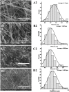
Fig. 3. Scanning electron microscopy images, captured at a magnification of 2500×, showing the morphology of electrospun PLGA/PMMA/col/gly/SCLE nanofibers: (A1) PLGA (15%)/PMMA (10%) (F1), (B1) PLGA/PMMA/col (1%) (F6), (C1) PLGA/PMMA/col/gly (0.1%) (F7), and (D1) PLGA/PMMA/col/gly (0.1%)/SCLE (1%) (F11). A2, B2, C2, and D2 represent diameter distribution histograms of F1, F6, F7, and F11, respectively.
Fig. 4
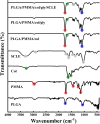
Fig. 4. FTIR spectra of individual components of the electrospun PLGA/PMMA/col/gly/SCLE nanofibers. The main characteristic peaks of each spectrum are highlighted to demonstrate changes in the formed nanofibers.
Fig. 5
Fig. 5. XRD patterns of components of electrospun PLGA/PMMA/col/gly nanofibers loaded with SCLE.
Fig. 6
Fig. 6. Swelling ratio of electrospun PLGA/PMMA/col/gly/SCLE nanofibers was measured at various time intervals between 0 and 12 hours. The results, presented as the mean ± standard deviation (n = 3). # refers to statistical difference compared to the untreated group (Group 1) (p < 0.0001).
Fig. 7
Fig. 7. Photographs and a chart show the measured inhibitory zone widths (mm ± SE) that were observed when our formulations (F6, F7, F8, F9, F10, F11, F12 (15% PLGA), F13 (10% PMMA), F14 (1% collagen), F15, and F16) were applied to inhibit the growth of human infections; (A) Salmonella paratyphi, (B) Escherichia coli, (C) Staphylococcus aureus, (D) Bacillus cereus, (E) Candida glabrata, and (F) Candida albicans. SE = standard error.
Fig. 8
Fig. 8. Cytotoxic assay of the prepared nanofibers against vero cells after treatment for 72 h. Cytotoxicity of the initial ingredients of SCLE, collagen, PLGA, and PMMA were also evaluated.
Fig. 9
Fig. 9. Representative pictures of skin wounds at various time points (days 0–14) illustrating the effects of nanofibers (NFs) on in vivo wound healing. The groups examined include group 1 (control), group 2 (PLGA/PMMA/col/gly (F7)), and group 3 (PLGA/PMMA/col/gly/SCLE (F11)).
Fig. 10
Fig. 10. In vivo wound healing results. (A) Wound diameter measurements at 0, 3, 6, 9, 12, and 14 days for different experimental groups. (B) Percentage healing of in vivo wounds at 0, 3, 6, 9, 12, and 14 days after creating a full-thickness skin wound. *, **, # refer to statistical differences compared to the untreated group (Group 1) at p < 0.01, p < 0.001 and p < 0.0001, respectively.
Fig. 11
Fig. 11. Histopathology of wound healing in control and NFs-treated skin wounds. (A) Section in skin wound in control group showing complete epithelial ulceration covered by fibrinopurulent exudate (black arrows) with underlying granulation tissue and neovascularization (*) with few scattered collagen fibers (red arrows) H&E × 200. (B) Section in skin wound in control group showing complete epithelial ulceration covered by fibrinopurulent exudate (black arrows) with underlying granulation tissue and neovascularization (*) with no evidence of collagen fibers. H&E × 200. (C) Section from wound in control group covered by regenerating attenuated squamous epithelium (black arrows) with underlying granulation and neovascularization (*). No evidence of collagen bands. H&E × 200. (D) Section from wound in control group covered by regenerating attenuated squamous epithelium (black arrows) with underlying maturing granulation tissue showing prominent fibroblasts (red arrows). Creeping collagen fibers with haphazard orientation are noted from deep dermis (*). H&E × 200. (E) Section in wound of nanofiber treated with F11 group showing complete epithelial recovery (black arrows). The underlying dermis shows well-formed collagen bands with organized parallel orientation to the surface epithelium and scar formation (red arrows) with complete replacement of granulation tissue indicating complete recovery. H&E × 200. (F) Section in wound of nanofiber treated with F11 group showing complete epithelial recovery (black arrows). The underlying dermis shows well-formed collagen bands with organized parallel orientation to the surface epithelium (red arrows) with complete replacement of granulation tissue indicating complete recovery. H&E × 200.

References

    1. Alabdali A. Y. M. Khalid R. Kzar M. Ezzat M. O. Huei G. M. Hsia T. W. Mogana R. Rahman H. Razik B. M. A. Issac P. K. Chinnappan S. Khalivulla S. I. Design, synthesis, in silico and antibacterial evaluation of curcumin derivatives loaded nanofiber as potential wound healing agents. J. King Saud Univ., Sci. 2022;34:102205. doi: 10.1016/j.jksus.2022.102205. - DOI
    1. Dias J. R. Granja P. L. Bártolo P. J. Advances in electrospun skin substitutes. Prog. Mater. Sci. 2016;84:314–334. doi: 10.1016/j.pmatsci.2016.09.006. - DOI
    1. Elsabahy M. Heo G. S. Lim S. M. Sun G. Wooley K. L. Polymeric Nanostructures for Imaging and Therapy. Chem. Rev. 2015;115:10967–11011. doi: 10.1021/acs.chemrev.5b00135. - DOI - PMC - PubMed
    1. Yao C. H. Chen K. Y. Chen Y. S. Li S. J. Huang C. H. Lithospermum radix extract-containing bilayer nanofiber scaffold for promoting wound healing in a rat model. Mater. Sci. Eng., C. 2019;96:850–858. doi: 10.1016/j.msec.2018.11.053. - DOI - PubMed
    1. Elsabahy M. Zhang M. Gan S. M. Waldron K. C. Leroux J. C. Synthesis and enzymatic stability of PEGylated oligonucleotide duplexes and their self-assemblies with polyamidoamine dendrimers. Soft Matter. 2008;4:294. doi: 10.1039/b714221h. - DOI - PubMed

LinkOut - more resources