

Non-invasive differentiation of hepatic steatosis and steatohepatitis in a mouse model using nitroxyl radical as an MRI-contrast agent
- PMID: 38173653
- PMCID: PMC10758964
- DOI: 10.1016/j.toxrep.2023.12.002
Non-invasive differentiation of hepatic steatosis and steatohepatitis in a mouse model using nitroxyl radical as an MRI-contrast agent
Abstract
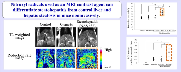
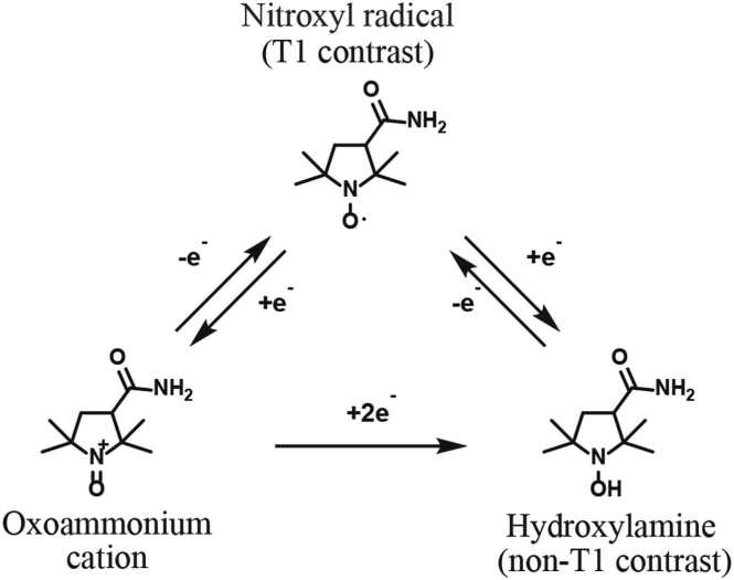
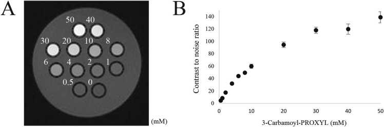
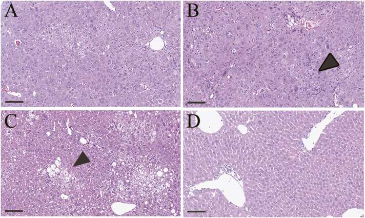
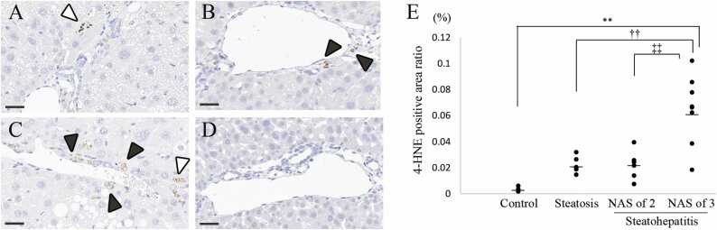
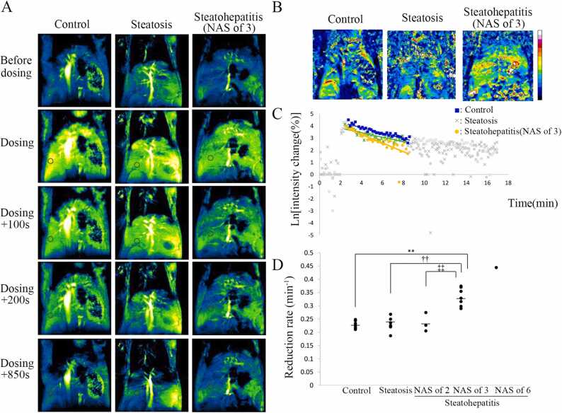
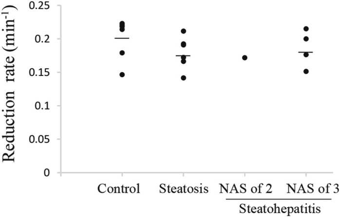
Drug-induced steatohepatitis is considered more serious than drug-induced hepatic steatosis, so that differentiating between the two is crucial in drug development. In addition, early detection of drug-induced steatohepatitis is considered important since recovery is possible with drug withdrawal. However, no method has been established to differentiate between the two. In the development of drug-induced steatohepatitis, reactive oxygen species (ROS) is excessively generated in the liver. It has been reported that ROS can be monitored with electron spin resonance (ESR) and dynamic nuclear polarization-magnetic resonance imaging (DNP-MRI) by using nitroxyl radicals, which are known to participate in various in vivo redox reactions. The decay/reduction rate, which is an index for monitoring nitroxyl radicals, has been reported to be increased in tissues with excessive ROS levels other than liver, but decreased in methionine choline deficient (MCD) diet-induced steatohepatitis with excess ROS. Therefore, looking to differentiate between drug-induced hepatic steatosis and steatohepatitis, we examined whether the reduction rate decreases in steatohepatitis other than the MCD-diet induced disease and whether the decrease could be detected by MRI. We used STAM™ mice in which hepatic steatosis and steatohepatitis developed sequentially under diabetic conditions. 3-carbamoyl-PROXYL (CmP), one of the nitroxyl radicals, was injected intravenously during the MRI procedure and the reduction rate was calculated. The reduction rate was significantly higher in early steatohepatitis than in hepatic steatosis and the control. Excess ROS in early steatohepatitis was detected by an immunohistochemical marker for ROS. Therefore, it was indicated that the increase or decrease in the reduction rate in steatohepatitis differs depending on the model, and early steatohepatitis could be noninvasively differentiated from hepatic steatosis using CmP in MRI. Since the change in direction of the reduction rate in steatohepatitis in clinical studies could be predicted by confirming the reduction rate in preclinical studies, the present method, which can be used consistently in clinical and preclinical studies, warrants consideration as a candidate monitoring method for differentiating between early drug-induced steatohepatitis and hepatic steatosis in drug development.
Keywords: Magnetic resonance imaging; Nitroxyl radicals; Reactive oxygen species; Steatohepatitis.
© 2023 The Authors. Published by Elsevier B.V.
Conflict of interest statement
The authors declare that they have no known competing financial interests or personal relationships that could have appeared to influence the work reported in this paper.
Figures
Similar articles
-
Redox imaging of skeletal muscle using in vivo DNP-MRI and its application to an animal model of local inflammation.Free Radic Biol Med. 2015 Dec;89:1097-104. doi: 10.1016/j.freeradbiomed.2015.10.418. Epub 2015 Oct 23. Free Radic Biol Med. 2015. PMID: 26505925
-
Spatiotemporal imaging of redox status using in vivo dynamic nuclear polarization magnetic resonance imaging system for early monitoring of response to radiation treatment of tumor.Free Radic Biol Med. 2022 Feb 1;179:170-180. doi: 10.1016/j.freeradbiomed.2021.12.311. Epub 2021 Dec 27. Free Radic Biol Med. 2022. PMID: 34968704
-
Development of 20 cm sample bore size dynamic nuclear polarization (DNP)-MRI at 16 mT and redox metabolic imaging of acute hepatitis rat model.Free Radic Biol Med. 2021 Jun;169:149-157. doi: 10.1016/j.freeradbiomed.2021.04.017. Epub 2021 Apr 15. Free Radic Biol Med. 2021. PMID: 33865961
-
[Novel redox molecular imaging "ReMI" with dual magnetic resonance].Yakugaku Zasshi. 2013;133(7):803-14. doi: 10.1248/yakushi.13-00139. Yakugaku Zasshi. 2013. PMID: 23811768 Review. Japanese.
-
In Vivo Dynamic Nuclear Polarization Magnetic Resonance Imaging for the Evaluation of Redox-Related Diseases and Theranostics.Antioxid Redox Signal. 2022 Jan;36(1-3):172-184. doi: 10.1089/ars.2021.0087. Epub 2021 Jul 7. Antioxid Redox Signal. 2022. PMID: 34015957 Review.
Cited by
-
Stable Nitroxide as Diagnostic Tools for Monitoring of Oxidative Stress and Hypoalbuminemia in the Context of COVID-19.Int J Mol Sci. 2024 Jul 24;25(15):8045. doi: 10.3390/ijms25158045. Int J Mol Sci. 2024. PMID: 39125614 Free PMC article. Review.
-
Novel non‑metal‑based contrast agents for MR imaging: Emerging approaches and clinical perspectives (Review).Int J Oncol. 2025 Aug;67(2):70. doi: 10.3892/ijo.2025.5776. Epub 2025 Jul 19. Int J Oncol. 2025. PMID: 40682851 Free PMC article. Review.
References
-
- Amacher D.E. Strategies for the early detection of drug-induced hepatic steatosis in preclinical drug safety evaluation studies. Toxicology. 2011;279(1–3):10–18. - PubMed
-
- Assayag M., Goldstein S., Samuni A., Berkman N. 3-Carbamoyl-proxyl nitroxide radicals attenuate bleomycin-induced pulmonary fibrosis in mice. Free Radic. Biol. Med. 2021;171:135–142. - PubMed
LinkOut - more resources
Full Text Sources
Miscellaneous
