

Peripapillary Choroidal Vascularity and Visual Correlates in Non-Arteritic Anterior Ischemic Optic Neuropathy Using Swept-Source Optical Coherence Tomography
- PMID: 38173700
- PMCID: PMC10764036
- DOI: 10.3389/fopht.2022.848040
Peripapillary Choroidal Vascularity and Visual Correlates in Non-Arteritic Anterior Ischemic Optic Neuropathy Using Swept-Source Optical Coherence Tomography
Abstract
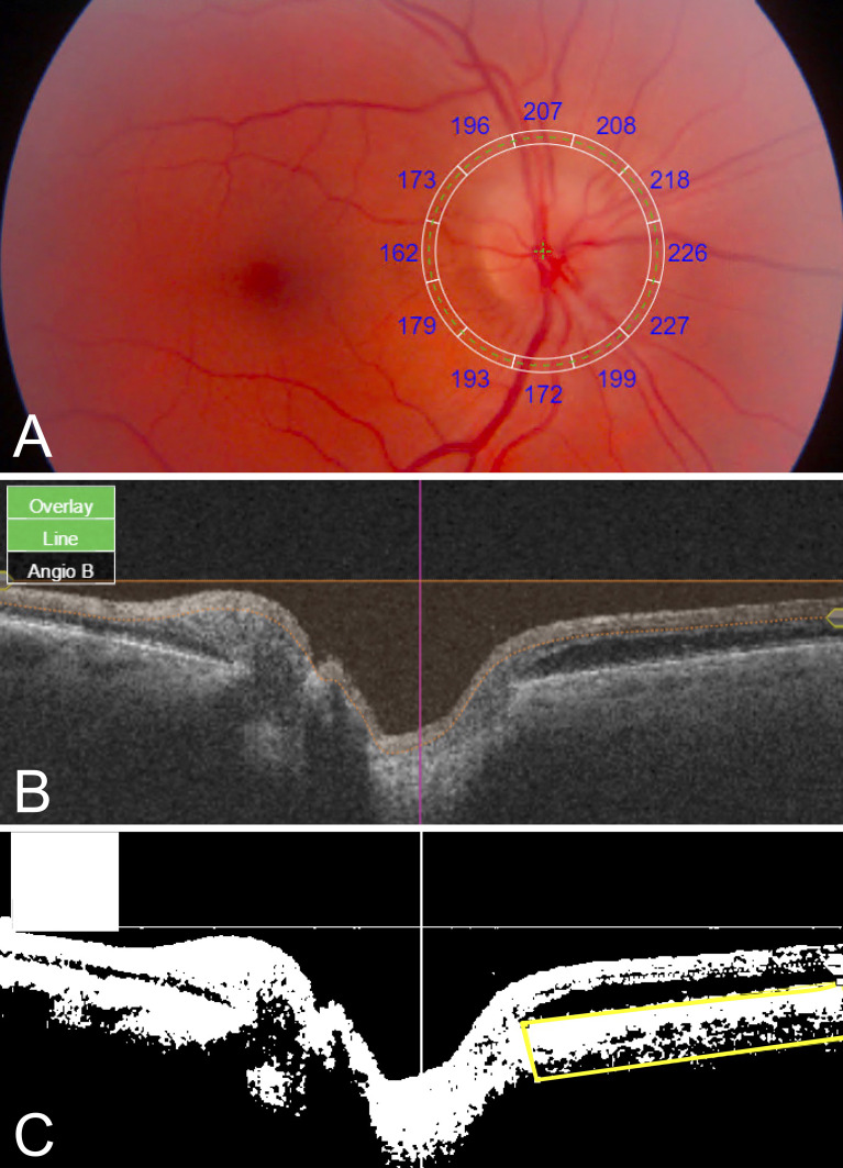
Introduction: The peripapillary choroid shares a blood supply with and is directly apposed to the optic nerve, and therefore may contribute to the pathogenesis of non-arteritic anterior ischemic optic neuropathy (NAION). Prior studies evaluating peripapillary choroidal thickness (PCT) or choroidal vascularity index (CVI; the ratio of the perfused area to total choroid area) have produced mixed results. None investigated the relationship between PCT and CVI or demonstrated functional correlates. We hypothesized that greater PCT and lower CVI would correlate with visual function in patients presenting with NAION.
Methods: Seventeen eyes with NAION (9 acute, 8 non-acute) and 6 unaffected "fellow" eyes in 13 patients, and 18 eyes in 18 age-matched control subjects were imaged using swept-source optical coherence tomography (SS-OCT) prospectively between 2017-2018. Mean PCT and CVI measurements were compared across groups and with respect to corresponding automated perimetric performance at the same visit.
Results: Analysis of variance showed significantly greater PCT (NAION: 278 ± 65 μm, Fellow: 221 ± 50 μm, Control: 158 ± 27 μm, p<0.001) and lower CVI (NAION: 0.35 ± 0.03, Fellow: 0.35 ± 0.04, Control: 0.38 ± 0.02, p<0.005) in patients with NAION compared to control subjects. Bonferroni-corrected pairwise comparisons showed greater PCT and lower CVI in NAION-affected eyes compared to control eyes (p values<0.008), and no significant differences in PCT or CVI between NAION and fellow eyes (p values>0.06). PCT was negatively correlated with CVI among unaffected fellow eyes (r=-0.8, p<0.05), but not among acute NAION eyes (r=-0.1, p>0.7), non-acute NAION eyes (r=0.1, p>0.7), or controls (r=-0.3, p>0.2). Nasal CVI was positively correlated with mean deviation scores in non-acute NAION (r=0.8, p<0.02), but not among fellow unaffected eyes (r=0.8, p>0.05) or acutely affected NAION eyes (r=-0.3, p>0.4). Mean and temporal PCT correlated with pattern standard deviation scores among unaffected fellow eyes (r=0.8, p<0.04; r=0.9, p<0.03), but not among acute NAION eyes (r=-0.2, p>0.5; r=-0.1, p>0.7) or non-acute NAION eyes (r=0.1, p>0.7; r=0.05, p>0.9).
Conclusion: NAION and unaffected fellow eyes demonstrate increased choroidal thicknesses and reduced vascular density. Perimetric performance is directly associated with vascular density among non-acutely affected eyes with NAION. Ongoing work will provide further insights into these structure-function relationships with pathogenic and pathophysiologic relevance.
Keywords: non-arteritic anterior ischemic optic neuropathy (NAION); peripapillary choroidal thickness; peripapillary choroidal vascularity; swept-source OCT (SS-OCT); visual correlation.
Conflict of interest statement
Conflict of Interest: JM: Alcon, Zeiss, Sunovion, Allergan, Genentech. EG: Luminopia, Inc (scientific advisor, equity, patent), Stoke Therapeutics, Inc (consultant). The remaining authors declare that the research was conducted in the absence of any commercial or financial relationships that could be construed as a potential conflict of interest.
Figures


References
Grants and funding
LinkOut - more resources
Full Text Sources