

Biomechanical analysis of lower limbs during stand-to-sit tasks in patients with early-stage knee osteoarthritis
- PMID: 38173868
- PMCID: PMC10763667
- DOI: 10.3389/fbioe.2023.1330082
Biomechanical analysis of lower limbs during stand-to-sit tasks in patients with early-stage knee osteoarthritis
Abstract
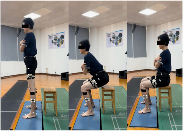
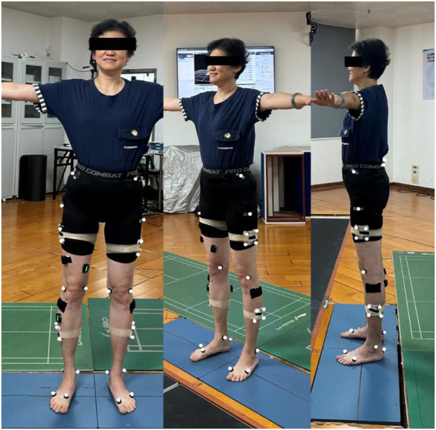
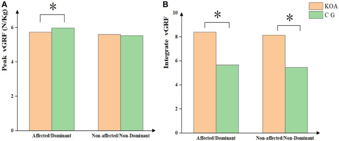
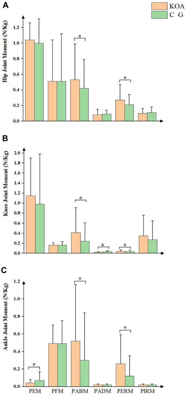
Background: Knee osteoarthritis (KOA) is a common degenerative disease among the older people that severely affects their daily life. Previous studies have confirmed that movement biomechanics are altered in patients with KOA during task performance. However, changes that occur in lower limb joints and muscles in the three planes during stand-to-sit (STS) tasks in patients with early-stage KOA are unclear. Method: Of the 36 participants recruited in this study, 24 (8 males and 16 females) and 12 (4 males and 8 females) were added to the KOA and control groups, respectively. The Nexus Vicon motion capture system along with Delsys wireless surface electromyography devices and plantar pressure measurement mat was used to record test data. A Visual 3D software was used to process the data and calculate the biomechanical and electromyographic parameters during STS tasks. Results: There was no significant difference in task duration between the two groups. Patients with KOA could perform a greater range of pelvic motion and smaller range of hip and knee joint motion with a lower maximum hip joint angular acceleration in the sagittal plane and greater knee and ankle joint motion in the coronal plane. There was no significant difference in the motion range in the horizontal plane. During the STS task, patients in the KOA group had a lower vertical ground reaction force (GRF) amplitude on the injured side but a higher integrated GRF on both sides than those in the control group. Moreover, patients with KOA demonstrated higher PERM and PABM of the lower limb joints and smaller knee PADM and ankle PEM. Additionally, maximum activation levels of GMed muscle, affected-side gluteus medius (GM), ST, rectus femoris (RF), and tibialis anterior (TA) muscles were lower in patients with KOA than in controls. Conversely, the activation level of biceps femoris (BF) was higher. Furthermore, the integral EMG values of GMed, GM, ST, VL, RF, vastus medialis VM, and TA muscles on the affected side were lower, except for the BF muscle, in patients with KOA. Conclusion: Compared with the participants in the control group, patients with early-stage KOA exhibited consistent changes in sEMG parameters and biomechanical alterations in the sagittal plane, as observed in previous studies. However, differences in parameters were observed in the coronal and transverse planes of these patients. The noninvasive analysis of the 3D parameters of the involved motion patterns may lead to the early detection of KOA.
Keywords: biomechanics; early-stage; knee osteoarthritis (KOA); osteoarthritis; sEMG; stand-to-sit task.
Copyright © 2023 Pan, Huang, Huang, Luan, Zhang and Liao.
Conflict of interest statement
The authors declare that the research was conducted in the absence of any commercial or financial relationships that could be construed as a potential conflict of interest.
Figures
Similar articles
-
Factors, characteristics and influences of the changes of muscle activation patterns for patients with knee osteoarthritis: a review.J Orthop Surg Res. 2025 Jan 30;20(1):112. doi: 10.1186/s13018-025-05484-x. J Orthop Surg Res. 2025. PMID: 39885604 Free PMC article. Review.
-
Biomechanics of the lower limb in patients with mild knee osteoarthritis during the sit-to-stand task.BMC Musculoskelet Disord. 2024 Apr 6;25(1):268. doi: 10.1186/s12891-024-07388-z. BMC Musculoskelet Disord. 2024. PMID: 38582828 Free PMC article.
-
Postural Balance in Individuals With Knee Osteoarthritis During Stand-to-Sit Task.Front Hum Neurosci. 2021 Nov 3;15:760960. doi: 10.3389/fnhum.2021.760960. eCollection 2021. Front Hum Neurosci. 2021. PMID: 34803639 Free PMC article.
-
Kinetics, kinematics, and knee muscle activation during sit to stand transition in unilateral and bilateral knee osteoarthritis.Gait Posture. 2021 May;86:38-44. doi: 10.1016/j.gaitpost.2021.02.023. Epub 2021 Feb 22. Gait Posture. 2021. PMID: 33677177
-
Contributions to the understanding of gait control.Dan Med J. 2014 Apr;61(4):B4823. Dan Med J. 2014. PMID: 24814597 Review.
Cited by
-
The effect of mild to moderate knee osteoarthritis on gait and three-dimensional biomechanical alterations.Front Bioeng Biotechnol. 2025 Apr 3;13:1562936. doi: 10.3389/fbioe.2025.1562936. eCollection 2025. Front Bioeng Biotechnol. 2025. PMID: 40248645 Free PMC article.
-
Artificial intelligence-based analysis of lower limb muscle mass and fatty degeneration in patients with knee osteoarthritis and its correlation with Knee Society Score.Int J Comput Assist Radiol Surg. 2025 Apr;20(4):635-642. doi: 10.1007/s11548-024-03284-y. Epub 2024 Nov 3. Int J Comput Assist Radiol Surg. 2025. PMID: 39489851 Free PMC article.
-
Tuina on knee pain and functional decline of lower limbs for patients with mild-to-moderate knee osteoarthritis in Shanghai: protocol for a multicentre, assessor-blinded, randomised controlled trial.BMJ Open. 2024 Jun 12;14(6):e083440. doi: 10.1136/bmjopen-2023-083440. BMJ Open. 2024. PMID: 38866576 Free PMC article.
-
Factors, characteristics and influences of the changes of muscle activation patterns for patients with knee osteoarthritis: a review.J Orthop Surg Res. 2025 Jan 30;20(1):112. doi: 10.1186/s13018-025-05484-x. J Orthop Surg Res. 2025. PMID: 39885604 Free PMC article. Review.
-
The Impact of Excessive Muscle Co-Contraction on Sit-To-Stand Performance in High-Heeled Footwear.Healthc Technol Lett. 2025 May 9;12(1):e70011. doi: 10.1049/htl2.70011. eCollection 2025 Jan-Dec. Healthc Technol Lett. 2025. PMID: 40352139 Free PMC article.
References
LinkOut - more resources
Full Text Sources
Research Materials
Miscellaneous