. 2023 Dec 19:14:1252470.
doi: 10.3389/fphys.2023.1252470. eCollection 2023.

Cysteine and glycine-rich protein 3 (Crp3) as a critical regulator of elastolysis, inflammation, and smooth muscle cell apoptosis in abdominal aortic aneurysm development

Affiliations

Cysteine and glycine-rich protein 3 (Crp3) as a critical regulator of elastolysis, inflammation, and smooth muscle cell apoptosis in abdominal aortic aneurysm development

Ana Barbosa Marcondes de Mattos et al. Front Physiol. .

Abstract

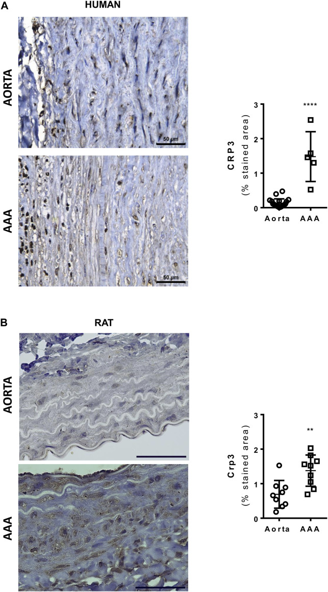
Abdominal aortic aneurysm (AAA) is a life-threatening vascular disease for which surgical or endovascular repair are the only currently available therapeutic strategies. The development of AAA involves the breakdown of elastic fibers (elastolysis), infiltration of inflammatory cells, and apoptosis of smooth muscle cells (SMCs). However, the specific regulators governing these responses remain unknown. We previously demonstrated that Cysteine and glycine-rich protein 3 (Crp3) sensitizes SMCs to apoptosis induced by stretching. Building upon this finding, we aimed to investigate the influence of Crp3 on elastolysis and apoptosis during AAA development. Using the elastase-CaCl2 rat model, we observed an increase in Crp3 expression, aortic diameter, and a reduction in wall thickness in wild type rats. In contrast, Crp3-/- rats exhibited a decreased incidence of AAA, with minimal or no changes in aortic diameter and thickness. Histopathological analysis revealed the absence of SMC apoptosis and degradation of elastic fibers in Crp3-/- rats, accompanied by reduced inflammation and diminished proteolytic capacity in Crp3-/- SMCs and bone marrow-derived macrophages. Collectively, our findings provide evidence that Crp3 plays a crucial role in AAA development by modulating elastolysis, inflammation, and SMC apoptosis. These results underscore the potential significance of Crp3 in the context of AAA progression and offer new insights into therapeutic targets for this disease.

Keywords: abdominal aortic aneurysm; apoptosis; cysteine and glycine-rich protein-3; elastolysis; smooth muscle cell.


Conflict of interest statement

The authors declare that the research was conducted in the absence of any commercial or financial relationships that could be construed as a potential conflict of interest.

Figures

FIGURE 1
FIGURE 1
Crp3 expression is induced in human and rat abdominal aortic aneurysm (AAA). (A) Representative images of Crp3 staining (brown) in human AAA. The quantification shows modulation of Crp3 in human AAA (n = 5) compared to normal aorta (n = 12). (B) Representative images of Crp3 staining (brown) in rat AAA. Similarly, Crp3 expression increases after 14 days of elastase-calcium chloride induction of AAA in rats (n = 9). The black scale bar represents 50 μm. **** indicates p < 0.0001 and ** indicates p = 0.0036.
FIGURE 2
FIGURE 2
Crp3 absence prevents AAA development. (A) Aortic diameter analysis performed 3 days (n = 10–14), 7 days (n = 5–9) and 14 days (n = 26–28) after elastase-CaCl2-AAA induction, showing increase in aortic diameter in AAA compared to saline in WT rats, which was attenuated in Crp3−/− AAA. * indicates p = 0.020. (B) Depiction of the increase in aortic lumen diameter 14 days after AAA induction in WT and Crp3−/− rats and (C) graphical representation of the lowered AAA incidence in Crp3−/− rats in comparison with WT AAA. WT (n = 26) and Crp3−/− (n = 27) *** indicates p < 0.001 and **** indicates p < 0.0001. (D) AAA development in WT rats was associated with reduction in intima: media thickness, while no change was observed upon AAA induction in Crp3−/− rats. WT saline (n = 25), Crp3−/− saline (n = 15), WT AAA (n = 6), Crp3−/− AAA (n = 15). * indicates p = 0.014.
FIGURE 3
FIGURE 3
Absence of elastolysis in response to AAA induction in Crp3−/− rats. Verhoeff’s van Gieson staining depicting elastic fibers degradation in WT AAA vs. saline but no difference between saline and AAA in Crp3−/− animals, as shown in the quantification. WT saline (n = 13), Crp3−/− saline (n = 15), WT AAA (n = 18), Crp3−/− AAA (n = 14). The black scale bar represents 500 μm (images at lower magnification or 30 μm (images at higher magnification). *** indicates p = 0.0002.
FIGURE 4
FIGURE 4
Absence of collagen deposition in response to AAA induction in Crp3−/− rats. Collagen amount evaluation via Picrosirius Red staining, demonstrating that WT AAA development increases collagen, while Crp3−/− remains unchanged. WT saline (n = 11), Crp3−/− saline (n = 8), WT AAA (n = 11), Crp3−/− AAA (n = 10). The black scale bar represents 500 μm (images at lower magnification or 30 μm (images at higher magnification). ** indicates p = 0.0093.
FIGURE 5
FIGURE 5
Absence of Crp3 protects SMCs from apoptosis. (A) Representative images of TUNEL assay demonstrating the presence of apoptosis in WT AAA but not in Crp3−/−. The white scale bar represents 50 μm. (B) Quantitative analysis of TUNEL staining performed in saline and elastase-CaCl2 aortas of WT and Crp3−/− rats for detection of apoptotic nuclei. AAA development was accompanied of apoptosis in WT AAA but not in Crp3−/− AAA. WT saline (n = 6), Crp3−/− saline (n = 7), WT AAA (n = 4), Crp3−/− AAA (n = 4). ** indicates p = 0.043 and *** indicates p = 0.0002. (C) Quantification of apoptosis in isolated aortic SMCs. While WT cells (n = 3) showed apoptotic response to 100 µM ceramide, little or no apoptosis is detected in Crp3−/− SMC (n = 3). **** indicates p < 0.0001.
FIGURE 6
FIGURE 6
The absence of Crp3 blunts the proteolytic profile of aortic SMCs. (A) Quantitative analysis of the secretion and activation of Mmp-2 in the media of control and IL-1β-stimulated aortic SMCs. IL-1β increased Mmp-2 activation, a response that was attenuated in Crp3−/− SMCs (WT n = 4, Crp3−/− n = 5). ** indicates p = 0.027 (B) Total Mmp-2 levels remained unchanged (WT n = 6, Crp3−/− n = 7). (C) The secretion of Mmp-9 was induced by IL-1β in WT, but not in Crp3−/− SMCs (WT n = 8, Crp3−/− n = 5). ** indicates p = 0.0048, *** indicates p < 0.001 (D) Secreted Timp-2 levels remained unchanged in response to IL-1β in WT and Crp3−/− SMCs (WT n = 7, Crp3−/− n = 7). (E) The collagenase activity was induced by IL-1β in WT, but not in Crp3−/− SMC (WT n = 17, Crp3−/− n = 17). *** indicates p = 0.0009. (F) The elastolytic activity was induced by IL-1β in both WT and Crp3−/− SMC, but in a higher level in WT (WT n = 11, Crp3−/− n = 11). * indicates p = 0.0208, and **** indicates p < 0.0001.
FIGURE 7
FIGURE 7
Impact of Crp3 knockout in the inflammatory response. Gene expression analysis showing (A) increased expression of Mmp-9 only by wild type macrophages in response to M2 polarization, while (B) the expression of Timp-2 is upregulated in Crp3−/−, but not in wild type macrophages. (C) Mmp-2 expression levels are similar in both groups (WT n = 6, Crp3−/− n = 5). ** indicates p = 0.0017.

References

    1. Alexander M. R., Owens G. K. (2012). Epigenetic control of smooth muscle cell differentiation and phenotypic switching in vascular development and disease. Annu. Rev. Physiol. 74, 13–40. 10.1146/annurev-physiol-012110-142315 - DOI - PubMed
    1. Bennett M. R., Sinha S., Owens G. K. (2016). Vascular smooth muscle cells in atherosclerosis. Circ. Res. 118, 692–702. 10.1161/CIRCRESAHA.115.306361 - DOI - PMC - PubMed
    1. Campos L. C. G., Miyakawa A. A., Barauna V. G., Cardoso L., Borin T. F., Dallan L. A. D. O., et al. (2009). Induction of CRP3/MLP expression during vein arterialization is dependent on stretch rather than shear stress. Cardiovasc. Res. 83, 140–147. 10.1093/cvr/cvp108 - DOI - PubMed
    1. Campos L. C. G., Ribeiro-Silva J. C., Menegon A. S., Barauna V. G., Miyakawa A. A., Krieger J. E. (2018). Cyclic stretch-induced Crp3 sensitizes vascular smooth muscle cells to apoptosis during vein arterialization remodeling. Clin. Sci. 132, 449–459. 10.1042/CS20171601 - DOI - PubMed
    1. Cao X., Cai Z., Liu J., Zhao Y., Wang X., Li X., et al. (2017). miRNA-504 inhibits p53-dependent vascular smooth muscle cell apoptosis and may prevent aneurysm formation. Mol. Med. Rep. 16, 2570–2578. 10.3892/mmr.2017.6873 - DOI - PMC - PubMed

LinkOut - more resources