

Ultrasound-guided needle tracking with deep learning: A novel approach with photoacoustic ground truth
- PMID: 38174105
- PMCID: PMC10761306
- DOI: 10.1016/j.pacs.2023.100575
Ultrasound-guided needle tracking with deep learning: A novel approach with photoacoustic ground truth
Abstract
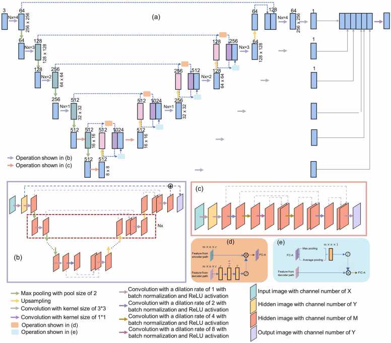
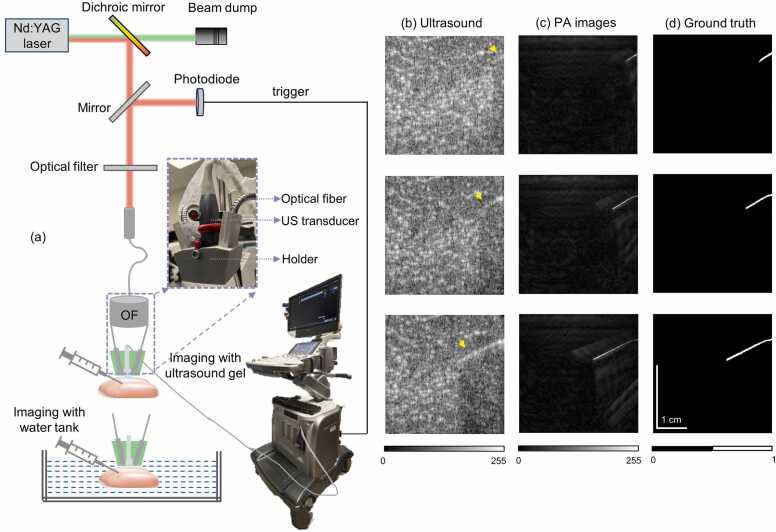
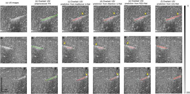
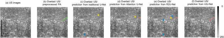
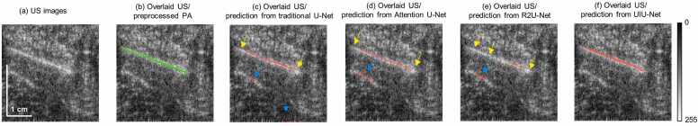
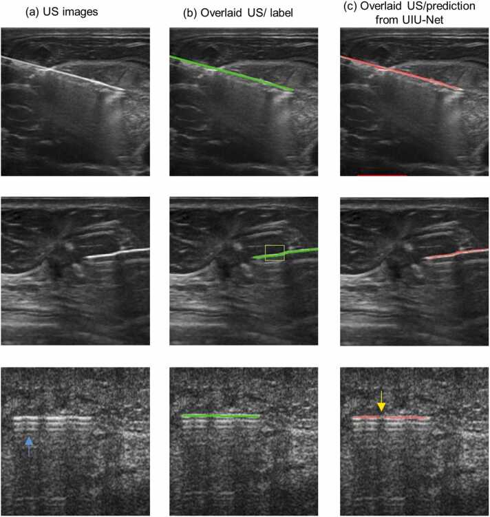
Accurate needle guidance is crucial for safe and effective clinical diagnosis and treatment procedures. Conventional ultrasound (US)-guided needle insertion often encounters challenges in consistency and precisely visualizing the needle, necessitating the development of reliable methods to track the needle. As a powerful tool in image processing, deep learning has shown promise for enhancing needle visibility in US images, although its dependence on manual annotation or simulated data as ground truth can lead to potential bias or difficulties in generalizing to real US images. Photoacoustic (PA) imaging has demonstrated its capability for high-contrast needle visualization. In this study, we explore the potential of PA imaging as a reliable ground truth for deep learning network training without the need for expert annotation. Our network (UIU-Net), trained on ex vivo tissue image datasets, has shown remarkable precision in localizing needles within US images. The evaluation of needle segmentation performance extends across previously unseen ex vivo data and in vivo human data (collected from an open-source data repository). Specifically, for human data, the Modified Hausdorff Distance (MHD) value stands at approximately 3.73, and the targeting error value is around 2.03, indicating the strong similarity and small needle orientation deviation between the predicted needle and actual needle location. A key advantage of our method is its applicability beyond US images captured from specific imaging systems, extending to images from other US imaging systems.
Keywords: Deep learning; Needle tracking; Photoacoustic imaging; Ultrasound imaging.
© 2023 The Authors.
Conflict of interest statement
The authors declare that they have no known competing financial interests or personal relationships that could have appeared to influence the work reported in this paper.
Figures
Similar articles
-
Improving needle visibility in LED-based photoacoustic imaging using deep learning with semi-synthetic datasets.Photoacoustics. 2022 Apr 7;26:100351. doi: 10.1016/j.pacs.2022.100351. eCollection 2022 Jun. Photoacoustics. 2022. PMID: 35495095 Free PMC article.
-
Multi-needle Localization with Attention U-Net in US-guided HDR Prostate Brachytherapy.Med Phys. 2020 Jul;47(7):2735-2745. doi: 10.1002/mp.14128. Epub 2020 Apr 3. Med Phys. 2020. PMID: 32155666 Free PMC article.
-
Automatic prostate segmentation using deep learning on clinically diverse 3D transrectal ultrasound images.Med Phys. 2020 Jun;47(6):2413-2426. doi: 10.1002/mp.14134. Epub 2020 Apr 8. Med Phys. 2020. PMID: 32166768
-
Handheld interventional ultrasound/photoacoustic puncture needle navigation based on deep learning segmentation.Biomed Opt Express. 2023 Oct 26;14(11):5979-5993. doi: 10.1364/BOE.504999. eCollection 2023 Nov 1. Biomed Opt Express. 2023. PMID: 38021141 Free PMC article.
-
Enhancement of needle visualization and localization in ultrasound.Int J Comput Assist Radiol Surg. 2021 Jan;16(1):169-178. doi: 10.1007/s11548-020-02227-7. Epub 2020 Sep 30. Int J Comput Assist Radiol Surg. 2021. PMID: 32995981 Review.
Cited by
-
MSD-Net: Multi-scale dense convolutional neural network for photoacoustic image reconstruction with sparse data.Photoacoustics. 2024 Dec 12;41:100679. doi: 10.1016/j.pacs.2024.100679. eCollection 2025 Feb. Photoacoustics. 2024. PMID: 39802237 Free PMC article.
-
A water cushion improved needle visualization by artificial beam steering during ultrasound-guided in-plane technique: an in vitro simulation study.Quant Imaging Med Surg. 2025 May 1;15(5):4791-4795. doi: 10.21037/qims-24-1995. Epub 2025 Apr 14. Quant Imaging Med Surg. 2025. PMID: 40384640 Free PMC article.
-
Needle Tip Tracking through Photoluminescence for Minimally Invasive Surgery.Biosensors (Basel). 2024 Sep 30;14(10):470. doi: 10.3390/bios14100470. Biosensors (Basel). 2024. PMID: 39451683 Free PMC article.
References
-
- Heslin M.J., Lewis J.J., Woodruff J.M., Brennan M.F. Core needle biopsy for diagnosis of extremity soft tissue sarcoma. Ann. Surg. Oncol. 1997;4:425–431. - PubMed
-
- Amedee R.G., Dhurandhar N.R. Fine‐needle aspiration biopsy. Laryngoscope. 2001;111(9):1551–1557. - PubMed
-
- Chapman G.A., Johnson D., Bodenham A.R. Visualisation of needle position using ultrasonography. Anaesthesia. 2006;61:148–158. - PubMed
-
- Fischer G.S., Deguet A., Csoma C., Taylor R.H., Fayad L., Carrino J.A., Zinreich S.J., Fichtinger G. MRI image overlay: application to arthrography needle insertion. Comput. Aided Surg. 2007;12(1):2–14. - PubMed
-
- Orebaugh S.L., McFadden K., Skorupan H., Bigeleisen P.E. Subepineurial injection in ultrasound-guided interscalene needle tip placement. Reg. Anesth. Pain. Med. 2010;35(5):450–454. 450-454. - PubMed
LinkOut - more resources
Full Text Sources
Research Materials
