. 2023 Dec 9;4(1):oead129.
doi: 10.1093/ehjopen/oead129. eCollection 2024 Jan.

Pericyte loss initiates microvascular dysfunction in the development of diastolic dysfunction

Affiliations

Pericyte loss initiates microvascular dysfunction in the development of diastolic dysfunction

Steven J Simmonds et al. Eur Heart J Open. .

Abstract

Aims: Microvascular dysfunction has been proposed to drive heart failure with preserved ejection fraction (HFpEF), but the initiating molecular and cellular events are largely unknown. Our objective was to determine when microvascular alterations in HFpEF begin, how they contribute to disease progression, and how pericyte dysfunction plays a role herein.

Methods and results: Microvascular dysfunction, characterized by inflammatory activation, loss of junctional barrier function, and altered pericyte-endothelial crosstalk, was assessed with respect to the development of cardiac dysfunction, in the Zucker fatty and spontaneously hypertensive (ZSF1) obese rat model of HFpEF at three time points: 6, 14, and 21 weeks of age. Pericyte loss was the earliest and strongest microvascular change, occurring before prominent echocardiographic signs of diastolic dysfunction were present. Pericytes were shown to be less proliferative and had a disrupted morphology at 14 weeks in the obese ZSF1 animals, who also exhibited an increased capillary luminal diameter and disrupted endothelial junctions. Microvascular dysfunction was also studied in a mouse model of chronic reduction in capillary pericyte coverage (PDGF-Bret/ret), which spontaneously developed many aspects of diastolic dysfunction. Pericytes exposed to oxidative stress in vitro showed downregulation of cell cycle-associated pathways and induced a pro-inflammatory state in endothelial cells upon co-culture.

Conclusion: We propose pericytes are important for maintaining endothelial cell function, where loss of pericytes enhances the reactivity of endothelial cells to inflammatory signals and promotes microvascular dysfunction, thereby accelerating the development of HFpEF.

Keywords: HFpEF; Metabolic comorbidities; Microvascular dysfunction; Pericytes.


Conflict of interest statement

Conflict of interest: None.

Figures

Graphical Abstract
Graphical Abstract
Figure 1
Figure 1
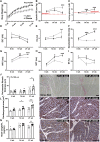
Metabolic risk factors develop early in obese ZSF1 rats, while many histological and echocardiographic signs of heart failure with preserved ejection fraction are not present until 21 weeks of age. Body weight (A; n = 11/group), systolic blood pressure (B, n = 7/group), and fasting glucose levels (C; n = 12/group) were analysed to establish when metabolic risk factors for heart failure with preserved ejection fraction develop in the ZSF1 rat model. E/E′ ratio (D), E/A ratio (E), deceleration time (F), isovolumetric relaxation time (G), non-flow time (H), and ejection fraction (I) were used to establish when heart failure with preserved ejection fraction could be detected by echocardiography in lean (n = 9) and obese (n = 11) ZSF1 rats. Cardiac Sirius Red staining to assess perivascular fibrosis in large (76–100 µm) vessels with representative image at 21 weeks (J and K; n = 7/group). Cardiac laminin staining was used to measure cardiomyocyte cross-sectional area with representative image at 14 and 21 weeks (L and M; n = 7/group). The heart weight to TL in obese and lean ZSF1 rats was also evaluated as a sign of hypertrophy (N; n = 7/group). Scale bars are 100 μm (K and M) and apply to all images within a panel. Values are presented as mean ± SEM. Significance is assessed by a non-paired two-way ANOVA followed by Sidak’s multiple comparisons test with *P < 0.05, **P < 0.01, and ***P < 0.001. A, late mitral inflow peak velocity; E, early mitral inflow peak velocity; E′, early diastolic annulus peak velocity; EF, ejection fraction; IVRT, isovolumetric relaxation time; NFT, non-flow time; TL, tibia length.
Figure 2
Figure 2
Pericyte loss is present in ZSF1 rats before diastolic dysfunction develops. Quantification of vascular density, based on VE-Cadherin staining indicates that vascular density is only decreased at the 21-week stage in ZSF1 obese rats as compared with lean ZSF1 rats (A; n = 7/group). Pericyte coverage, assessed by NG2+ cells localized to IB4+ capillary-sized vessels, was reduced from the 14-week stage onwards (B and C; n = 7/group). Representative images are shown at 14 and 21 weeks. Average number of capillary vessels per cardiomyocyte (CM) was calculated by multiplying the capillary density by the average cardiomyocyte size per heart (D; n = 7/group).
Figure 3
Figure 3
Fourteen-week-old obese ZSF1 rats show a reduction in pericyte proliferation, altered pericyte morphology, increased capillary luminal diameter, and endothelial junctional remodelling. Proliferation of cardiac pericytes and endothelial cells in ZSF1 rat hearts at 14 (top) and 21 (bottom) weeks was assessed by Ki67 staining (A; n = 6–7/group) and representative images are shown [B]. Representative transmission electron microscopy images of the endothelial cell—pericyte interface in capillaries of ZSF1 rat hearts at 14 weeks (C; n = 3/group). The endothelial cell layer is shown in pink, red blood cells in red, orange arrowheads indicate pericyte body, and blue arrowheads indicate pericyte extensions. Capillary diameter was measured on isolectinB4+ capillary-sized vessels in ZSF1 rat hearts at 14 weeks (D; n = 6–7/group) and representative images are shown (E; blue arrowheads indicate capillary lumen). Endothelial junctional remodelling was assessed by VE-Cadherin staining and blindly scored whether junctions were jagged, mixed, or straight at all stages in both lean and obese ZSF1 rats (F and G; n = 3–4/group, grey indicates lean and black indicates obese ZSF1 rats, and blue arrowheads indicate area of jagged endothelium). Quantification and representative transmission electron microscopy images of the endothelial cell junctions in ZSF1 rat hearts at 14 weeks (H and I; n = 3/group; blue arrowheads indicate endothelial junction). Scale bars are 30 µm (B, E, and G), 500 nm (C), and 200 nm (I) and apply to all images within a panel. Values are presented as mean ± SEM. Significance is assessed by an unpaired Student’s t-test (A, D, and H) or by a Pearson’s chi-squared test (F) with *P < 0.05, **P < 0.01, and ***P < 0.001. NG2, neural/glial antigen 2; IsoB4, isolectinB4.
Figure 4
Figure 4
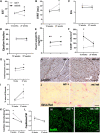
Twenty-seven-week-old female PDGF-Bret/ret mice show echocardiographic signs of diastolic dysfunction and cardiac fibrosis. Diastolic function was assessed using E/E′ ratio (A), isovolumetric relaxation time (B), E/A (C) while normal systolic function is shown with the preserved ejection fraction (D). The heart weight to tibia length was evaluated as a sign of hypertrophy (E). Cardiac CD45 staining was used to measure immune cell infiltration (F). Cardiac laminin staining was used to measure cardiomyocyte cross-sectional area (G and H). Cardiac Sirius Red staining was used to assess the presence of total fibrosis (I and J). Capillary density of PDGF-Bret/ret 12-week- and 27-week-old mice was assessed via isolectinB4 staining (K and M) and also normalized to cardiomyocyte size to give the average number of capillaries per cardiomyocyte (L). Representative images are at 27 weeks. Scale bars are 30 μm (H and M) and 50 µm (J) and apply to all images within a panel. n = 3 ret/+ and 5 ret/ret at 12 weeks and n = 9 ret/+ and 9 ret/ret at 27 weeks, for PDGF-Bret/+ and PDGF-Bret/ret, respectively. Values are presented as mean ± SEM. Significance is assessed by a non-paired two-way analysis of variance followed by Sidak’s multiple comparisons test with *P < 0.05, **P < 0.01, and ***P < 0.001. CM, cardiomyocyte; E, early mitral inflow peak velocity; E′, early diastolic annulus peak velocity; EF, ejection fraction; HW heart weight; IsoB4, isolectinB4; IVRT, isovolumetric relaxation time; PDGF-B, platelet-derived growth factor B; TL, tibia length.
Figure 5
Figure 5
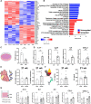
Pericytes exposed to oxidative stress are more inflammatory and make endothelial cells more reactive to inflammatory signals. Human pericytes were exposed to oxidative stress (100 µM H2O2) for 24 h. Four replicates per condition were processed for bulk RNA-seq followed by Reactome pathway analysis on the differentially expressed genes. Hierarchical clustering and heatmaps of upregulated and downregulated differentially expressed genes (2619 differentially expressed genes, adjusted P < 0.05) after oxidative stress (OX) vs. non-oxidative conditions (CTL) (A). The scale bar represents the gene expression difference between the maximum and minimum values for each gene by z-score. Graphs represent the top 10 enriched pathways associated with downregulated (blue) and upregulated (red) genes in oxidative stress-treated pericytes vs. controls (B). Multiple cell cycle pathways were overrepresented in genes downregulated by oxidative stress vs. controls, whereas p53-mediated cell cycle arrest, ER stress, and interleukins pathways were enriched in oxidative stress-upregulated genes. Pericytes treated with oxidative stress for 24 h show reduced proliferation as assessed by a Ki67 staining (C). Pericytes treated with oxidative stress for 24 h upregulate the expression of interleukin-1 alpha, interleukin-1 beta, interleukin 6, and C-X-C motif chemokine ligand 2 (B, n = 8) (D). Quantitative polymerase chain reaction on the hearts of 21-week-old obese and lean ZSF1 rats for interleukin-1 beta and interleukin 6 (E). Detection of circulating levels of interleukin-1 beta and interleukin 6 in plasma of lean and obese ZSF1 rats at 21 weeks by ELISA (F). Oxidative stress-treated and control pericytes were co-cultured with endothelial cells for 2 days and then treated with TNFα (1 ng/mL for 8 h) (G). Low TNFα concentration was chosen such that they did not induce inflammation in control conditions. Endothelial cells cultured with oxidative stress-treated pericytes upregulated interleukin-1 alpha, interleukin 8, C-C motif chemokine ligand 2, and C-X-C motif chemokine ligand 1 (G, n = 8 per condition). Values are presented as mean ± SEM. Significance is assessed by an unpaired Student’s t-test (C–F) or a two-way analysis of variance (G) with Sidak’s post hoc test with *P < 0.05, **P < 0.01, and ***P < 0.001. CCL2, C-C motif chemokine ligand 2; CXCL1, C-X-C motif chemokine ligand 1; CXCL2, C-X-C motif chemokine ligand 2; EC, endothelial cell; IL1A, interleukin-1 alpha; IL1B, interleukin-1 beta; IL6, interleukin 6; IL8, interleukin 8; OX, oxidative stress; PC, pericyte.

References

    1. Borlaug BA, Paulus WJ. Heart failure with preserved ejection fraction: pathophysiology, diagnosis, and treatment. Eur Heart J 2011;32:670–679. - PMC - PubMed
    1. Paulus WJ, Tschope C. A novel paradigm for heart failure with preserved ejection fraction: comorbidities drive myocardial dysfunction and remodeling through coronary microvascular endothelial inflammation. J Am Coll Cardiol 2013;62:263–271. - PubMed
    1. Sinha A, Rahman H, Webb A, Shah AM, Perera D. Untangling the pathophysiologic link between coronary microvascular dysfunction and heart failure with preserved ejection fraction. Eur Heart J 2021;42:4431–4441. - PMC - PubMed
    1. Rush CJ, Berry C, Oldroyd KG, Rocchiccioli JP, Lindsay MM, Touyz RM, Murphy CL, Ford TJ, Sidik N, McEntegart MB, Lang NN, Jhund PS, Campbell RT, McMurray JJV, Petrie MC. Prevalence of coronary artery disease and coronary microvascular dysfunction in patients with heart failure with preserved ejection fraction. JAMA Cardiol 2021;6:1130–1143. - PMC - PubMed
    1. Ahmad A, Corban MT, Toya T, Verbrugge FH, Sara JD, Lerman LO, Borlaug BA, Lerman A. Coronary microvascular dysfunction is associated with exertional haemodynamic abnormalities in patients with heart failure with preserved ejection fraction. Eur J Heart Fail 2021;23:765–772. - PubMed