. 2024 Jan 23;98(1):e0161823.
doi: 10.1128/jvi.01618-23. Epub 2024 Jan 4.

Within-host evolutionary dynamics and tissue compartmentalization during acute SARS-CoV-2 infection

Affiliations

Within-host evolutionary dynamics and tissue compartmentalization during acute SARS-CoV-2 infection

Mireille Farjo et al. J Virol. .

Abstract

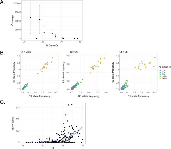
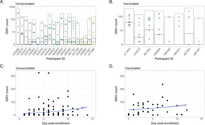
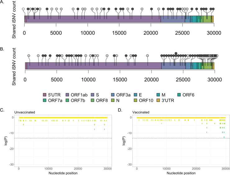
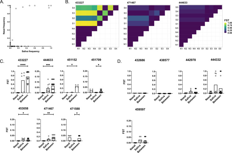
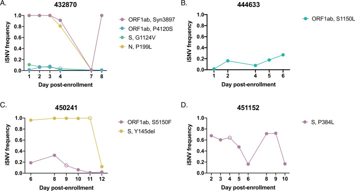
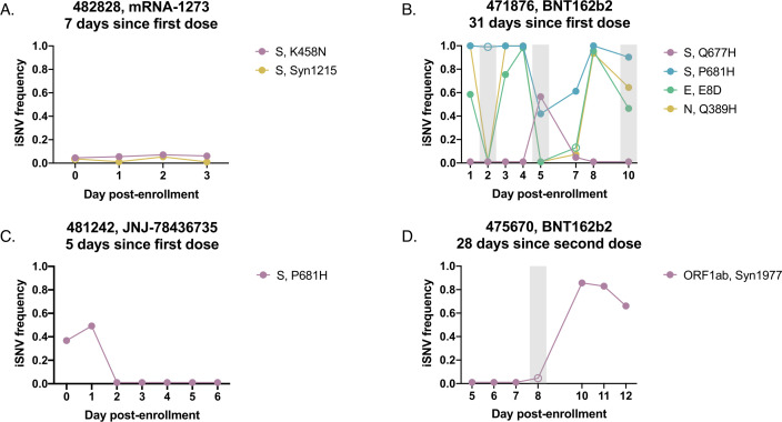
The global evolution of SARS-CoV-2 depends in part upon the evolutionary dynamics within individual hosts with varying immune histories. To characterize the within-host evolution of acute SARS-CoV-2 infection, we sequenced saliva and nasal samples collected daily from vaccinated and unvaccinated individuals early during infection. We show that longitudinal sampling facilitates high-confidence genetic variant detection and reveals evolutionary dynamics missed by less-frequent sampling strategies. Within-host dynamics in both unvaccinated and vaccinated individuals appeared largely stochastic; however, in rare cases, minor genetic variants emerged to frequencies sufficient for forward transmission. Finally, we detected significant genetic compartmentalization of viral variants between saliva and nasal swab sample sites in many individuals. Altogether, these data provide a high-resolution profile of within-host SARS-CoV-2 evolutionary dynamics.IMPORTANCEWe detail the within-host evolutionary dynamics of SARS-CoV-2 during acute infection in 31 individuals using daily longitudinal sampling. We characterized patterns of mutational accumulation for unvaccinated and vaccinated individuals, and observed that temporal variant dynamics in both groups were largely stochastic. Comparison of paired nasal and saliva samples also revealed significant genetic compartmentalization between tissue environments in multiple individuals. Our results demonstrate how selection, genetic drift, and spatial compartmentalization all play important roles in shaping the within-host evolution of SARS-CoV-2 populations during acute infection.

Keywords: SARS-CoV-2; genetic compartmentalization; viruses; within-host evolution.


Conflict of interest statement

The authors declare no conflict of interest.

Figures

Fig 1
Fig 1
Relationship between saliva sample Ct values and sequence quality. (A) Linear regression between Ct values of nucleocapsid (N) gene and mean sequence coverage depth. Error bars represent standard deviation. (B) Frequencies of characteristic B.1.1.7 SNPs at Ct values of 23.6, 26, and 28. B.1.1.7 RNA was spiked into B.1.2 RNA at final percentages ranging from 1% to 50% and divided between two replicates (R1 and R2). (C) Relationship between Ct of the SARS-CoV-2 nucleocapsid (N) gene and total iSNV count of the associated sample (Poisson regression, regression coefficient = 0.321, P < 0.001).
Fig 2
Fig 2
Intra-host single nucleotide variant (iSNV) diversity compared between samples and individuals. (A) Total iSNV counts for each sample from each unvaccinated participant. Light gray boxes indicate total discrete iSNV count for all samples, and horizontal black lines indicate number of shared iSNVs for each participant. (B) iSNV counts for vaccinated participants. (C) iSNV counts for individual samples from unvaccinated participants as a function of number of days post enrollment (Poisson regression, reg. coef. = 0.117, P < 0.001). (D) iSNV counts for individual samples from vaccinated participants as a function of number of days post enrollment (Poisson regression, reg. coef. = 0.0546, P < 0.001).
Fig 3
Fig 3
Locations of shared iSNVs across the SARS-CoV-2 genome. Genome locations of shared iSNVs detected in unvaccinated (A) and vaccinated (B) participants. Number of dots at a locus indicates number of participants in which the shared iSNV was detected. Light gray dots indicate synonymous mutations, dark gray dots indicate nonsynonymous mutations, and white dots indicate UTR mutations. (C) Log(P-values) for shared iSNV counts within 100-nt genomic windows, based on a Poisson distribution derived from the average shared iSNV count for all genomic windows. Plot shows shared iSNV counts in unvaccinated individuals. Dashed line marks significance threshold of 1.68e-06. (D) Log(P-values) for shared iSNV counts within 100-nt genomic windows, in vaccinated individuals.
Fig 4
Fig 4
Quantification of genetic compartmentalization of virus between sample sites. (A) Comparison of iSNV frequencies between matched samples in saliva and nasal environments (Pearson correlation, r = 0.615, P < 0.001). (B) Representative heatmaps exemplifying compartmentalization. Maps show FST values between pairs of samples from nasal (“N”) and/or saliva (“S”) environments (numbered by order of sampling). (C) Participants exhibiting compartmentalization (one set of within-environment FST values is lower than between-environment FST values). (D) Participants exhibiting no significant compartmentalization (neither set of within-environment FST values is lower than between-environment FST values). Scales range from 0 to 1 or 0 to 0.2 depending on the spread of the data. Asterisks indicate levels of significance (*P < .05, **P < .01, ***P < .001, ****P < .0001). P-values are derived from Monte Carlo permutation tests.
Fig 5
Fig 5
iSNV dynamics over time in saliva from unvaccinated individuals. Frequency tracking of selected iSNVs from unvaccinated participants (A) 432870, (B) 444633, (C) 450241, and (D) 451152. Unfilled points mark iSNVs with read depths below the threshold of 1,000 reads.
Fig 6
Fig 6
iSNV dynamics over time in saliva from vaccinated individuals. Frequency tracking of selected iSNVs from vaccinated participants (A) 482828 (newly vaccinated), (B) 471876 (partially vaccinated), (C) 481242 (newly vaccinated), and (D) 475670 (fully vaccinated). Unfilled points mark iSNVs with read depths below the threshold of 1,000 reads. Gray boxes mark samples with mean per-nucleotide coverages below 1,000 reads. Panel headings indicate vaccine received and time between enrollment and last vaccine dose.

References

    1. Baang JH, Smith C, Mirabelli C, Valesano AL, Manthei DM, Bachman MA, Wobus CE, Adams M, Washer L, Martin ET, Lauring AS. 2021. Prolonged severe acute respiratory syndrome Coronavirus 2 replication in an immunocompromised patient. J Infect Dis 223:23–27. doi:10.1093/infdis/jiaa666 - DOI - PMC - PubMed
    1. Corey L, Beyrer C, Cohen MS, Michael NL, Bedford T, Rolland M. 2021. SARS-CoV-2 variants in patients with immunosuppression. N Engl J Med 385:562–566. doi:10.1056/NEJMsb2104756 - DOI - PMC - PubMed
    1. Avanzato VA, Matson MJ, Seifert SN, Pryce R, Williamson BN, Anzick SL, Barbian K, Judson SD, Fischer ER, Martens C, Bowden TA, de Wit E, Riedo FX, Munster VJ. 2020. Case study: prolonged infectious SARS-CoV-2 shedding from an asymptomatic immunocompromised individual with cancer. Cell 183:1901–1912. doi:10.1016/j.cell.2020.10.049 - DOI - PMC - PubMed
    1. Kemp SA, Collier DA, Datir RP, Ferreira I, Gayed S, Jahun A, Hosmillo M, Rees-Spear C, Mlcochova P, Lumb IU, et al. . 2021. SARS-CoV-2 evolution during treatment of chronic infection. Nature 592:277–282. doi:10.1038/s41586-021-03291-y - DOI - PMC - PubMed
    1. Choi B, Choudhary MC, Regan J, Sparks JA, Padera RF, Qiu X, Solomon IH, Kuo H-H, Boucau J, Bowman K, et al. . 2020. Persistence and evolution of SARS-CoV-2 in an immunocompromised host. N Engl J Med 383:2291–2293. doi:10.1056/NEJMc2031364 - DOI - PMC - PubMed

Publication types

LinkOut - more resources