. 2024 Jan 4;147(1):8.
doi: 10.1007/s00401-023-02658-x.

Cortical matrix remodeling as a hallmark of relapsing-remitting neuroinflammation in MR elastography and quantitative MRI

Affiliations

Cortical matrix remodeling as a hallmark of relapsing-remitting neuroinflammation in MR elastography and quantitative MRI

Rafaela V Silva et al. Acta Neuropathol. .

Abstract

Multiple sclerosis (MS) is a chronic neuroinflammatory disease that involves both white and gray matter. Although gray matter damage is a major contributor to disability in MS patients, conventional clinical magnetic resonance imaging (MRI) fails to accurately detect gray matter pathology and establish a clear correlation with clinical symptoms. Using magnetic resonance elastography (MRE), we previously reported global brain softening in MS and experimental autoimmune encephalomyelitis (EAE). However, it needs to be established if changes of the spatiotemporal patterns of brain tissue mechanics constitute a marker of neuroinflammation. Here, we use advanced multifrequency MRE with tomoelastography postprocessing to investigate longitudinal and regional inflammation-induced tissue changes in EAE and in a small group of MS patients. Surprisingly, we found reversible softening in synchrony with the EAE disease course predominantly in the cortex of the mouse brain. This cortical softening was associated neither with a shift of tissue water compartments as quantified by T2-mapping and diffusion-weighted MRI, nor with leukocyte infiltration as seen by histopathology. Instead, cortical softening correlated with transient structural remodeling of perineuronal nets (PNNs), which involved abnormal chondroitin sulfate expression and microgliosis. These mechanisms also appear to be critical in humans with MS, where tomoelastography for the first time demonstrated marked cortical softening. Taken together, our study shows that neuroinflammation (i) critically affects the integrity of PNNs in cortical brain tissue, in a reversible process that correlates with disease disability in EAE, (ii) reduces the mechanical integrity of brain tissue rather than leading to water accumulation, and (iii) shows similar spatial patterns in humans and mice. These results raise the prospect of leveraging MRE and quantitative MRI for MS staging and monitoring treatment in affected patients.

Keywords: Cerebral cortex; Experimental autoimmune encephalomyelitis; Magnetic resonance elastography; Multiple sclerosis; Perineuronal nets; Tomoelastography.


Figures

Fig. 1
Fig. 1
Experimental design and in vivo MRE in the EAE mouse model. a Technical setup. A customized animal holder inserted into a 7-T preclinical MRI scanner generates mechanical waves encoded by a single-shot spin-echo MRE sequence via a piezo actuator. The waves are transmitted into the mouse skull via a transducer rod. b Study design. Serial in vivo MRE imaging of EAE mice starts prior to disease induction (baseline scan, day − 7), followed by EAE induction with PLP139–151 and adjuvants (day 0) and imaging and tissue sampling at different time points of the disease course: pre-onset, onset, peak, remission, and relapse. c Wave displacement images from one slice of an EAE mouse are shown to illustrate the propagation of shear waves through the brain using frequencies of 1000, 1100, 1200, 1300, and 1400 Hz (thru-plane component). d Representative group-averaged stiffness maps (SWS, viridis colormap, n = 15 baseline, onset, peak; n = 14 pre-onset; n = 12 remission; n = 7 relapse) at different disease phases
Fig. 2
Fig. 2
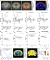
Stiffness measured in the cortical region reflects EAE progression more reliably than in other brain regions. a Segmentation of the brain into regions according to the Allen mouse brain atlas overlaid on group-averaged SWS maps (n = 15). b Effect map of percentage changes in SWS between peak and baseline shows softening throughout the brain including gray matter regions. cf Temporal changes in brain stiffness (SWS) in regions significantly affected during EAE (for detailed statistics see Supplementary Table 1); n = 15 baseline, onset, peak; n = 14 pre-onset; n = 12 remission; n = 7 relapse. gj Disability scores inversely correlate with regional stiffness in the cortex and caudoputamen, but not in hippocampus or deep gray matter; (n = 15). k A relapsing–remitting paradigm (left-hand side) used for pixel-wise correlation analysis identifies cortical tissue as the best fitting area for assessment of disease progression, as indicated by a high correlation coefficient (l) and low p values (m). n Compared with deep gray matter regions, the change in stiffness (ΔSWS) from baseline to peak is significantly greater in the cortex (n = 15). *P < 0.05, **P < 0.01, **P < 0.001, ****P < 0.0001
Fig. 3
Fig. 3
Detection of disseminated water biophysical properties by quantitative MRI. a Longitudinal variations in cortical T2 RT in ms (n = 9 onset; n = 8 baseline, pre-onset; n = 7 peak; n = 6 remission n = 5 relapse); b ADC in × 106 mm2/s (n = 12 baseline, pre-onset, peak; n = 11 remission; n = 6 relapse). **P < 0.01. c, d Effect maps of percentage changes in T2 RT and ADC from peak to baseline reveal a focal reduction in T2 RT and a disseminated reduction in ADC in the cortex. eh A relapsing–remitting paradigm applied to T2 RT and ADC maps demonstrates a lack of specificity for cortical alterations
Fig. 4
Fig. 4
Cortical inflammation occurs independently of leukocyte infiltration and is characterized by microgliosis. a HE stain shows lesions in the corpus callosum (arrowhead) and cortical parenchyma (upper panel) (scale bar 500 µm). b Quantification of cortical lesions indicates that disease activity is independent of magnitude leukocyte infiltration. c Cortical microgliosis is seen at peak EAE, based on higher Iba1+ cell counts (arrowheads), compared with a naive brain. d Quantification of microgliosis by Iba1+ cell counts; n = 7 naive, peak; n = 6 remission; n = 2 relapse *P < 0.05, ***P < 0.001; scale bar 100 µm. e Iba1 immunofluorescence displays distribution and morphological differences between healthy (naïve; white arrow) and EAE (white arrowhead); scale bar 50 µm
Fig. 5
Fig. 5
Remodeling of WFA+-PNNs in the cortex of EAE mice follows the course of the disease and is correlated with stiffness. a Representative immunofluorescence images of WFA+-PNNs in the mouse cortex. Remodeling of PNNs is seen as a decrease in green fluorescent signal (baseline: arrows; peak: arrowheads). b Quantification of WFA+ cortical PNNs in cross-sectional groups in the course of EAE shows a reduction during inflammatory phases, which is partially reversed during remission. c Cortical stiffness in cross-sectional groups of mice used for histological analysis shows similar behavior as seen in the longitudinal group; n = 8 baseline; n = 10 peak; n = 11 remission; n = 6 relapse. d WFA+-PNN remodeling correlates with the disease course. e, f Changes in cortical stiffness correlate with the disease course and WFA signal intensity; n = 10 peak; n = 11 remission; n = 6 relapse *P < 0.05, **P < 0.01, ***P < 0.001
Fig. 6
Fig. 6
Cortical softening as a signature of MS tissue remodeling. a Shear wave propagation through the human brain at 20, 25, 30, and 35 Hz (one central slice, thru-plane wave component). b Group-averaged SWS maps (rainbow color scale) of HCs (n = 14, left-hand side) and MS patients (n = 12, right) show visually apparent disseminated softening throughout the brain and marked effect sizes within cortical gray matter areas in the MS group. c Examples of whole brain (WB), cortical gray matter (CGM); and deep gray matter (DGM) masks. Group statistics of MS-related tissue softening in whole brain d, cortical gray matter e, and deep gray matter f; *P < 0.05, **P < 0.01
Fig. 7
Fig. 7
Overview of cortical microstructural remodeling behind transient softening during EAE. In homeostatic conditions, ECM molecules are constantly produced by neurons and glial cells. Cortical microglia are found in low numbers surveilling the tissue. As stiffness transiently drops in the inflammatory phases, a reversible neuroinflammatory process affecting tissue microstructure drives the mechanical changes. The proposed hypothesis involves the activation and proliferation of microglial cells in the cortex via interaction with meninges-derived soluble factors, i.e., cytokines, diffusing through the parenchyma. Activated microglia release matrix-degrading elements, such as proteases and reactive oxygen species (ROS), to induce matrix degradation as reflected by remodeled PNNs. In the recovery phase, lower inflammatory activity leads to a decrease in the number of microglia, allowing restoration of normal matrix and PNN composition. In this phase, the tissue microenvironment does not return to its original state, still displaying stiffness clearly distinct from healthy tissue. Thus, an interplay between PNN remodeling as well as overall matrix alterations and local cellular composition produces the observed transitory cortical softening, which can thus be used as a quantitative marker of neuroinflammation. Created with BioRender.com

Similar articles

Cited by

References

    1. Ashburner J, Friston KJ. Voxel-based morphometry—the methods. Neuroimage. 2000;11:805–821. doi: 10.1006/nimg.2000.0582. - DOI - PubMed
    1. Batzdorf CS, Morr AS, Bertalan G, Sack I, Silva RV, Infante-Duarte C. Sexual dimorphism in extracellular matrix composition and viscoelasticity of the healthy and inflamed mouse brain. Biology. 2022;11:230. doi: 10.3390/biology11020230. - DOI - PMC - PubMed
    1. Bertalan G, Becker J, Tzschätzsch H, Morr A, Herthum H, Shahryari M, Greenhalgh RD, Guo J, Schröder L, Alzheimer C, et al. Mechanical behavior of the hippocampus and corpus callosum: An attempt to reconcile ex vivo with in vivo and micro with macro properties. J Mech Behav Biomed Mater. 2023;138:105613. doi: 10.1016/j.jmbbm.2022.105613. - DOI - PubMed
    1. Bertalan G, Guo J, Tzschätzsch H, Klein C, Barnhill E, Sack I, Braun J. Fast tomoelastography of the mouse brain by multifrequency single-shot MR elastography. Magn Reson Med. 2019;81:2676–2687. doi: 10.1002/mrm.27586. - DOI - PubMed
    1. Bertalan G, Klein C, Schreyer S, Steiner B, Kreft B, Tzschätzsch H, de Schellenberger AA, Nieminen-Kelhä M, Braun J, Guo J, et al. Biomechanical properties of the hypoxic and dying brain quantified by magnetic resonance elastography. Acta Biomater. 2020;101:395–402. doi: 10.1016/j.actbio.2019.11.011. - DOI - PubMed

Publication types