. 2024 Jan 5;383(6678):101-108.
doi: 10.1126/science.adj3347. Epub 2024 Jan 4.

Molecular insights into atypical modes of β-arrestin interaction with seven transmembrane receptors

Affiliations

Molecular insights into atypical modes of β-arrestin interaction with seven transmembrane receptors

Jagannath Maharana et al. Science. .

Abstract

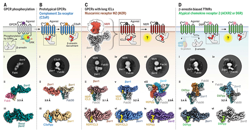
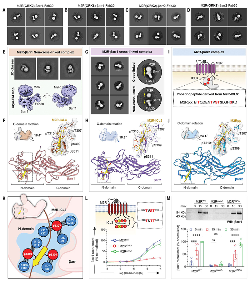
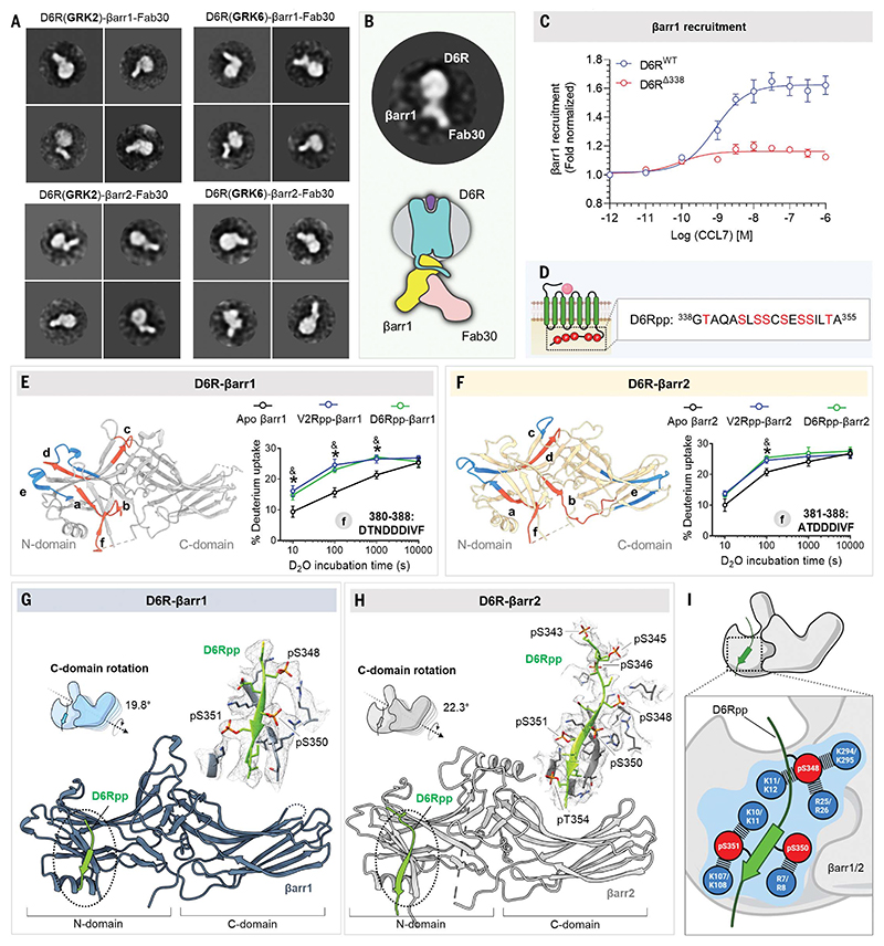
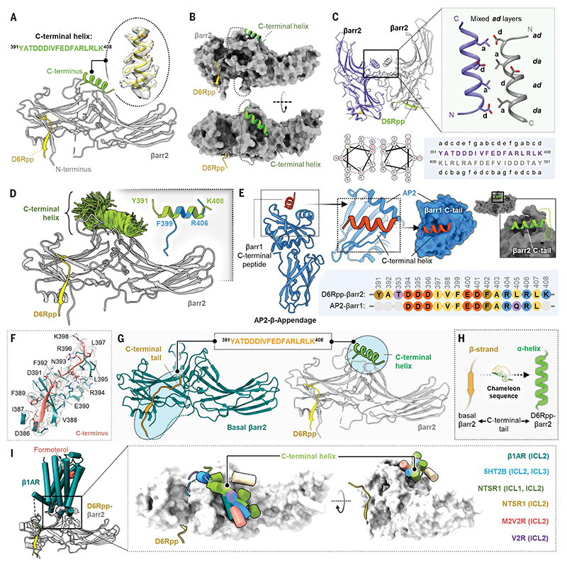
β-arrestins (βarrs) are multifunctional proteins involved in signaling and regulation of seven transmembrane receptors (7TMRs), and their interaction is driven primarily by agonist-induced receptor activation and phosphorylation. Here, we present seven cryo-electron microscopy structures of βarrs either in the basal state, activated by the muscarinic receptor subtype 2 (M2R) through its third intracellular loop, or activated by the βarr-biased decoy D6 receptor (D6R). Combined with biochemical, cellular, and biophysical experiments, these structural snapshots allow the visualization of atypical engagement of βarrs with 7TMRs and also reveal a structural transition in the carboxyl terminus of βarr2 from a β strand to an α helix upon activation by D6R. Our study provides previously unanticipated molecular insights into the structural and functional diversity encoded in 7TMR-βarr complexes with direct implications for exploring novel therapeutic avenues.


Conflict of interest statement

Competing interests: The authors declare that they have no competing interests.

Figures

Fig. 1
Fig. 1. A structural approach to understand the atypical modes of βarr interaction with 7TMRs.
(A) Cryo-EM structure of full-length βarr2 sheds light on its basal-state conformation. 2D class average (i), overall 3D map of βarr2 bound to Fab6 (ii), and structure of βarr2 alone (iii). (B) b-arrestins adopt two distinct modes of interaction with phosphorylated typical GPCRs. The phosphorylation pattern of complement receptor C3aR was used to delineate the “hanging” mode of βarr interaction. 2D class average (i), overall dimeric 3D map (ii), and structure of C3aRpp-βarr1 (iii). (C) A 3D reconstruction (left) showing a “hanging” mode of complex organization in M2R. High-resolution structures of M2R-ICL3–bound βarr1/2 are shown below. 2D class average (i), overall 3D map (ii), and structure of M2R-βarr1 (iii); M2R-βarr1 of cross-linked complex (iv, v, and vi); and M2Rpp-βarr2 (vii, viii, and ix). (D) 2D class average (i), overall dimeric 3D map (ii), and structure (iii) of D6Rpp-βarr1, and D6Rpp-βarr2 (iv, v, and vi). The estimated resolutions for all the structures are shown next to each map.
Fig. 2
Fig. 2. Structural insights into ICL3-driven βarr interaction with M2R.
(A to D) Negative-staining EM class averages of M2R, endogenously phosphorylated by GRK2/6 in complex with βarr1 or βarr2. (E) Cryo-EM 2D classes, 3D reconstruction of “hanging” M2R-βarr1-Fab30 complex. (F) Structure of βarr1 bound to phosphorylated M2R-ICL3. The EM density of ICL3 and surrounding residues within 4 Å are shown in the inset. βarr1 attains an active conformation with a C-domain rotation of 18.4° with respect to the N-domain. (G) Representative negative-staining EM 2D classes depicting the effect of cross-linking. Yellow arrows show potential transition of the complex subunits. (H) Structure of the crosslinked M2R-βarr1 complex. The EM density of ICL3 and surrounding residues within 4 Å are shown in the inset. C-domain rotation value with respect to N-domain is 18.6°. (I) Sequence of phosphopeptide derived from the ICL3 of M2R. (J) Structure of M2Rpp-βarr2 in ribbon representation. M2Rpp is shown in yellow and βarr2 in blue. Density map of phosphopeptide and surrounding residues within 4Å are displayed to the right. βarr2 attains an active conformation with 23.4° rotation of C-domain upon activation with M2Rpp. (K) The phosphorylated residues from ICL3 making critical contacts with Lys and Arg residues of βarr1 (upper) and βarr2 (lower) are highlighted in blue. (L) Cartoon representation illustrating the presence of possible phosphorylation clusters in the ICL3 of M2R. Mutations of the two phosphor-motifs: TVST and TNTT were generated to assess the βarr recruitment measured by bystander NanoBiT assay (receptor+SmBiT-βarr1+LgBiT-CAAX). Substitution of phosphosites of TVST to AVAA leads to abrupt reduction in βarr recruitment, whereas TNTT to ANAA substitution maintains βarr recruitment, suggesting a critical role played by TVST on βarr recruitment to M2R (mean ± SEM; n = 3 independent experiments; normalized with respect to highest ligand concentration signal for M2RWT as 100%). (M) Role of TVST in βarr recruitment is further corroborated by coimmunoprecipitation assay. On carbachol stimulation, M2RAVAA showed drastic reduction in βarr1 recruitment. A representative blot and densitometry-based quantification are presented (mean ± SEM; n = 4 independent experiments; normalized with M2R 30-min stimulation condition signal as 100%; two-way analysis of variance, Tukey’s multiple comparisons test). The exact P values are as follows: M2RWT 0 min versus 15 min, P = 0.0006;M2RWT 0 min versus 30 min, P = <0.0001; M2RANAA 0 min versus 15 min, P = 0.0008;M2RANAA 0 min versus 30 min, P = <0.0001 (***P = 0.0001; ****P < 0.0001; ns, nonsignificant). Single-letter abbreviations for the amino acid residues are as follows: A, Ala; C, Cys; D, Asp; E, Glu; F, Phe; G, Gly; H, His; I, Ile; K, Lys; L, Leu; M, Met; N, Asn; P, Pro; Q, Gln; R, Arg; S, Ser; T, Thr; V, Val; W, Trp; and Y, Tyr.
Fig. 3
Fig. 3. Structural insights into D6R-βarr complex interaction and activation.
(A) Negative-staining EM 2D class averages of D6R-βarr1/2 complexes endogenously phosphorylated with GRK2/6. (B) A representative 2D class average highlighting the “hanging” mode of βarr1 interaction with the receptor. (C) Dose response curve for CCL7-induced βarr1 recruitment for the mentioned D6R constructs using NanoBiT assay (Receptor-SmBiT+LgBiT-βarr1) (mean ± SEM; n = 3 independent experiments; normalized with respect to the lowest ligand concentration signal as 1). (D) Design of selected phosphopeptide derived from the C terminus of D6R. (E and F) HDX-MS plots to show the potential of generated phosphopeptides from D6R to activate βarr1 and βarr2, respectively. Among regions (a to f) showing significant changes upon deuterium exchange, the fragment at the C terminus (f) has been demonstrated to show activation of βarrs upon D6Rpp binding. (G) Structure of D6Rpp-βarr1 complex in ribbon representation. The density map of D6Rpp and surrounding residues within 4 Å are shown to the left. C-domain rotation of βarr1 bound to D6Rpp is 19.8°. (H) Structure of D6Rpp-βarr2 complex in ribbon representation. The density map of D6Rpp and surrounding residues within 4 Å are shown in the inset. C-domain rotation of βarr2 bound to D6Rpp was calculated to be 22.3°. (I) The phosphorylation pattern from D6Rpp engages with a network of Lys and Arg residues present on the N-domains of βarrs. Residues highlighted with blue circles show the Lys and Arg residues in βarr1 (upper) and βarr2 (lower).
Fig. 4
Fig. 4. Discovery of a C-terminal helix in D6R-activated βarr2.
(A) Cartoon representation of βarr2 bound to D6R phosphopeptide. βarr2 and D6Rpp are presented in gray and yellow, respectively, and the sequence of the C-terminal helix is shown in the inset. (B) D6Rpp-βarr2 structure displayed in surface representation in two different views to highlight the pose of the helix. The C-terminal helix (green) and D6Rpp (yellow) are shown as ribbon diagrams. (C) Dimeric organization of D6Rpp-βarr2 structure shown in ribbon representation (top left). Formation of antiparallel coiled-coil by the C-terminal helix of βarr2 at the dimeric interface (top right) shown as cartoon representation. The antiparallel coiled-coil exhibits mixed ad layers. Helical wheel representation of the antiparallel coiled-coil shows Asp at position d of one helix, which forms a salt bridge with Arg at position g in the other helix (bottom left). Heptad helical representation of the antiparallel coiled-coil residues in the βarr2 sequence (bottom right). (D) MD simulations confirm stability of the distal C-terminal helix/βarr2 interface. Structural snapshots (one snapshot every 10 ns, 7 × 250 ns of simulation time) presented here are of the position of the C-tail during simulation. For each residue, frames where it assembles an α-helical conformation are colored green. Fragments of the C-terminal helix can spontaneously assemble an a-helical conformation (right corner, blue cartoon) in three out of four independent MD simulations (each 2 μs) which is overlayed with the crystallized C-tail for comparison (green cartoon). For each residue, frames where it assembles a helical conformation are colored green. Comparison of a spontaneously assembled helical conformation of the βarr2 C-tail (blue) with that present in the structure (gray). (E) Structure of AP2 b-appendage protein in complex with βarr1 C-terminal peptide (PDB ID 2IV8) is shown as cartoon representation (left). The βarr1 C-terminal peptide can be seen to adopt similar helical conformation as the C-terminal helix in the D6Rpp-bound βarr2 structure (right). The sequence alignment of the C-terminal stretches of βarr1 and βarr2 are shown in the inset. (F) Cryo-EM density map of the isolated C terminus of βarr2 and surrounding residues within 4Å. (G) The peptide stretch sequence (top) of the C-tail in basal βarr2 transforms into a helical conformation in the D6Rpp-bound state (highlighted in cyan circles). (H) The C-tail of βarr2 exhibits a chameleon-like property, adopting a helical conformation in the active state from a β strand in the basal state. (I) Ribbon representation of the β1AR-βarr1 structure superimposed with D6Rpp-βarr2 on βarrs (left) shows positioning of the C-terminal helix on the central crest of βarrs. Upon structural superimposition with all reported GPCR-βarr1 structures, ICL1/2/3 of various receptors reside on the central crest as a C-terminal helix on D6Rpp-βarr2 (right).

References

    1. Kang DS, Tian X, Benovic JL. Curr Opin Cell Biol. 2014;27:63–71. - PMC - PubMed
    1. Gurevich VV, Gurevich EV. Mol Cell Endocrinol. 2019;484:34–41. - PMC - PubMed
    1. Ahn S, Shenoy SK, Luttrell LM, Lefkowitz RJ. Cell. 2020;182:1362–1362.e1. - PubMed
    1. Maharana J, Banerjee R, Yadav MK, Sarma P, Shukla AK. Curr Opin Struct Biol. 2022;75:102406. - PMC - PubMed
    1. Ranjan R, Dwivedi H, Baidya M, Kumar M, Shukla AK. Trends Cell Biol. 2017;27:851–862. - PubMed

MeSH terms

Substances