. 2024 Feb:100:104952.
doi: 10.1016/j.ebiom.2023.104952. Epub 2024 Jan 4.

Lactobacillus acidophilus suppresses non-alcoholic fatty liver disease-associated hepatocellular carcinoma through producing valeric acid

Affiliations

Lactobacillus acidophilus suppresses non-alcoholic fatty liver disease-associated hepatocellular carcinoma through producing valeric acid

Harry Cheuk-Hay Lau et al. EBioMedicine. 2024 Feb.

Abstract

Background: Gut probiotic depletion is associated with non-alcoholic fatty liver disease-associated hepatocellular carcinoma (NAFLD-HCC). Here, we investigated the prophylactic potential of Lactobacillus acidophilus against NAFLD-HCC.

Methods: NAFLD-HCC conventional and germ-free mice were established by diethylnitrosamine (DEN) injection with feeding of high-fat high-cholesterol (HFHC) or choline-deficient high-fat (CDHF) diet. Orthotopic NAFLD-HCC allografts were established by intrahepatic injection of murine HCC cells with HFHC feeding. Metabolomic profiling was performed using liquid chromatography-mass spectrometry. Biological functions of L. acidophilus conditional medium (L.a CM) and metabolites were determined in NAFLD-HCC human cells and mouse organoids.

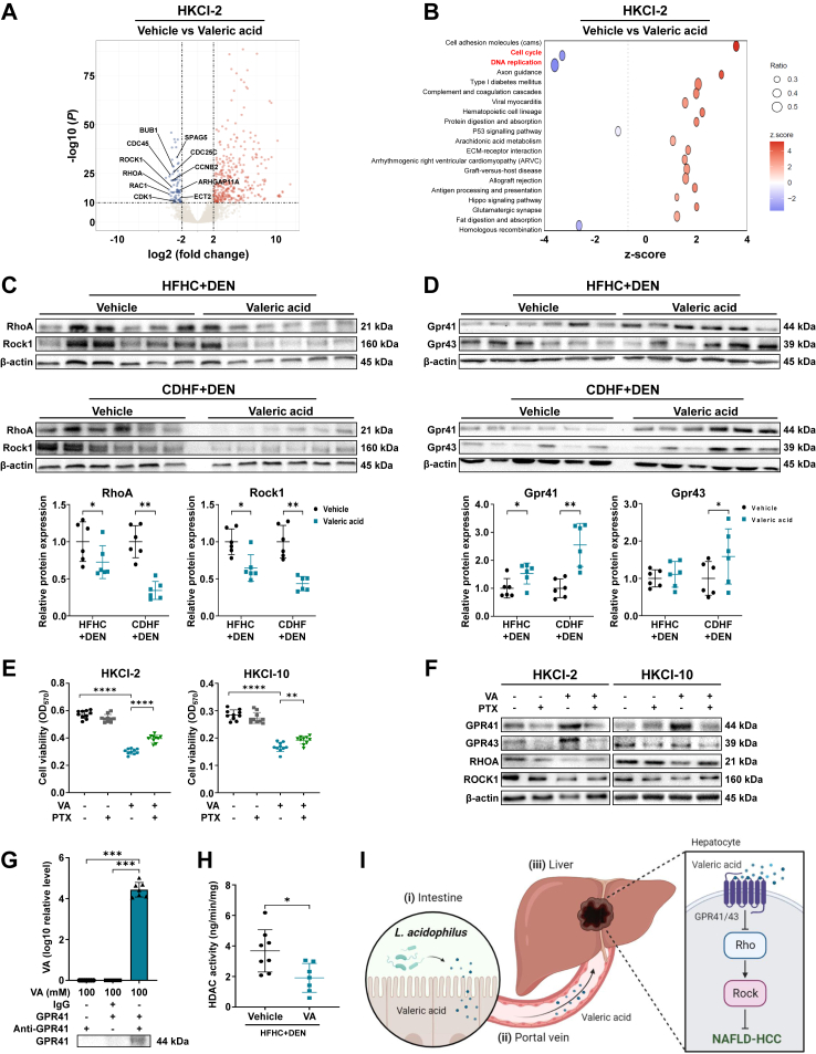
Findings: L. acidophilus supplementation suppressed NAFLD-HCC formation in HFHC-fed DEN-treated mice. This was confirmed in orthotopic allografts and germ-free tumourigenesis mice. L.a CM inhibited the growth of NAFLD-HCC human cells and mouse organoids. The protective function of L. acidophilus was attributed to its non-protein small molecules. By metabolomic profiling, valeric acid was the top enriched metabolite in L.a CM and its upregulation was verified in liver and portal vein of L. acidophilus-treated mice. The protective function of valeric acid was demonstrated in NAFLD-HCC human cells and mouse organoids. Valeric acid significantly suppressed NAFLD-HCC formation in HFHC-fed DEN-treated mice, accompanied by improved intestinal barrier integrity. This was confirmed in another NAFLD-HCC mouse model induced by CDHF diet and DEN. Mechanistically, valeric acid bound to hepatocytic surface receptor GPR41/43 to inhibit Rho-GTPase pathway, thereby ablating NAFLD-HCC.

Interpretation: L. acidophilus exhibits anti-tumourigenic effect in mice by secreting valeric acid. Probiotic supplementation is a potential prophylactic of NAFLD-HCC.

Funding: Shown in Acknowledgments.

Keywords: Cancer prevention; Lactobacillus acidophilus; Non-alcoholic fatty liver disease-associated hepatocellular carcinoma; Rho-GTPase pathway; Valeric acid.


Conflict of interest statement

Declaration of interests The authors disclose no conflicts of interest.

Figures

Fig. 1
Fig. 1
L. acidophilus suppresses NAFLD-HCC development in tumourigenesis mouse model. (A) Heatmap of faecal metagenomic sequencing on NAFLD-HCC mice showing differential microbes (arranged by relative abundance). Differential analysis was performed by comparing HFHC-fed mice (n = 10) to HFLC-fed mice (n = 8) using Wilcoxon rank-sum test. (B) Experimental schematic of HFHC-fed DEN-treated mice and representative images of liver tumours (10 mice per group). Mice without any treatment (NC) were compared to HFHC-fed DEN-treated mice with PBS control to confirm the establishment of NAFLD-HCC mouse model. (C) Measurement of tumour parameters. (D) Representative H&E images and histological scoring of non-tumour liver tissues. (E) Measurement of serum makers. (F) Representative Ki-67 images and scoring of liver tissues. HFLC, high-fat low-cholesterol; L.a, L. acidophilus; NC, normal chow; NT, non-tumour; T, tumour. ∗P < 0.05, ∗∗∗P < 0.001, ∗∗∗∗P < 0.0001.
Fig. 2
Fig. 2
L. acidophilus suppresses NAFLD-HCC development in orthotopic allografts and germ-free mice. (A) Experimental schematic of orthotopic NAFLD-HCC allografts and bioluminescent imaging of tumours (6 mice per group). (B) Macroscopic pictures and representative H&E images of liver tumours. (C) Measurement of tumour parameters. (D) Experimental schematic of HFHC-fed DEN-treated germ-free mice and representative images of liver tumours (PBS, n = 8; L.a, n = 10). (E) Measurement of tumour parameters. (F) Representative H&E images and histological scoring of non-tumour liver tissues. (G) Measurement of serum markers. (H) Representative Ki-67 images and scoring of liver tissues. L.a, L. acidophilus; NT, non-tumour; T, tumour. ∗P < 0.05, ∗∗P < 0.01, ∗∗∗P < 0.001.
Fig. 3
Fig. 3
L. acidophilus secretes non-protein small molecules against NAFLD-HCC. (A) Cell viability at OD570 of human NAFLD-HCC (HKCI-2, HKCI-10) or normal hepatocyte (MIHA) cell lines treated with bacterial conditional medium. (B) Representative images and size of mouse organoids after treatment. Scale bar = 100 μm. (C) Cell viability at OD570 after treatment of heated conditional medium. (D–H) Cell viability at OD570(D), colony formation assay (E), Ki-67 immunofluorescent staining with a scale bar of 25 μm (F), cell apoptosis assay (G), and cell cycle assay (H) after treatment of conditional medium with different molecular weights. (I) Western blot of protein markers related to cell proliferation and apoptosis. The used concentration of conditional medium was 7.5% in cell culture medium. P values in (A, C) were calculated by comparing L.a CM (A) or L.a CM <3 kDa (C) to every other groups using repeated measures two-way ANOVA.∗P < 0.05, ∗∗P < 0.01, ∗∗∗P < 0.001, ∗∗∗∗P < 0.0001.
Fig. 4
Fig. 4
Valeric acid is the major metabolite produced by L. acidophilus. (A) Principal component analysis on the metabolomic profile of bacterial conditional medium. (B) Heatmap of differentially enriched metabolites in L.a CM compared to broth control and E. coli CM. (C) Level of major SCFAs in bacterial conditional medium detected by targeted metabolomics. (D, E) Valeric acid level in liver and portal vein serum of HFHC-fed DEN-treated conventional (10 mice per group) (D) or germ-free (PBS, n = 8; L.a, n = 10) (E) mice. L.a, L. acidophilus; VA, valeric acid. ∗P < 0.05, ∗∗P < 0.01.
Fig. 5
Fig. 5
Valeric acid exhibits protective function against NAFLD-HCC in vitro. (A) Cell viability at OD570 of human NAFLD-HCC (HKCI-2, HKCI-10) or normal hepatocyte (MIHA) cell lines after treatment of valeric acid for 3 days. P values were calculated by comparing each valeric acid concentration to vehicle control (0 μM) using repeated measures two-way ANOVA. (B) Representative images and size of mouse organoids after treatment. Scale bar = 100 μm. (C–G) Colony formation assay (C), Ki-67 immunofluorescent staining with a scale bar of 25 μm (D), cell apoptosis assay (E), and cell cycle assay (F) after treatment. (G) Western blot of protein markers related to cell proliferation, apoptosis, and cell cycle. The used concentration of valeric acid was 500 μM. VA, valeric acid. ∗P < 0.05, ∗∗P < 0.01, ∗∗∗P < 0.001, ∗∗∗∗P < 0.0001.
Fig. 6
Fig. 6
Valeric acid suppresses NAFLD-HCC development in tumourigenesis mouse models. (A) Experimental schematic of HFHC-fed DEN-treated mice and representative images of liver tumours (10 mice per group). (B) Measurement of tumour parameters. (C) Representative H&E images and histological scoring of non-tumour liver tissues. (D) Measurement of serum markers. (E) Representative Ki-67 images and scoring of liver tissues. (F) Experimental schematic of CDHF-fed DEN-treated mice and representative images of liver tumours (PBS, n = 12; valeric acid, n = 10). (G) Measurement of tumour parameters. (H) Representative H&E images and histological scoring of non-tumour liver tissues. (I) Measurement of serum markers. (J) Representative Ki-67 images and scoring of liver tissues. NT, non-tumour; T, tumour; VA, valeric acid. ∗P < 0.05, ∗∗P < 0.01.
Fig. 7
Fig. 7
Valeric acid binds to hepatocytic surface receptor GPR41/43 to inhibit oncogenic Rho-GTPase signalling pathway. (A) Volcano plot of differential genes (log2 fold change ≥2, −log(P) ≥ 10) in HKCI-2 cell line with or without valeric acid treatment. Top depleted genes related to Rho-GTPase pathway are indicated. (B) Pathway analysis of differential genes. (C, D) Hepatic protein expression of markers related to Rho-GTPase pathway (C) or GPRs (D) in mice. (E) Cell viability at OD570 of human NAFLD-HCC (HKCI-2, HKCI-10) cell lines after treatment of valeric acid and/or PTX for 3 days. (F) Protein expression in cells treated with valeric acid and/or PTX. (G) Direct pull-down assay of valeric acid using GPR41 recombinant proteins. (H) Hepatic HDAC activity in HFHC-fed DEN-treated mice. (I) Overview schematic of the study (created by BioRender.com). The used concentration of valeric acid was 500 μM. VA, valeric acid. ∗P < 0.05, ∗∗P < 0.01, ∗∗∗P < 0.001, ∗∗∗∗P < 0.0001.

References

    1. Powell E.E., Wong V.W., Rinella M. Non-alcoholic fatty liver disease. Lancet. 2021;397:2212–2224. - PubMed
    1. Huang D.Q., El-Serag H.B., Loomba R. Global epidemiology of NAFLD-related HCC: trends, predictions, risk factors and prevention. Nat Rev Gastroenterol Hepatol. 2021;18:223–238. - PMC - PubMed
    1. Pfister D., Nunez N.G., Pinyol R., et al. NASH limits anti-tumour surveillance in immunotherapy-treated HCC. Nature. 2021;592:450–456. - PMC - PubMed
    1. Foerster F., Gairing S.J., Muller L., et al. NAFLD-driven HCC: safety and efficacy of current and emerging treatment options. J Hepatol. 2022;76:446–457. - PubMed
    1. Tripathi A., Debelius J., Brenner D.A., et al. The gut-liver axis and the intersection with the microbiome. Nat Rev Gastroenterol Hepatol. 2018;15:397–411. - PMC - PubMed

MeSH terms

LinkOut - more resources