. 2024 Jan 18;187(2):360-374.e19.
doi: 10.1016/j.cell.2023.11.031. Epub 2024 Jan 3.

Structural and functional basis of VLDLR usage by Eastern equine encephalitis virus

Affiliations

Structural and functional basis of VLDLR usage by Eastern equine encephalitis virus

Lucas J Adams et al. Cell. .

Abstract

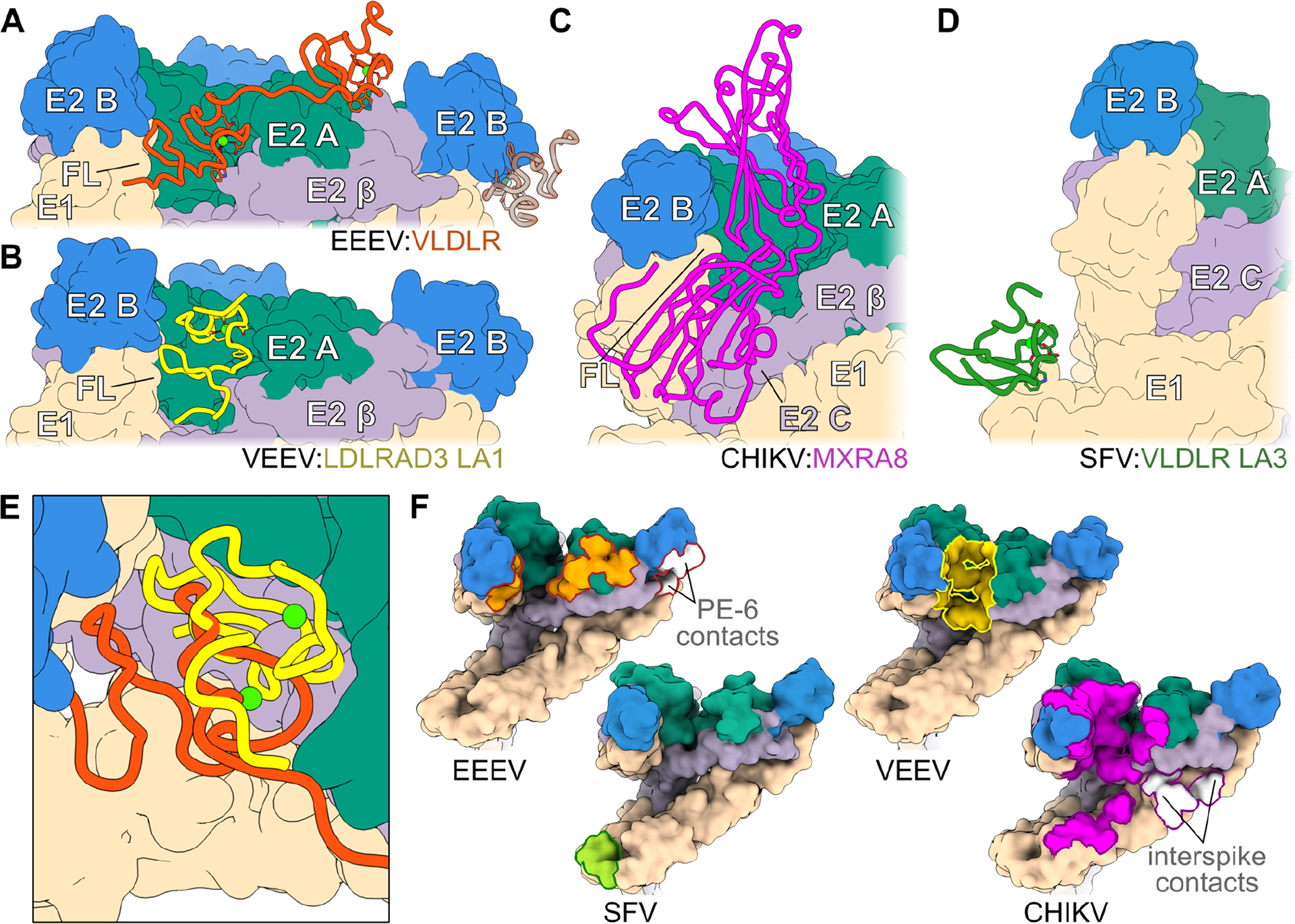
The very-low-density lipoprotein receptor (VLDLR) comprises eight LDLR type A (LA) domains and supports entry of distantly related alphaviruses, including Eastern equine encephalitis virus (EEEV) and Semliki Forest virus (SFV). Here, by resolving multiple cryo-electron microscopy structures of EEEV-VLDLR complexes and performing mutagenesis and functional studies, we show that EEEV uses multiple sites (E1/E2 cleft and E2 A domain) to engage more than one LA domain simultaneously. However, no single LA domain is necessary or sufficient to support efficient EEEV infection. Whereas all EEEV strains show conservation of two VLDLR-binding sites, the EEEV PE-6 strain and a few other EEE complex members feature a single amino acid substitution that enables binding of LA domains to an additional site on the E2 B domain. These structural and functional analyses informed the design of a minimal VLDLR decoy receptor that neutralizes EEEV infection and protects mice from lethal challenge.

Keywords: alphavirus; cryo-electron microscopy; encephalitis; mice; pathogenesis; receptor; therapeutic.


Conflict of interest statement

Declaration of interests M.S.D. is a consultant or advisor for Inbios, Ocugen, Vir Biotechnology, Topspin Therapeutics, Moderna, Merck, and Immunome. The Diamond laboratory has received funding support from Emergent BioSolutions, Moderna, and Vir Biotechnology. D.H.F. is a founder of Courier Therapeutics and has received funding support from Emergent BioSolutions and Mallinckrodt Pharmaceuticals.

Figures

Figure 1.
Figure 1.. Cryo-EM structure of EEEV PE–6 in complex with VLDLR.
(A) Icosahedral reconstructions of EEEV PE–6 VLP alone (upper panel) or in complex with full-length VLDLR (lower panel) with 2-fold (i2), 5-fold (i5), 3-fold (i3), and quasi-3-fold (q3) axes designated. Central sections are shown in round insets. Proteins are differentially colored with E1 in tan, E2 A domain in sea green, E2 B domain in blue, the remainder of E2 colored purple, and VLDLR shown in orange. (B) Focused reconstructions of the EEEV asymmetric unit alone (upper panel) or in complex with full-length VLDLR (lower panel), colored as in (A). (C) An atomic model of a single E1/E2 heterodimer with non-descript LA domains docked into the experimental electron density map, colored as in (A) with capsid shown in navy and the lipid bilayer depicted as dashed lines. Interfacial lysines are highlighted in yellow, with the region near K156 magnified in the inset. See also Fig S1 and S2.
Figure 2.
Figure 2.. No single LA domain of the VLDLR LBD is required to support EEEV.
(A) Scheme of LDLRAD3-LA1 domain replacement of VLDLR LA domains. (B, D, F, H) SINV-EEEV-GFP PE–6 infection of K562 cells transduced with indicated N-terminal Flag-tagged constructs as quantified by flow cytometry. (C) Scheme of N-terminal VLDLR LA domain truncation constructs. (E) Scheme of single LA domain constructs in the context of the VLDLRΔLBD backbone. (G) BLI sensorgrams of biosensors coated with indicated Fc-fusion proteins following incubation with EEEV PE–6 VLPs (left panel) or biosensors coated with EEEV PE–6 VLPs following incubation with Fc-fusion proteins in solution (right panel). (I) Scheme of VLDLR with Trp (W) to Ala (A) mutations in LA1, LA2, LA3, LA5, and LA6 (left) VLDLR (WA), and also VLDLR (WA) with LA4 (F171W), LA7 (R295W), LA8 (K336W) residues changed to Trp. (J-K) SINV-EEEV-GFP PE–6 infection of K562 cells transduced with variants of VLDLR (WA) in which the indicated single (J) or two (K) LA domain(s) has been reverted to Trp as indicated. (L) SINV-EEEV-GFP PE–6 infection of K562 cells transduced with the indicated tandem LA domain constructs in the context of the VLDLRΔLBD backbone. Data in (B, D, F, I-L) are pooled from two to six experiments. Data in G are representative of two experiments. *p < 0.05, ****p < 0.0001, n.s., not significant; one-way ANOVA with Dunnett’s post-test. See also Fig S3, S4, and S8.
Figure 3.
Figure 3.. Multiple LA domains mediate the neutralization of EEEV by VLDLR receptor decoys.
(A, C) Infection of 293T cells by SINV-EEEV-GFP PE–6 following pre-incubation with the indicated Fc-fusion proteins (10 μg/mL) prior to inoculation. GFP expression was measured by flow cytometry. (B, D) Dose-response curves of neutralization by Fc-fusion proteins against SINV-EEEV-GFP PE–6. Data in (A, C) are pooled from three to six experiments. **p < 0.01, **** p < 0.0001, n.s., not significant by one-way ANOVA with Dunnett’s post-test. Data in (B, D) are representative of three experiments with mean half-maximal effective inhibitory concentrations (EC50 values) calculated. (E–J) Steady-state BLI curves of the indicated monovalent VLDLR domains bound to EEEV PE–6 VLP coated biosensors. Data are pooled from three experiments. See also Fig S8.
Figure 4.
Figure 4.. Cryo-EM structure of EEEV PE–6 VLPs in complex with VLDLR LA(1–2).
(A) Focused reconstruction of the EEEV PE–6 asymmetric unit in complex with VLDLR-LA(1–2). Proteins are differentially colored: E1, tan; E2 A domain, sea green; E2 B domain, blue; remainder of E2, purple; and VLDLR, orange. (B) Individual E1/E2 heterodimers at the binding interface, illustrating conventional “wrapped” and “intraspike” contacts, colored as in (A)., (C) Ribbon diagram of VLDLR LA(1–2) (orange) overlaying a surface representation of neighboring E1/E2 heterodimers, colored as in (A). Domains of E2 are labeled, as is the fusion loop of E1. (D-F) Magnified regions from boxes in panel (C). Interface details between VLDLR LA1 and the E1 fusion loop (pale green) (D), LA1 and E2 (E), and LA2 and E2 (F). VLDLR residues, in white or orange; EEEV residues, in black. Predicted salt bridges (interatomic distance ≤ 4.0 Å) and cation-π interactions (≤ 6.0 Å from aromatic plane) are demarcated by white or yellow dashed lines, respectively. (G) Binding of LA(1–2)-Fc to captured wild-type (WT) and mutant EEEV FL93–939 VLPs. Biosensors were coated with WT or mutant VLPs followed by incubation with 1 μM of LA(1–2)-Fc for 300 sec. Binding was calculated as percent signal (Rmax) relative to WT VLPs. (H) Infection of K562 cells expressing WT and mutant constructs of VLDLR LA(1–2) by SINV-EEEV PE–6 as measured by flow cytometry. Data in G-H are pooled from two to four experiments. **p < 0.01, ****p < 0.0001, n.s., not significant by one-way ANOVA with Dunnett’s post-test. See also Fig S8, and Table S1 and S2.
Figure 5.
Figure 5.. Mapping of LA domain and EEEV E2 binding sites.
(A) Cryo-EM reconstructions of EEEV PE–6 or FL93–939 VLP in complex with different VLDLR fragments. VLDLR constructs include full-length VLDLR; LA(1–2); LA(1–3); LA(1–5mut3); LA(1–6mut3,5); LBDmut3,5,6; LA(3–8); LA(1–6mut2); and LA(1–6). EEEV E1, tan; E2 A domain, sea green; E2 B domain, blue; remainder of E2, purple; and VLDLR, orange. (B) Ribbon diagram depicting the interaction of VLDLR LA6 with EEEV E1 and E2, colored as in (A) with the fusion loop (FL) shown in pale green. Predicted salt bridges (interatomic distance ≤ 4.0 Å) are demarcated by white dashed lines. The fit of this model within the experiment density is pictured within the inset. (C, D, F) Infection of SINV-EEEV-GFP PE–6 WT and mutants in K562 cells expressing WT VLDLR as quantified by flow cytometry. (E) Dose-response neutralization (10, 1, and 0.1 μg/mL) of indicated Fc-fusion proteins against SINV-EEEV PE–6 HKR→AAA virus in 293T cells. Data in (C, D, F) are pooled from three to six experiments. Data in E are representative of two experiments. ***p < 0.001, ****p < 0.0001; one-way ANOVA with Dunnett’s post-test. See also Fig S5, S8–S10, and Tables S1–S3.
Figure 6.
Figure 6.. Comparative analysis of virus-receptor complexes.
(A-D) Structures of alphavirus-receptor complexes, with VLDLR (orange; strain-specific site shown as transparent), LDLRAD3 LA1 (yellow, PDB 7N1H), MXRA8 (magenta, PDB 6NK7), and VLDLR LA3 (green, PDB 8IHP) displayed as ribbon diagrams, overlaying surface representations of EEEV, VEEV, CHIKV, or SFV structural proteins. E1, tan; E2 A domain, sea green; E2 B domain, blue; and remainder of E2, pale purple. (E) Magnified top view showing overlay of VLDLR LA1 (orange) and LDLRAD3 LA1 (yellow) within the EEEV/VEEV receptor binding cleft. (F) Surface representations of neighboring E1/E2 heterodimers, colored as in (A-D), with receptor binding interfaces (determined by PISA) highlighted on their respective alphaviruses. See also Fig S6, S7, S9, and S10.
Figure 7.
Figure 7.. VLDLR LA(1–2)-Fc protects against EEEV FL93–939 challenge.
(A-C) Survival (A), weight change (B), and clinical scores (C) of CD-1 mice administered 100 μg of indicated Fc-fusion protein prior to subcutaneous challenge with EEEV FL93–939. The scoring system is described in STAR Methods. (D) Survival of mice treated as in (A) following aerosol EEEV FL93–939 challenge. Two experiments with n = 10 mice per group. (A) log-rank test with Bonferroni correction; (D) log-rank test: **p < 0.01, ****p < 0.0001.

Update of

References

    1. Levinson RS, Strauss JH, and Strauss EG (1990). Complete sequence of the genomic RNA of O’nyong-nyong virus and its use in the construction of alphavirus phylogenetic trees. Virology 175, 110–123. 10.1016/0042-6822(90)90191-s. - DOI - PubMed
    1. Strauss JH, and Strauss EG (1994). The alphaviruses: gene expression, replication, and evolution. Microbiol Rev 58, 491–562. 10.1128/mr.58.3.491-562.1994. - DOI - PMC - PubMed
    1. Suhrbier A, Jaffar-Bandjee M-C, and Gasque P (2012). Arthritogenic alphaviruses--an overview. Nat Rev Rheumatol 8, 420–429. 10.1038/nrrheum.2012.64. - DOI - PubMed
    1. Zacks MA, and Paessler S (2010). Encephalitic alphaviruses. Vet Microbiol 140, 281–286. 10.1016/j.vetmic.2009.08.023. - DOI - PMC - PubMed
    1. Silverman MA, Misasi J, Smole S, Feldman HA, Cohen AB, Santagata S, McManus M, and Ahmed AA (2013). Eastern equine encephalitis in children, Massachusetts and New Hampshire,USA, 1970–2010. Emerg Infect Dis 19, 194–201; quiz 352. 10.3201/eid1902.120039. - DOI - PMC - PubMed

LinkOut - more resources