Deep Circumflex Iliac Artery-vascularized Iliac Bone Graft for Femoral Head Osteonecrosis: Computed Tomography Anatomical Study
- PMID: 38176431
- PMCID: PMC11309803
- DOI: 10.1055/a-2238-7798
Deep Circumflex Iliac Artery-vascularized Iliac Bone Graft for Femoral Head Osteonecrosis: Computed Tomography Anatomical Study
Abstract
Background: Deep circumflex iliac artery (DCIA)-vascularized iliac graft transposition is a method for treating femoral head osteonecrosis but with inconsistent efficacy. We aim to improve the method of this surgery by recommending the optimal location of the iliac pedicle to satisfy the vascular length for transposition and the blood supply of the vascularized iliac graft.
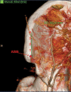
Methods: The DCIA and its surrounding tissues were assessed on computed tomography angiography images for 100 sides (left and right) of 50 patients. The length of the vascular pedicle required for transposition and the length of the pedicle at different iliac spine positions were compared. The diameter and cross-sectional area of the DCIA and the distance between the DCIA and iliac spine were measured at different points to assess blood supply. We also compared differences in sex and left-right position.
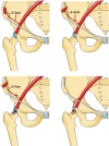
Results: The diameter and cross-sectional area of the DCIA gradually decreased after crossing the anterior superior iliac spine (ASIS), and it approached the iliac bone. However, when the DCIA was 4 cm behind the ASIS (54 sides, 54%), it coursed posteriorly and superiorly away from the iliac spine. The vascular length of the pedicle was insufficient to transpose the vascularized iliac graft to the desired position when it was within 1 cm of the ASIS. The vascular length requirement was satisfied, and the blood supply was sufficient when the pedicle was positioned at 2 or 3 cm.
Conclusion: To obtain a satisfactory pedicle length and sufficient blood supply, the DCIA pedicle of the vascularized iliac graft should be placed 2 to 3 cm behind the ASIS. The dissection of DCIA has slight differences in sex and left-right position due to anatomical differences.
The Author(s). This is an open access article published by Thieme under the terms of the Creative Commons Attribution-NonDerivative-NonCommercial License, permitting copying and reproduction so long as the original work is given appropriate credit. Contents may not be used for commercial purposes, or adapted, remixed, transformed or built upon. (https://creativecommons.org/licenses/by-nc-nd/4.0/).
Conflict of interest statement
None declared.
Figures


References
-
- Sodhi N, Acuna A, Etcheson Jet al.Management of osteonecrosis of the femoral head Bone Joint J 2020102-B(7_Supple_B)122–128. - PubMed
-
- Kaneko S, Takegami Y, Seki T et al.Surgery trends for osteonecrosis of the femoral head: a fifteen-year multi-centre study in Japan. Int Orthop. 2020;44(04):761–769. - PubMed
-
- Kuijpers M FL, Colo E, Schmitz M WJL, Hannink G, Rijnen W HC, Schreurs B W. The outcome of subsequent revisions after primary total hip arthroplasty in 1,049 patients aged under 50 years : a single-centre cohort study with a follow-up of more than 30 years. Bone Joint J. 2022;104-B(03):368–375. - PubMed
MeSH terms
LinkOut - more resources
Full Text Sources
Medical

