. 2024 Jan 4;15(1):221.
doi: 10.1038/s41467-023-43554-y.

15-Lipoxygenase promotes resolution of inflammation in lymphedema by controlling Treg cell function through IFN-β

Affiliations

15-Lipoxygenase promotes resolution of inflammation in lymphedema by controlling Treg cell function through IFN-β

A Zamora et al. Nat Commun. .

Abstract

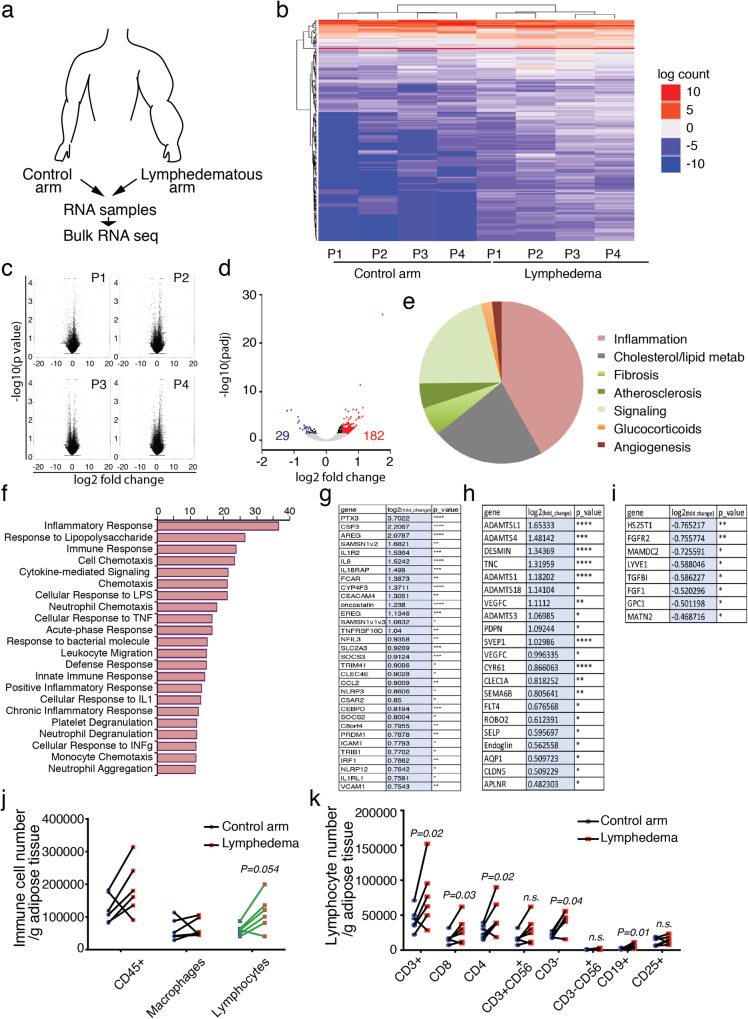
Lymphedema (LD) is characterized by the accumulation of interstitial fluid, lipids and inflammatory cell infiltrate in the limb. Here, we find that LD tissues from women who developed LD after breast cancer exhibit an inflamed gene expression profile. Lipidomic analysis reveals decrease in specialized pro-resolving mediators (SPM) generated by the 15-lipoxygenase (15-LO) in LD. In mice, the loss of SPM is associated with an increase in apoptotic regulatory T (Treg) cell number. In addition, the selective depletion of 15-LO in the lymphatic endothelium induces an aggravation of LD that can be rescued by Treg cell adoptive transfer or ALOX15-expressing lentivector injections. Mechanistically, exogenous injections of the pro-resolving cytokine IFN-β restores both 15-LO expression and Treg cell number in a mouse model of LD. These results provide evidence that lymphatic 15-LO may represent a therapeutic target for LD by serving as a mediator of Treg cell populations to resolve inflammation.


Conflict of interest statement

The authors have submitted a patent application (application numbers EP22305165, EP22305165.7, inventor B. Garmy-Susini) based on the results reported in this study and the authors declare no other competing interests.

Figures

Fig. 1
Fig. 1. Human lymphedema exhibits an inflammatory profile.
a Schematic representation of the experimental procedure. b Heatmap of hierarchical clustering analysis of genes upregulated (red) and downregulated (blue). c Volcano plots highlighting the genes that differ between LD/control in each patient. d Volcano plot showing the upregulated and downregulated genes in lipodermectomies. e Comparison of pathways regulated in LD. f Gene ontology of immune regulations in LD. g Upregulated expressed genes associated with immune response. h Upregulated expressed genes associated with lymphangiogenic response. i Downregulated expressed genes associated with lymphangiogenic response j Flow cytometry analysis of adipose tissue from LD dermolipectomies compared to the normal arm on the same patient. k Flow cytometry analysis of T cells in LD dermolipectomies. For bi, n = 4 women with LD (normal arm and LD arm from the same patient). For j and k n = 5 women with LD (tissue biopsies from the normal arm and LD arm from the same patient). Data are shown as mean ± s.e.m.; gi Wald test was used to generate p-values and log2 fold-changes (*p < 0.05, **p < 0.01, ***p < 0.005, ****p < 0.001). j, k p-values are derived from multiple t-tests. Source data are provided as a Source data file.
Fig. 2
Fig. 2. Downregulation of lipid mediators in human LD.
a Schematic representation of the experimental procedure. b Heatmap of lipid mediators derived from arachidonic acid (AA), Docosahexaenoic acid (DHA), and Eicosapentaenoic acid (EPA) analysis in lymphedematous skin. c Quantification of 15HETE and 17HDOHE, respectively, AA- and DHA-derived lipid mediators generated by 15-LO conversion. d ALOX15 mRNA expression in LD. e 15-LO protein expression in LD. f Immunodetection of 15-LO in lymphedematous skin (scale bar: 50 μm). g Quantification of 15-LO-positive cells in lymphedematous skin. h Immunodetection of 15-LO (red) and podoplanin (green) in normal and lymphedematous skin (Scale bar: 50 μm). i Quantification of 15-LO-positive lymphatic vessels in normal and lymphedematous skin. For b, c n = 10 women with LD (6 normal and 10 LD arm tissue biopsies). For di n = 4 women tissue biopsies with LD (normal arm and LD arm from the same patient). Data are shown as mean ± s.e.m.; b, c p-values are derived from the Mann–Whitney test. d, g, i p-values are derived from unpaired t-tests. Source data are provided as a Source data file.
Fig. 3
Fig. 3. Lymphatic endothelial ALOX15 controls T cells function in LD.
a Immunodetection of Foxp3 (green) and Lyve-1 (red) in LD skin (Scale bar: 50 μm). b Quantification of Foxp3-positive cells in LD from mice treated with PD146176. c Schematic representation of experimental procedure in Foxp3-GFP transgenic mice. d, e Flow cytometry analysis and quantification of CD4+GFP+ (Foxp3) Treg cell populations in LD adipose tissue. f Quantification of the limb diameter in ALOX15LECKO mice. g Lymphography of the limb from ALOX15LECKO mice (Scale bar: 1 mm). h Quantification of lymphatic branch point in ALOX15LECKO mice. i Quantification of lymph dermal backflow in ALOX15LECKO mice. j Quantification of the number of Treg cells (Foxp3+) in the limb from ALOX15LECKO mice or control littermates. k Immunodetection of apoptotic (TUNEL+) Treg cells (Foxp3+) in the limb from ALOX15LECKO mice (Scale bar: 50 μm). l Quantification of apoptotic Treg cells in the limb from ALOX15LECKO mice. m Quantification of PPARγ-positive Treg cells in the limb from ALOX15LECKO mice. Data are shown as mean ± s.e.m.; For b, n = 5 mice per group. For d, e n = 4 mice per group per experiment, 2 independent experiments. For fm n = 4 mice per group per experiment, 3 independent experiments. b, e p-values are derived from one-way ANOVA. f, h, I, j, l, m p-values are derived from Two-way ANOVA. Source data are provided as a Source data file.
Fig. 4
Fig. 4. 15-LO treatment restores lymphatic function in LD.
a Quantification of the limb diameter in LD from mice treated with three intradermal injections of ALOX15 lentivectors. b Quantification of ALOX15-positive lymphatic endothelial cells in the skin from mice treated with intradermal injections of ALOX15 lentivectors. c Lymphography of the limb from ALOX15-treated mice (Scale bar: 1 mm). d Quantification of lymphatic branch point in ALOX15-treated mice. e Masson’s trichrome coloration of the lymphedematous skin. f Quantification of dermis thickness in mice treated with ALOX15 lentivector. g Skin thickness repartition related to collagen (Coll) vs. adipose tissue (AT). h Hair follicle quantification in LD skin. i Quantification of Foxp3-positive pixels in the limb from mice treated with ALOX15 lentivector. j Quantification of the number of PPARγ-positive Treg cells in the limb from mice treated with ALOX15 lentivector. k Quantification of the limb diameter in LD from mice treated with Resolvin D1 (RvD1), Lipoxin A4 (LxA4), and 15-HETE. l Capillary electrophoresis and its quantification showing ALOX15 knock down in LEC. m Immunodetection of 15-LO (green) and VE-Cadherin (red) in LEC (Scale bar: 25 μm). n Quantitative RT-qPCR analysis of CCL21, SPHK1, SPHK2, LTBR, S1PR1, ICAM1, and VCAM1 in LEC after ALOX15 knock down. For an n = 4 mice per group, 2 independent experiments. Data are shown as mean ± s.e.m.; (a, b, d, f, k) p-values are derived from two-way ANOVA. i, j, l, n p-values are derived from unpaired t-tests. Source data are provided as a Source data file.
Fig. 5
Fig. 5. Interferon-beta protects from LD.
a IFN-α mRNA expression in LD skin from ALOX15LECKO mice. b IFN-β mRNA expression in LD skin from ALOX15LECKO mice. c ALOX12/15 gene expression in LD skin from ALOX15LECKO mice and control littermates (Cre-) treated with IFN-β (100 ng IP, every 2 days for 10 days). d Limb lymphography from Cre- (left panel) and ALOX15LECKO (right panel) mice with LD treated with IFN-β (Scale bar: 1 mm). e Quantification of lymphatic branch point in IFN-β-treated ALOX15LECKO mice and control littermates. f, g Quantification of the limb diameter in Cre- (f) and ALOX15LECKO (g) mice with LD treated with IFN-β. h Quantification of Treg number in LD skin from ALOX15LECKO mice and control littermates (Cre-) treated with IFN-β. i CCL21 mRNA expression in LD skin from ALOX15LECKO mice treated with IFN-β. j LTBR mRNA expression in LD skin from ALOX15LECKO mice treated with IFN-β. Data are shown as mean ± s.e.m. For an, n = 4–8 mice per group. ac, ej p-values are derived from two-way ANOVA. Source data are provided as a Source data file.

References

    1. Alitalo K, Tammela T, Petrova TV. Lymphangiogenesis in development and human disease. Nature. 2005;438:946–953. doi: 10.1038/nature04480. - DOI - PubMed
    1. Mortimer PS, Rockson SG. New developments in clinical aspects of lymphatic disease. J. Clin. Investig. 2014;124:915–921. doi: 10.1172/JCI71608. - DOI - PMC - PubMed
    1. Serhan CN, et al. Resolution of inflammation: state of the art, definitions and terms. FASEB J. 2007;21:325–332. doi: 10.1096/fj.06-7227rev. - DOI - PMC - PubMed
    1. Serhan CN. Pro-resolving lipid mediators are leads for resolution physiology. Nature. 2014;510:92–101. doi: 10.1038/nature13479. - DOI - PMC - PubMed
    1. Gimbrone MA, Jr., Garcia-Cardena G. Endothelial Cell Dysfunction and the Pathobiology of Atherosclerosis. Circ. Res. 2016;118:620–636. doi: 10.1161/CIRCRESAHA.115.306301. - DOI - PMC - PubMed

Publication types