. 2024 Jan 4;15(1):285.
doi: 10.1038/s41467-023-44534-y.

Cleavage-intermediate Lassa virus trimer elicits neutralizing responses, identifies neutralizing nanobodies, and reveals an apex-situated site-of-vulnerability

Affiliations

Cleavage-intermediate Lassa virus trimer elicits neutralizing responses, identifies neutralizing nanobodies, and reveals an apex-situated site-of-vulnerability

Jason Gorman et al. Nat Commun. .

Abstract

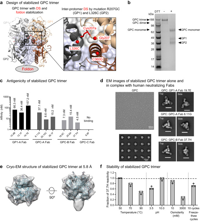
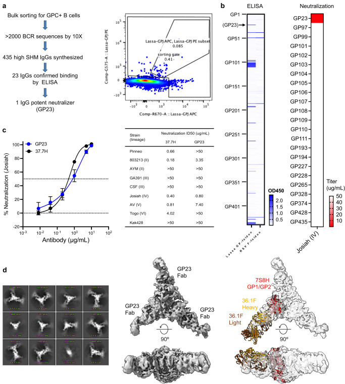
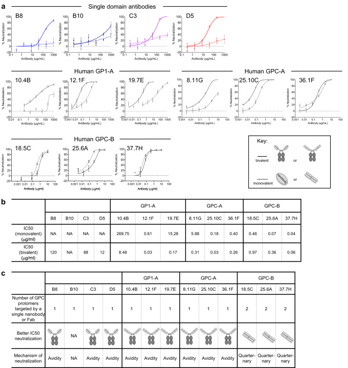
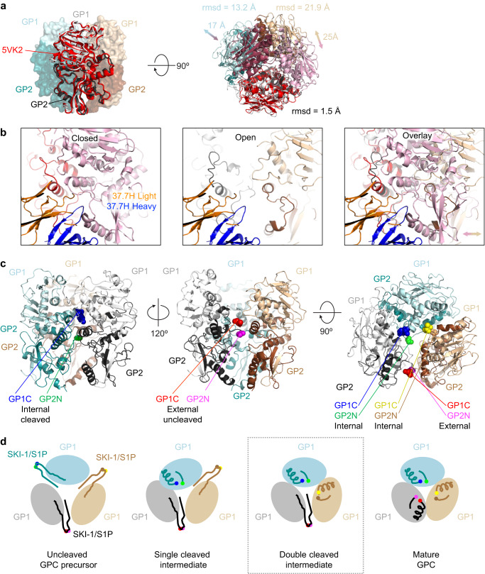
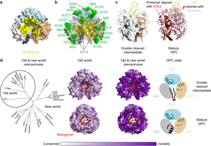
Lassa virus (LASV) infection is expanding outside its traditionally endemic areas in West Africa, posing a pandemic biothreat. LASV-neutralizing antibodies, moreover, have proven difficult to elicit. To gain insight into LASV neutralization, here we develop a prefusion-stabilized LASV glycoprotein trimer (GPC), pan it against phage libraries comprising single-domain antibodies (nanobodies) from shark and camel, and identify one, D5, which neutralizes LASV. Cryo-EM analyses reveal D5 to recognize a cleavage-dependent site-of-vulnerability at the trimer apex. The recognized site appears specific to GPC intermediates, with protomers lacking full cleavage between GP1 and GP2 subunits. Guinea pig immunizations with the prefusion-stabilized cleavage-intermediate LASV GPC, first as trimer and then as a nanoparticle, induce neutralizing responses, targeting multiple epitopes including that of D5; we identify a neutralizing antibody (GP23) from the immunized guinea pigs. Collectively, our findings define a prefusion-stabilized GPC trimer, reveal an apex-situated site-of-vulnerability, and demonstrate elicitation of LASV-neutralizing responses by a cleavage-intermediate LASV trimer.


Conflict of interest statement

NIH has submitted a patent application for GPC trimer and GPC trimer nanoparticle on which J.G., C.S-F.C., L.O., M.W., C.C., J.R.M., D.D.H, and P.D.K. are co-inventors. NIH has also submitted a patent application for neutralizing nanobodies on which J.G., C.S-F.C., Z.D., Y.S., M.H. and P.D.K. are co-inventors. The other authors declare no competing interests.

Figures

Fig. 1
Fig. 1. Design and characterization of stabilized soluble Lassa GPC trimer.
a Structure-based design of stabilized soluble Lassa GPC trimer. An inter-protomer disulfide (DS) bond linked GP1 of one protomer to GP2 of a neighboring protomer and a foldon domain was appended to the C-terminus of GP2. Inset shows a view of the inter-protomer DS shown as spheres. The two protomers are shown as ribbons in light gray (GP1) and dark gray (GP2), and wheat (GP1) and brown (GP2). b Representative SDS-PAGE of stabilized Lassa trimer under non-reducing and reducing conditions. A high molecular weight band three times the size of the monomeric form was observed under non-reducing conditions. Three repeats show the same results. Source image is provided in the Source Data file. c Binding affinity of the stabilized Lassa GPC trimer towards Fabs of four groups of human Lassa-neutralizing antibodies, GP1-A, GPC-A, GPC-B, and GPC-C. d A representative negative-stain EM micrograph (top left) and 2D class averages (bottom left) are shown of the stabilized Lassa GPC trimer, along with 2D class averages of the GPC trimer in complex with human neutralizing Fabs (right panels). At least 25 micrographs were recorded in each case. The 2D class averages of the complexes include representative top and side views. For right panels, the scale bars represent 10 nm. e Cryo-EM structure of the stabilized Lassa GPC at 5.8 Å resolution confirmed its trimeric association. f Physical properties of the stabilized Lassa GPC trimer. Stability of the stabilized trimer was assessed by fractional binding reactivity to antibody 37.7H after treatments under various temperatures, pHs, osmolarities, and freeze-thaw cycles. Triplicate measurements were made, and results are shown as mean ± SEM. The dotted line shows the antibody reactivity of trimer prior to physical stress.
Fig. 2
Fig. 2. Potent neutralizing response elicited by Lassa GPC trimer and nanoparticle immunization in guinea pigs.
a Preparation of GPC nanoparticle. Lassa GPC nanoparticle profiles are shown from three individual size exclusion chromatographs. A representative SDS PAGE gel is shown from two repeats, source image is provided in the Source Data file. b CryoEM structure of Nanoparticle Lassa GPC-Enc. c Schematic representation of the immunization schedule. d Immune responses against to Lassa GPC trimer, GPC nanoparticle and nanoparticle alone were measured by ELISA. Initial dilution is shown as a horizontal dotted line. Data are presented as mean values +/- SEM from n = 10 independent biological replicates. e Lassa GPC trimer immunizations followed by multiple nanoparticles boosts elicited high neutralizing responses in guinea pigs. Source data are provided as a Source Data file.
Fig. 3
Fig. 3. Potent Lassa virus neutralizing antibody elicited by stabilized GPC trimer—trimer nanoparticle immunization.
a Workflow and B-cell FACS analysis for antibodies isolated from guinea pigs. b ELISA screening of expressed guinea pig IgGs for Lassa GPC-specific binding (left). Selected IgGs with positive ELISA binding were further assessed Lassa Josiah neutralization. c Neutralization on Lassa Josiah strain (left) and diverse strains (right). Neutralization was determined by interpolation after fitting data globally to a 5-parameter dose-response curve. The IC50 (dot) and 95% CI (error bar) of a global fit of six serial dilutions. d Cryo-EM analysis shows GP23 binding to overlap with the epitope of 36.1 F from PDB ID 7S8H. Aggregation illustrated in the 2D class images of the GP23-GPC sample (left) did not allow for accurate model building despite a nominal resolution of 5.4 Å.
Fig. 4
Fig. 4. Neutralization of Lassa virus by single domain antibodies and most human neutralizing antibodies utilizes avidity.
a Neutralization of pseudotyped Josiah strain of Lassa virus by single domain antibodies and human Lassa neutralizing antibodies in both monovalent (dotted line) and bivalent (solid line) formats. Neutralization was determined by interpolation after fitting data globally to a five-parameter dose-response curve. The IC50 (dot) and 95% CI (error bar) of a global fit of six serial dilutions. b Summary of the IC50 values of single domain antibodies and human Lassa neutralizing antibodies in monovalent and bivalent formats. c Summary of the proposed neutralization mechanisms of single domain antibodies and human antibodies for Lassa virus.
Fig. 5
Fig. 5. Cryo-EM structure of D5 and 8.11 G with Lassa trimer reveals details of 8.11 G and D5 recognition.
a Cryo-EM density is shown for a complex of the stabilized GPC trimer bound to two Fabs of 8.11 G and a single D5. Density for the R207GC-L326C engineered disulfide is shown in the right panel. b The atomic model is shown in cartoon representation. Glycans for which density was observed are displayed as green spheres. A 90-degree view from the top is shown without glycans in the center. In the right panel is density around the D5 nanobody chain. c The glycosylated epitope of 8.11 G is highlighted. The structure is displayed in cartoon format with a transparent surface. The inset box shows a view rotated 90° along the axis of the 8.11 G Fab binding with Fab contact loops shown as cartoon and the GPC protein and glycans shown as spheres. The right panel shows representative density of the antibody epitope. d The 8.11 G epitope partially overlaps with that of 36.1 F (PDB ID: 7S8H). A 5 A footprint of each antibody is shown.
Fig. 6
Fig. 6. Analysis of asymmetric Lasa GPC trimer reveals a cleavage-intermediate site of vulnerability at the trimer apex.
a A single protomer of the GPC trimer shows a closely matching rmsd with that of the C3 symmetric GPC (PDB ID 5VK2, colored red, pink, and raspberry). The C1 symmetric GPC trimer observed here does not maintain the same quaternary assembly with the adjacent protomer oriented to accommodate the uncleaved protomer. After removing outlying residues the rmsd for one GP1/GP2 across 294 Ca atoms of the protomer is 1.5 Å, however the rmsd’s for the other two protomers are 13.2 and 21.9 Å across 296 and 294 Ca, respectively. Specific apex regions showing distances from 17-25 Å. b Explanation for the loss of one, two, or three of the 37.7H binding sites on a trimer with D5 stabilizing a more open interface between protomers. The open gap that accommodates uncleaved peptide is shown compared to the closed epitope interface. c The Lassa GPC is shown with focus on one of the two cleaved protomers (left) with the internal termini of GP1 (light cyan) and GP2 (teal) shown. Rotation by 120° shows the external location of the uncleaved peptide. d A schematic representation of the cleavage intermediates in the maturation of the GPC trimer is shown from a top view looking down the trimer axis. The SKI-1/S1P cleavage site must be cleaved on all three protomers to enable a tightly packed GPC trimer. The neutralizing nanobody D5 binds the first three populations (uncleaved, single-cleavage, and double-cleaved). Gray box highlights the intermediate corresponding to the structure shown above.
Fig. 7
Fig. 7. Apex site of nanobody vulnerability overlaps with the binding site for matriglycan receptor.
a A surface representation of the cleavage intermediate trimer is shown. Residues within a 5 Å footprint of the D5 nanobody are highlighted in yellow. b A transparent surface representation is shown as in panel A with cartoon representation underneath. Glycans are displayed as green spheres. c D5 fits into pocket at apex of double-cleaved intermediate, but not into the mature GPC, in which one protomer orientation shifts the apex region by >10 Å (pink) and the second protomer shifts at apex regions by >20 Å (raspberry), sterically blocking access to the the D5 binding site. d (left) A phylogenetic tree of select arenavirus GPC sequences is displayed to illustrate the overall sequence diversity and highlight the clustering of old world versus new world viruses. (right) Conservation of the GPC residues is shown for the old world viruses versus all arenaviruses with the double-cleavage intermediate on the top and the mature GPC on the bottom, the matriglycan (red) from PDB ID 7PVD is shown on all surfaces.

References

    1. Shaffer JG, et al. Lassa fever in post-conflict sierra leone. PLoS Negl. Trop. Dis. 2014;8:e2748. doi: 10.1371/journal.pntd.0002748. - DOI - PMC - PubMed
    1. Asogun DA, et al. Molecular diagnostics for lassa fever at Irrua specialist teaching hospital, Nigeria: lessons learnt from two years of laboratory operation. PLoS Negl. Trop. Dis. 2012;6:e1839. doi: 10.1371/journal.pntd.0001839. - DOI - PMC - PubMed
    1. Eberhardt KA, et al. Ribavirin for the treatment of Lassa fever: a systematic review and meta-analysis. Int J. Infect. Dis. 2019;87:15–20. doi: 10.1016/j.ijid.2019.07.015. - DOI - PubMed
    1. Carey DE, et al. Lassa fever. Epidemiological aspects of the 1970 epidemic, Jos, Nigeria. Trans. R. Soc. Trop. Med. Hyg. 1972;66:402–408. doi: 10.1016/0035-9203(72)90271-4. - DOI - PubMed
    1. Haas WH, et al. Imported Lassa fever in Germany: surveillance and management of contact persons. Clin. Infect. Dis. 2003;36:1254–1258. doi: 10.1086/374853. - DOI - PubMed

Publication types

Associated data