. 2024 Jan 4;14(1):568.
doi: 10.1038/s41598-023-49964-8.

A pilot optical coherence tomography angiography classification of retinal neovascularization in retinopathy of prematurity

Affiliations

A pilot optical coherence tomography angiography classification of retinal neovascularization in retinopathy of prematurity

Xi Chen et al. Sci Rep. .

Abstract

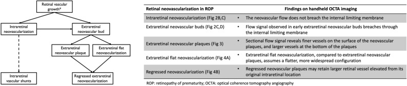
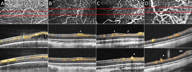
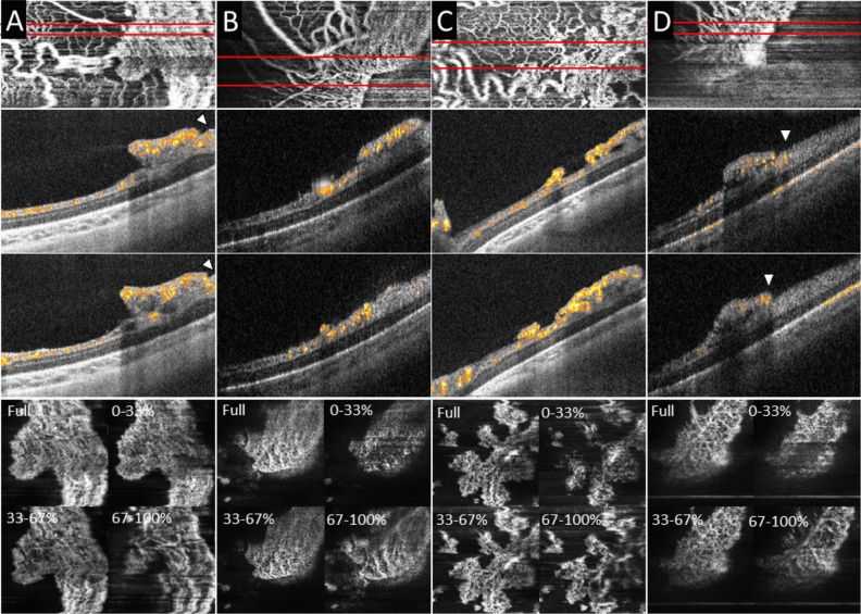
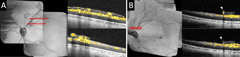
Extraretinal neovascularization is a hallmark of treatment-requiring retinopathy of prematurity (ROP). Optical coherence tomography angiography (OCTA) offers vascular flow and depth information not available from indirect ophthalmoscopy and structural OCT, but OCTA is only commercially available as a tabletop device. In this study, we used an investigational handheld OCTA device to study the vascular flow in and around retinal neovascularization in seven preterm infants with treatment-requiring ROP and contrasted them to images of vascular flow in six infants of similar age without neovascular ROP. We showed stages of retinal neovascularization visible in preterm infants from 32 to 47 weeks postmenstrual age: Intraretinal neovascularization did not break through the internal limiting membrane; Subclinical neovascular buds arose from retinal vasculature with active flow through the internal limiting membrane; Flat neovascularization in aggressive ROP assumed a low-lying configuration compared to elevated extraretinal neovascular plaques; Regressed neovascularization following treatment exhibited decreased vascular flow within the preretinal tissue, but flow persisted in segments of retinal vessels elevated from their original intraretinal location. These findings enable a pilot classification of retinal neovascularization in eyes with ROP using OCTA, and may be helpful in detailed monitoring of disease progression, treatment response and predicting reactivation.


Conflict of interest statement

Drs. Viehland, Izatt and Toth and Duke University have patent application pending related to the novel handheld probe used in the manuscript. Drs. Viehland and Toth have owner equity in Theia Imaging, LLC. No other authors have related conflict of interest.

Figures

Figure 1
Figure 1
Pilot Classification of the development and sequelae of intra- and extraretinal neovascularization in retinopathy of prematurity using optical coherence tomography angiography.
Figure 2
Figure 2
Bedside handheld optical coherence tomography angiography (OCTA) images of the preterm infant retinal vasculature in eyes without (A) and with (B and C, same infant, &D) neovascularization. The top row shows the vascular pattern en face, the bottom two rows are cross-sectional B-scans with flow overlay from the top row images at the site of the red lines. OCTA flow signal appears yellow and red in cross-section and sums to create the white vascular pattern of the en face images in the top row. (A) At 36 weeks postmenstrual age (PMA) in an infant who never developed severe ROP, retinal microvasculature was well-formed in the macula with flow signal present from the innermost border of the retina (internal limiting membrane) to the inner nuclear layer/inner plexiform layer junction. Macular edema was visible and common in preterm infants. (B) At 32 weeks PMA in an infant who later required treatment for severe ROP, prominent vascular flow was observed in the perifoveal region, with focal mild elevation of the inner retinal surface (asterisk). (C) Two weeks later (at 34 weeks PMA), the same eye as in (B) developed severe (aggressive) ROP that required treatment. The inner retinal neovascularization produced greater elevation and splitting of the internal limiting membrane / nerve fiber layer on either side of the fovea (F), and now breached the internal limiting membrane in the inferior macula (arrowhead), assuming an early extraretinal neovascular bud configuration. (D) At 34 weeks in an infant who required treatment of severe ROP one week later, an extraretinal neovascular bud (arrowhead) exhibited flow signal extending above the inner retinal surface over a larger retinal vessel posterior to the margin of vascularized retina. Neovascular flow signals also extended into the preretinal tissue at the vascular-avascular junction (open arrowhead).
Figure 3
Figure 3
Extraretinal neovascular plaques and distribution of vascular flow by axial location within the neovascular plaques on handheld optical coherence tomography angiography (OCTA). Top row: en face OCTA flow; Second and third rows: B-scans with flow overlay; Bottom row: en face sub-sectional flow (generated by the percentage of distance from the top to bottom of the neovascular plaque). In three eyes with treatment requiring-ROP, extraretinal neovascular plaques were found elevated from the retinal surface along the vascular-avascular junction. On OCT B-scans with flow overlay, the retinal vascular flow signal extends to the edge or beyond the edge of the neovascular plaque (arrowheads). The vascular flow signal on cross-section on B-scans with flow overlay and the sub-sectional flow analysis (Full, 0–33%, 33–67%, and 67–100%) showed finer capillaries at the surface of the plaque (vitreous side) and larger, more dilated capillaries at the bottom of the plaque (retina side).
Figure 4
Figure 4
Optical coherence tomography angiography of extraretinal flat neovascularization in an eye with aggressive retinopathy of prematurity (ROP), and regressed extraretinal neovascularization following intraretinal bevacizumab treatment. (A) In an eye with aggressive ROP at 36 weeks PMA, OCT B-scans with flow overlay near the posterior pole superior to the optic disc along the superotemporal arcade showed vascular flow in the extraretinal neovascular tissue appeared flatter and over a wider retinal surface area compared to the extraretinal neovascular plaques. (B) The same eye was imaged at 47 weeks PMA following bevacizumab treatment at 36 weeks PMA. The preretinal neovascular tissue was regressed and marked reduction of vascular flow within the preretinal tissue. However, one larger retinal vessel was seen (arrowheads) retained in the preretinal tissue elevated from its original intraretinal location.

References

    1. Hartnett ME, Penn JS. Mechanisms and management of retinopathy of prematurity. N. Engl. J. Med. 2012;367:2515–2526. doi: 10.1056/NEJMra1208129. - DOI - PMC - PubMed
    1. Klufas MA, et al. Influence of fluorescein angiography on the diagnosis and management of retinopathy of prematurity. Ophthalmology. 2015;122:1601–1608. doi: 10.1016/j.ophtha.2015.04.023. - DOI - PMC - PubMed
    1. Lepore D, et al. Early angiographic signs of retinopathy of prematurity requiring treatment. Eye (Lond) 2021;35:3094–3101. doi: 10.1038/s41433-020-01305-z. - DOI - PMC - PubMed
    1. Lepore D, et al. Atlas of fluorescein angiographic findings in eyes undergoing laser for retinopathy of prematurity. Ophthalmology. 2011;118:168–175. doi: 10.1016/j.ophtha.2010.04.021. - DOI - PubMed
    1. Maldonado RS, Toth CA. Optical coherence tomography in retinopathy of prematurity: Looking beyond the vessels. Clin. Perinatol. 2013;40:271–296. doi: 10.1016/j.clp.2013.02.007. - DOI - PMC - PubMed

MeSH terms