. 2024 Jan;16(1):132-157.
doi: 10.1038/s44321-023-00009-7. Epub 2024 Jan 2.

Versican accumulation drives Nos2 induction and aortic disease in Marfan syndrome via Akt activation

Affiliations

Versican accumulation drives Nos2 induction and aortic disease in Marfan syndrome via Akt activation

María Jesús Ruiz-Rodríguez et al. EMBO Mol Med. 2024 Jan.

Abstract

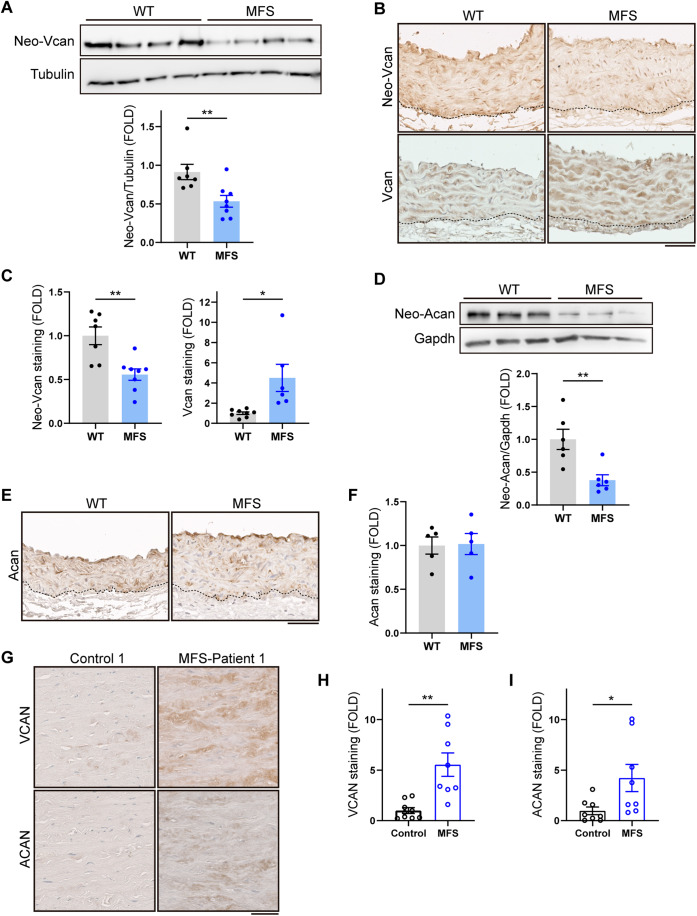
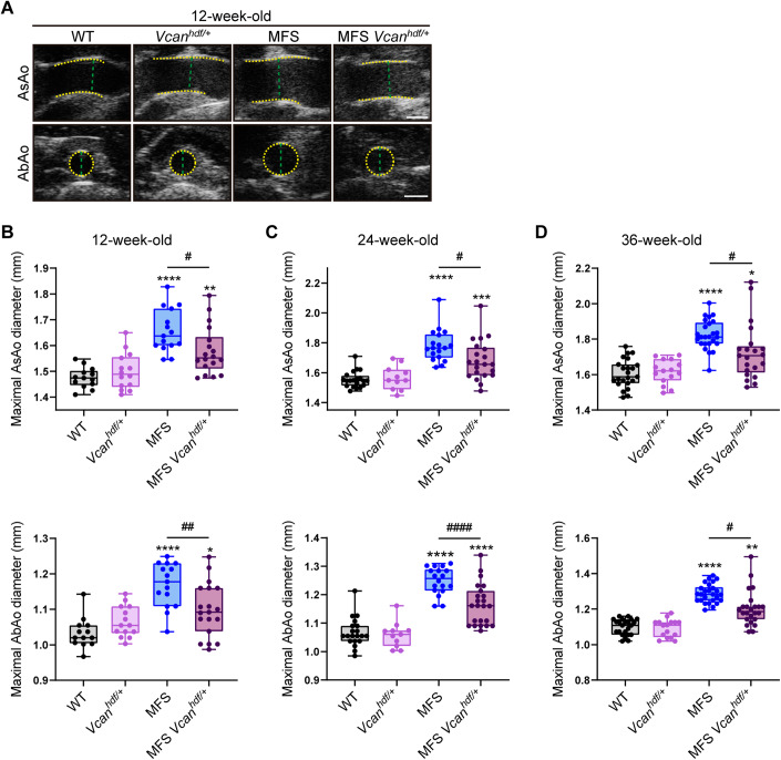
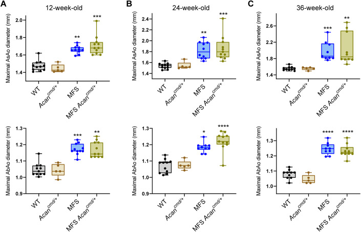
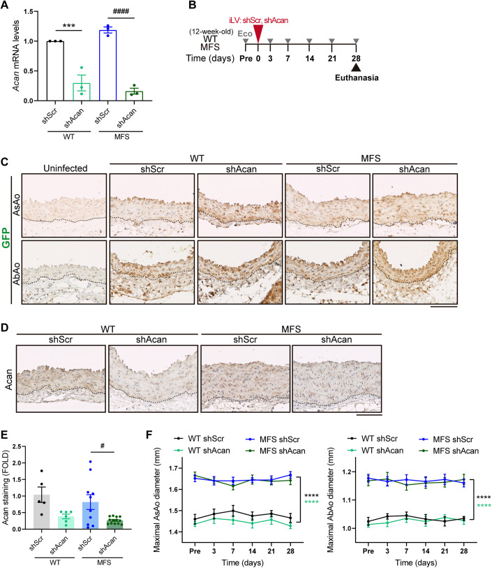
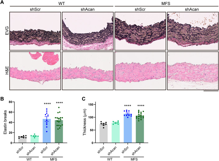
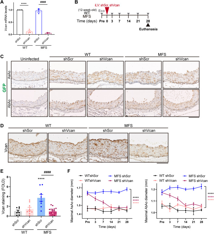
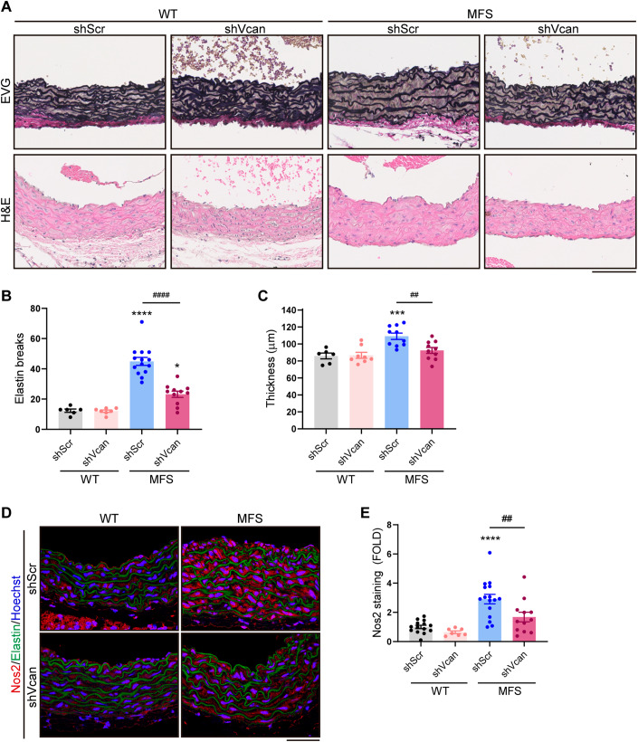
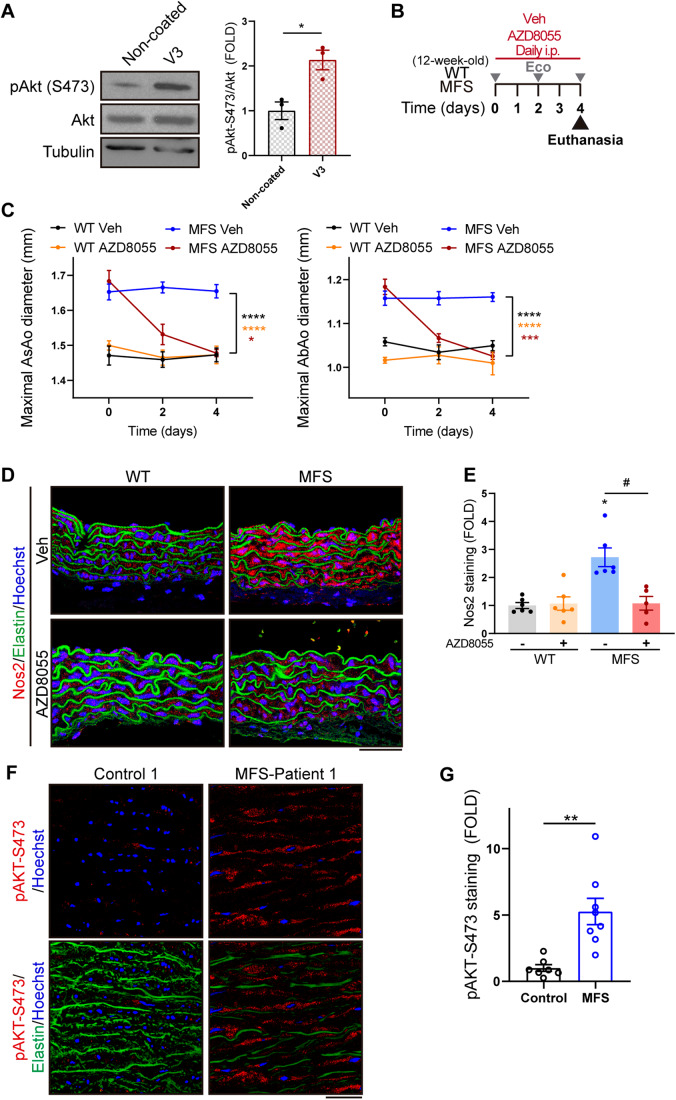
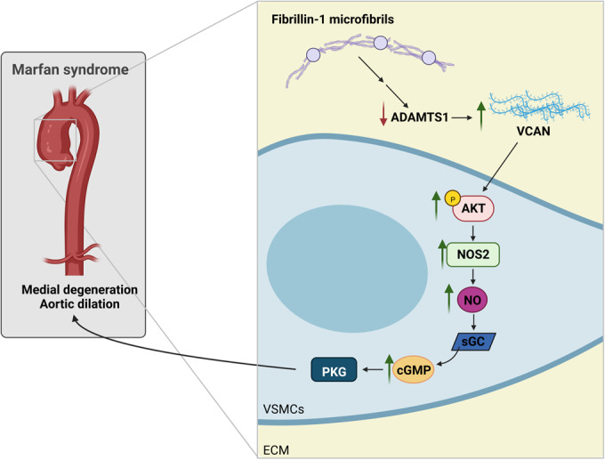
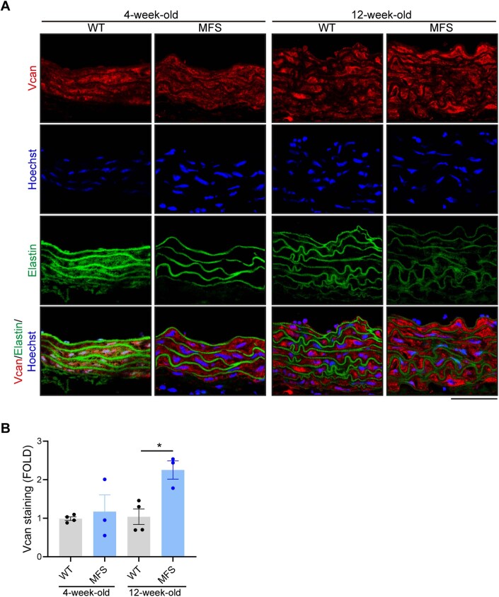
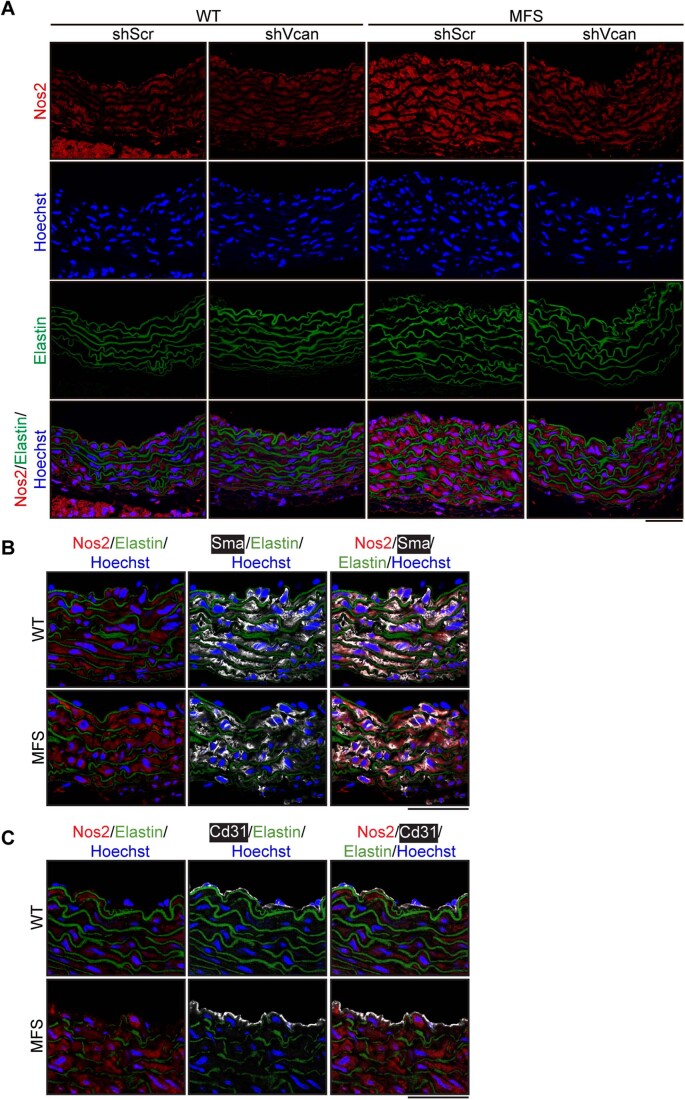
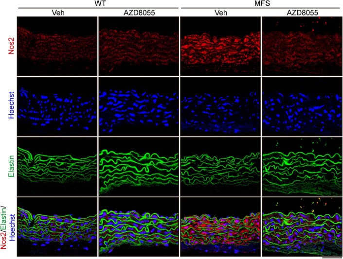
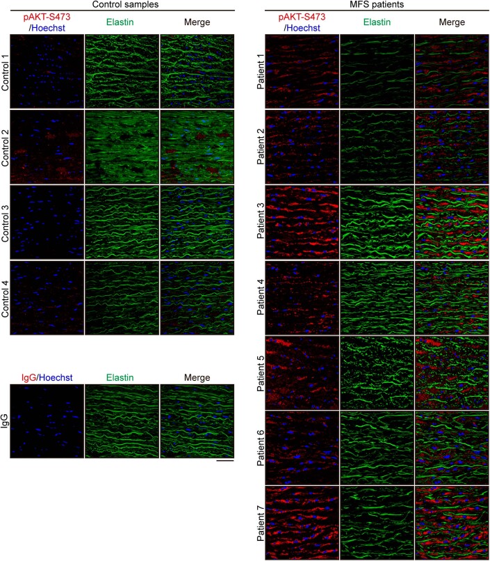
Thoracic aortic aneurysm and dissection (TAAD) is a life-threatening condition associated with Marfan syndrome (MFS), a disease caused by fibrillin-1 gene mutations. While various conditions causing TAAD exhibit aortic accumulation of the proteoglycans versican (Vcan) and aggrecan (Acan), it is unclear whether these ECM proteins are involved in aortic disease. Here, we find that Vcan, but not Acan, accumulated in Fbn1C1041G/+ aortas, a mouse model of MFS. Vcan haploinsufficiency protected MFS mice against aortic dilation, and its silencing reverted aortic disease by reducing Nos2 protein expression. Our results suggest that Acan is not an essential contributor to MFS aortopathy. We further demonstrate that Vcan triggers Akt activation and that pharmacological Akt pathway inhibition rapidly regresses aortic dilation and Nos2 expression in MFS mice. Analysis of aortic tissue from MFS human patients revealed accumulation of VCAN and elevated pAKT-S473 staining. Together, these findings reveal that Vcan plays a causative role in MFS aortic disease in vivo by inducing Nos2 via Akt activation and identify Akt signaling pathway components as candidate therapeutic targets.

Keywords: Akt; Aortic Aneurysm; Marfan Syndrome; Nos2; Versican.


Conflict of interest statement

The authors declare no competing interests.

Figures

Figure 1
Figure 1. Vcan accumulates in the aortas of Fbn1C1041G/+ MFS mice.
(A) Representative immunoblot analysis of neo-Vcan in aortic extracts from WT and MFS mice, with quantification of the immunoblot signal (n = 7–8 mice per group). (B) Representative immunohistochemistry images of neo-Vcan and Vcan in mouse AsAo. Dotted lines outline the boundaries between the media and the adventitia. Scale bar, 50 μm. (C) Quantification of neo-Vcan and Vcan immunostaining in aortic sections from WT and MFS mice (n = 6–8 mice). (D) Representative immunoblot analysis of neo-Acan in aortic extracts from WT and MFS mice, with quantification of the immunoblot signal (n = 6 mice per group). (E) Representative images of Acan immunohistochemistry in mouse AsAo. Dotted lines outline the boundaries between the media and the adventitia. Scale bar, 50 μm. (F) Quantification of Acan immunostaining in AsAo from WT (n = 5) and MFS (n = 5) mice. (G) Representative medial layer images of VCAN and ACAN immunohistochemistry in aortic cross sections of human samples from control donors and MFS patients. Scale bar, 50 μm. (H, I) Quantification of VCAN (H) and ACAN (I) immunohistochemistry in aortas from control donors and MFS patients (n = 8–9 samples per group). Data information: (A, C, D, F) data are shown relative to WT mice as mean ±  s.e.m. Each data point denotes an individual mouse. *P < 0.05, **P < 0.01 (Student t test or unpaired t test with Welch’s correction, as appropriate). (H, I) Data are shown relative to control donors as mean ± s.e.m. Each data point denotes an individual. *P < 0.05, **P < 0.01 (unpaired t test with Welch’s correction). Source data are available online for this figure.
Figure 2
Figure 2. Vcan haploinsufficiency protects MFS mice from aortic dilation.
(A) Representative in vivo ultrasound images of the AsAo and AbAo from 12-week-old WT, Vcanhfd/+, MFS, and MFS Vcanhdf/+ mice. Yellow dashed lines delineate lumen boundaries and green dashed lines denote lumen diameters. Scale bar, 1 mm. (BD) Maximal AsAo and AbAo diameters in Vcan haploinsufficient mice aged (B) 12, (C) 24, and (D) 36 weeks. Data information: (BD) data are shown as box-and-whisker plots. The box itself spans from the first quartile (25%) to the third quartile (75%), representing the interquartile range where the central 50% of data values fall. Inside the box, a line denotes the median value. The whiskers of the boxplot extend from the ends of the box to the minimum and maximum values. Each data point denotes an individual mouse (12-week-old mice: WT, n = 13; Vcanhdf/+, n = 14; MFS, n = 15; MFS;Vcanhdf/+, n = 18. 24-week-old mice: WT, n = 20; Vcanhdf/+, n = 11; MFS, n = 18; MFS;Vcanhdf/+, n = 21. 36-week-old mice: WT, n = 23; Vcanhdf/+, n = 15; MFS, n = 25; MFS;Vcanhdf/+, n = 21). *P < 0.05, **P < 0.01, ***P < 0.001, ****P < 0.0001 versus WT mice; #P < 0.05, ##P < 0.01, ####P < 0.0001 versus MFS mice (two-way ANOVA with Tukey’s post hoc test or the Kruskal–Wallis test with Dunn’s multiple comparison test, as appropriate). Source data are available online for this figure.
Figure 3
Figure 3. Acan haploinsufficiency does not prevent aortic enlargement in MFS mice.
(AC) Maximal AsAo and AbAo diameters in (A) 12-, (B) 24-, and (C) 36-week-old mice (WT, n = 10–11; Acancmd/+, n = 5; MFS, n = 9–11; MFS;Acancmd/+, n = 10–11). Data information: (AC) data are shown as box-and-whisker plots. The box itself spans from the first quartile (25%) to the third quartile (75%), representing the interquartile range where the central 50% of data values fall. Inside the box, a line denotes the median value. The whiskers of the boxplot extend from the ends of the box to the minimum and maximum values. Each data point denotes an individual mouse. *P < 0.05, **P < 0.01, ***P < 0.001, ****P < 0.0001 versus WT mice (two-way ANOVA with Tukey’s post hoc test or the Kruskal–Wallis test with Dunn’s multiple comparison test, as appropriate). Source data are available online for this figure.
Figure 4
Figure 4. Acan silencing does not reduce aortic dilation in MFS mice.
(A) Acan mRNA expression assessed by RT-qPCR in WT and MFS VSMCs transduced with lentivirus encoding shScr- or Acan-specific shRNA (n = 3 biological replicates). (B) Experimental design: 12-week-old WT and MFS mice were inoculated with lentivirus (iLV) expressing shScr or shAcan, monitored for aortic dilation (Eco), and euthanized at 28 days. (C) Representative images of GFP immunostaining in AsAo and AbAo of uninfected mice or mice expressing shScr or shAcan. Dotted lines outline the boundaries between the media and the adventitia. Scale bar, 100 μm. (D) Representative images of Acan immunohistochemistry in AsAo from shScr- or shAcan-transduced WT and MFS mice. Dotted lines outline the boundaries between the media and the adventitia. Scale bar, 100 μm. (E) Quantification of Acan immunostaining in aortic sections from the mouse cohorts shown in D (WT shScr, n = 5; WT shAcan, n = 6; MFS shScr, n = 10; MFS shAcan, n = 13 mice per group). (F) Maximal AsAo and AbAo diameters in WT shScr (n = 7), WT shAcan (n = 8), MFS shScr (n = 14), and MFS shAcan (n = 17) mice at the indicated time points. Data information: (A) ***P < 0.001 versus shScr-infected WT VSMCs; ####P < 0.0001 versus shScr-infected MFS VSMCs (two-way ANOVA with Tukey’s post hoc test). (E) Data are shown relative to shScr-transduced WT mice as mean ± s.e.m. Each data point denotes an individual mouse. #P < 0.05 versus shScr-transduced MFS mice (two-way ANOVA with Tukey’s post hoc test). (F) Data are shown as mean ± s.e.m. ****P < 0.0001 versus shScr-transduced MFS mice (repeated-measurements two-way ANOVA with Tukey’s post hoc test). Source data are available online for this figure.
Figure 5
Figure 5. Acan silencing does not regress medial degeneration in MFS mice.
(A) Representative images showing elastic van Gieson (EVG) and hematoxylin and eosin (H&E) staining in AsAo of the indicated mice (n = 6–17 mice per group). Scale bar, 100 µm. (B, C) Quantification of elastin breaks (B) and medial wall thickness (C) in AsAo from the mouse cohorts shown in (A). Data information: (B, C) data are shown as mean ± s.e.m. Each data point denotes an individual mouse. ****P < 0.0001 versus shScr- transduced WT mice (two-way ANOVA with Tukey’s post hoc test). Source data are available online for this figure.
Figure 6
Figure 6. Vcan silencing regresses aortic dilation in MFS mice.
(A) Vcan mRNA expression assessed by RT-qPCR in WT and MFS VSMCs transduced with lentivirus encoding shScr or Vcan-specific shRNA (n = 3 biological replicates). (B) Experimental design: 12-week-old WT and MFS mice were inoculated with lentivirus (iLV) expressing shScr or shVcan, monitored for aortic dilation (Eco), and euthanized at 28 days. (C) Representative images of GFP immunostaining in AsAo and AbAo of uninfected mice of mice expressing shScr- or shVcan. Dotted lines outline the boundaries between the media and the adventitia. Scale bar, 100 μm. (D) Representative images of Vcan immunohistochemistry in AsAo from shScr- or shVcan-transduced WT and MFS mice. Dotted lines outline the boundaries between the media and the adventitia. Scale bar, 100 μm. (E) Quantification of Vcan immunostaining in aortic sections from the mouse groups shown in (D) (WT shScr, n = 8; WT shVcan, n = 9; MFS shScr, n = 8; and MFS shVcan, n = 20 mice per group). (F) Maximal AsAo and AbAo diameters at the indicated time points in WT shScr (n = 6), WT shVcan (n = 9), MFS shScr (n = 13), and MFS shVcan (n = 15) mice. Data information: (A) ****P < 0.0001 versus shScr-infected WT VSMCs; ####P < 0.0001 versus shScr-infected MFS VSMCs (two-way ANOVA with Tukey’s post hoc test). (E) Data are shown relative to shScr-transduced WT mice as mean ± s.e.m. Each data point denotes an individual mouse. ****P < 0.0001 versus shScr-transduced WT mice; ####P < 0.0001 versus shScr-transduced MFS mice (two-way ANOVA with Tukey’s post hoc test). (F) Data are shown as mean ± s.e.m. ****P < 0.0001 versus shScr-transduced MFS mice (repeated-measurements two-way ANOVA with Tukey’s post hoc test). Source data are available online for this figure.
Figure 7
Figure 7. Vcan silencing decreases medial degeneration and Nos2 expression in MFS mice.
(A) Representative elastic van Gieson (EVG) and hematoxylin and eosin (H&E) staining in AsAo of the indicated mice (n = 6–14 mice per group). Scale bar, 100 µm. (B, C) Quantification of elastin breaks (B) and medial wall thickness (C) in AsAo from the mouse cohorts shown in (A). (D) Representative images of Nos2 immunofluorescence (red), elastin autofluorescence (green), and Hoechst-stained nuclei (blue) in mouse aortic sections. Scale bar, 50 μm. The same images are also presented in the bottom line of Fig. EV3A. (E) Quantification of Nos2 immunofluorescence in AsAo from WT shScr (n = 14), WT shVcan (n = 7), MFS shScr (n = 16), and MFS shVcan (n = 12) mice. Data information: (B, C) data are shown as mean ± s.e.m. Each data point denotes an individual mouse. ***P < 0.001, ****P < 0.0001 versus shScr- transduced WT mice; ##P < 0.01, ####P < 0.0001 versus shScr-transduced MFS mice (two-way ANOVA with Tukey’s post hoc test). (E) Data are shown relative to shScr-transduced WT mice as mean ± s.e.m. Each data point denotes an individual mouse. ****P < 0.0001 versus shScr- transduced WT mice; ##P < 0.01 versus shScr-transduced MFS mice (two-way ANOVA with Tukey’s post hoc test). Source data are available online for this figure.
Figure 8
Figure 8. VCAN triggers aortic dilation via Akt activation and Nos2 induction in MFS mice.
(A) Representative immunoblot analysis (left panel) and quantification of the immunoblot signal (right panel) of p-Akt-S473, total Akt, and tubulin in protein extracts from VSMCs grown in serum-starved conditions overnight and subsequently seeded for 1 h on plates precoated with 20 µg/ml VCAN V3 or treated with PBS as a control (n = 3 independent experiments). (B) Experimental design: 12-week-old WT and MFS mice were treated on 4 consecutive days with 20 mg/kg/day AZD8055. Longitudinal ultrasound (Eco) was performed at the indicated time points, and mice were euthanized after 4 days of treatment. (C) Maximal AsAo and AbAo diameters of WT Veh (n = 6), WT AZD8055 (n = 5), MFS Veh (n = 6), and MFS AZD8055 (n = 6) mice at the indicated time points. (D) Representative images of Nos2 immunofluorescence (red), elastin autofluorescence (green), and Hoechst-stained nuclei (blue) in mouse aortic sections. Scale bar, 50 μm. The same images are also presented in the bottom line of Fig. EV4. (E) Quantification of Nos2 immunofluorescence in AsAo from WT Veh (n = 6), WT AZD8055 (n = 6), MFS Veh (n = 6), and MFS AZD8055 (n = 5) mice. (F) Representative images of pAKT-S473 immunofluorescence (red), elastin autofluorescence (green), and Hoechst-stained nuclei (blue) in the medial layer of human aortic tissue from control donors and MFS patients. Scale bar, 50 µm. The same images are also presented in the first line of Fig. EV5. (G) Quantification of pAKT-S473 immunofluorescence in aortas from control donors (n = 7) and MFS patients (n = 8). Data information: (A) data are shown relative to non-coated plates (control) as mean ± s.e.m. Each data point denotes an independent experiment. *P < 0.05 (Student t test). (C)Data are shown as mean ± s.e.m. *P < 0.05, ***P < 0.001, ****P < 0.0001 versus MFS Veh mice (repeated-measurements two-way ANOVA with Tukey’s post hoc test). (E) Data are shown relative to Veh-treated WT mice as mean ± s.e.m. Each data point denotes an individual mouse. *p < 0.05 versus Veh-treated WT mice; #P < 0.05 versus Veh-treated MFS mice (Kruskal–Wallis test with Dunn’s multiple comparison test). (G) Data are shown relative to control donors as mean ± s.e.m. Each data point denotes an individual. **P < 0.01 (unpaired t test with Welch’s correction). Source data are available online for this figure.
Figure 9
Figure 9. Model depicting the contribution of VCAN and AKT to the aortic phenotype in MFS.
Mutations in the FBN1 gene lead to ADAMTS1 deficiency, resulting in VCAN accumulation, which activates the AKT signaling pathway. The resulting induction of NOS2 leads to the production of supraphysiological concentrations of NO and overactivation of the canonical NO signaling pathway, which ultimately leads to aortic dilation and medial degeneration.
Figure EV1
Figure EV1. Accumulation of Vcan in aortas of MFS mice at 12 weeks but not at 4 weeks of age.
(A) Representative images of Vcan immunofluorescence (red), elastin autofluorescence (green), and Hoechst-stained nuclei (blue) in aortic sections from 4- or 12-week-old WT and MFS mice. Scale bar, 50 μm. (B) Quantification of Vcan immunofluorescence in mouse AsAo (4-week-old: WT, n = 4; MFS, n = 3. 12-week-old: WT, n = 4; MFS, n = 3). Data information: (B) data are shown relative to 12-week-old WT mice as mean ± s.e.m. Each data point denotes an individual mouse. *P < 0.05 (two-way ANOVA with Tukey’s post hoc test). Source data are available online for this figure.
Figure EV2
Figure EV2. VCAN and ACAN protein expression in aortas of MFS patients.
(A, B) Representative images of (A) VCAN and (B) ACAN in the medial layer of aortic sections from 3 control donors and 6 MFS patients. Scale bar, 50 μm. Source data are available online for this figure.
Figure EV3
Figure EV3. Vcan silencing reduces Nos2 expression in aortas from MFS mice.
(A) Representative images of Nos2 immunofluorescence (red), Hoechst-stained nuclei (blue), and elastin autofluorescence (green) in aortic sections from 16-week-old MFS and WT mice. Individual channels are shown followed by a composite image. Scale bar, 50 μm. The 4 merged images are identical to those shown in Fig. 7D. (B, C) Representative images of (B) Sma or (C) Cd31 (pale gray), Nos2 immunofluorescence (red), elastin autofluorescence (green), and Hoechst-stained nuclei (blue) in aortic sections from 12-week-old WT and MFS mice. Scale bar, 50 μm. Source data are available online for this figure.
Figure EV4
Figure EV4. Pharmacological inhibition of Akt signaling decreases aortic Nos2 expression in MFS mice.
Representative images of Nos2 immunofluorescence (red), Hoechst-stained nuclei (blue), and elastin autofluorescence (green), in aortic sections from WT and MFS mice treated as indicated. Individual channels are shown followed by a composite image. Scale bar, 50 μm. The 4 merged images are identical to those shown in Fig. 8D. Source data are available online for this figure.
Figure EV5
Figure EV5. AKT is activated in the aortas of MFS patients.
Representative images of pAKT-S473 immunofluorescence (red), Hoechst-stained nuclei (blue), and elastin autofluorescence (green) in the medial layer of aortic sections from 4 control donors and 7 MFS patients. A representative image of the staining with a control IgG (red) is also shown. Scale bar, 50 µm. The pAKT-S473/Hoechst and merge images corresponding to Control 1 and Patient 1 are identical to the pAKT-S473/Hoechst and pAKT-S473/Elastin/Hoechst images shown in Fig. 8F. Source data are available online for this figure.

Similar articles

Cited by

References

    1. Alfranca A, Campanero MR, Redondo JM. New methods for disease modeling using lentiviral vectors. Trends Mol Med. 2018;24(10):825–837. doi: 10.1016/j.molmed.2018.08.001. - DOI - PubMed
    1. Asano K, Nelson CM, Nandadasa S, Aramaki-Hattori N, Lindner DJ, Alban T, Inagaki J, Ohtsuki T, Oohashi T, Apte SS, et al. Stromal versican regulates tumor growth by promoting angiogenesis. Sci Rep. 2017;7(1):17225. doi: 10.1038/s41598-017-17613-6. - DOI - PMC - PubMed
    1. Azeloglu EU, Albro MB, Thimmappa VA, Ateshian GA, Costa KD. Heterogeneous transmural proteoglycan distribution provides a mechanism for regulating residual stresses in the aorta. Am J Physiol Heart Circ Physiol. 2008;294(3):H1197–1205. doi: 10.1152/ajpheart.01027.2007. - DOI - PubMed
    1. Bossone E, Eagle KA. Epidemiology and management of aortic disease: Aortic aneurysms and acute aortic syndromes. Nat Rev Cardiol. 2021;18(5):331–348. doi: 10.1038/s41569-020-00472-6. - DOI - PubMed
    1. Carthy JM, Meredith AJ, Boroomand S, Abraham T, Luo Z, Knight D, McManus BM. Versican v1 overexpression induces a myofibroblast-like phenotype in cultured fibroblasts. PLoS ONE. 2015;10(7):e0133056. doi: 10.1371/journal.pone.0133056. - DOI - PMC - PubMed

MeSH terms