Case Reports
. 2024 Feb;25(2):902-926.
doi: 10.1038/s44319-023-00043-z. Epub 2024 Jan 2.

The NSP3 protein of SARS-CoV-2 binds fragile X mental retardation proteins to disrupt UBAP2L interactions

Affiliations
Case Reports

The NSP3 protein of SARS-CoV-2 binds fragile X mental retardation proteins to disrupt UBAP2L interactions

Dimitriya H Garvanska et al. EMBO Rep. 2024 Feb.

Abstract

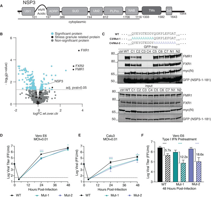
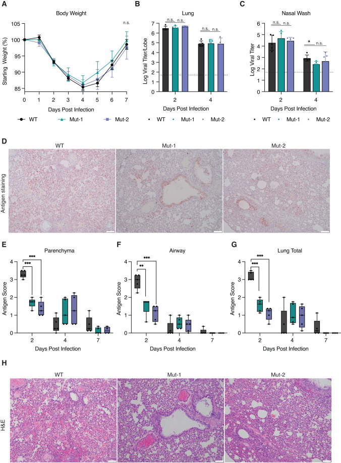
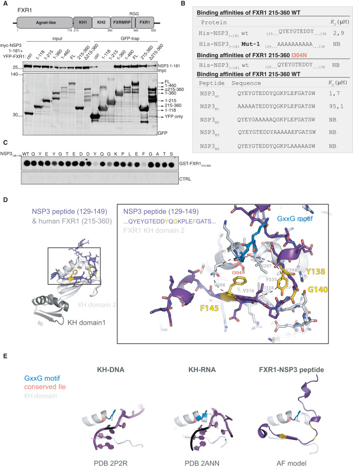
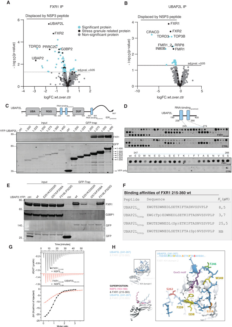
Viruses interact with numerous host factors to facilitate viral replication and to dampen antiviral defense mechanisms. We currently have a limited mechanistic understanding of how SARS-CoV-2 binds host factors and the functional role of these interactions. Here, we uncover a novel interaction between the viral NSP3 protein and the fragile X mental retardation proteins (FMRPs: FMR1, FXR1-2). SARS-CoV-2 NSP3 mutant viruses preventing FMRP binding have attenuated replication in vitro and reduced levels of viral antigen in lungs during the early stages of infection. We show that a unique peptide motif in NSP3 binds directly to the two central KH domains of FMRPs and that this interaction is disrupted by the I304N mutation found in a patient with fragile X syndrome. NSP3 binding to FMRPs disrupts their interaction with the stress granule component UBAP2L through direct competition with a peptide motif in UBAP2L to prevent FMRP incorporation into stress granules. Collectively, our results provide novel insight into how SARS-CoV-2 hijacks host cell proteins and provides molecular insight into the possible underlying molecular defects in fragile X syndrome.

Keywords: Fragile X Syndrome; NSP3; SARS-CoV-2; Stress Granules; UBAP2L.


Conflict of interest statement

VDM has filed a patent on the reverse genetic system and reporter SARS-CoV-2. The remaining authors declare no competing interests.

Figures

Figure 1
Figure 1. An NSP3–FMRPs interaction is required for efficient SARS-CoV-2 replication.
(A) Schematic of NSP3 protein with distinct domains indicated. (B) Interactome of the cytoplasmic domains of NSP3 in HeLa cells. Data from four technical replicates. (C) Interaction of NSP3 mutants with FXR1, FMR1 and myc-tagged N protein to map binding sites. Each variant has ten amino acids mutated to Alanine. Representative of two biological replicates. (D, E) VeroE6 cells or Calu3 cells were infected with the indicated SARS-CoV-2 viruses and viral titers measured at 24 and 48 h post infection (n = 6 from two experiments each with three biological replicates). (F) VeroE6 cells were pretreated with control (solid) or 100 unit of type I IFN (hashed) for 16 h and then infected with the indicated SARS-CoV-2 viruses and viral titers measured after 48 h (n = 6 from two experiments each with three biological replicates). The fold change relative to control is shown. Data information: In (B), a two-sided unpaired t test was used for statistical analysis. (DF) Statistical analysis measured by two-tailed Student’s t test: ****P < 0.0001,***P < 0.001, *P < 0.05. (DF) Data are presented as mean with SD. Source data are available online for this figure.
Figure 2
Figure 2. In vivo characterization of SARS-CoV-2 virus unable to bind FMRPs.
(A) Golden Syrian hamsters were infected with 105 plaque-forming units (PFU) of WT SARS-CoV-2 (n = 15), NSP3 mutants (n = 15), or mock (PBS, n = 15) and monitored for weight loss and signs of disease over a 7 day time course. (B, C) At days 2, 4, and 7 post infection, hamsters (n = 5 individual hamsters) were nasal washed and subsequently euthanized and tissue collected to assay viral titers from (B) lung or (C) nasal wash. (D) Representative lung tissue sections stained for viral antigen (nucleocapsid) at day 2 from WT, NSP3 Mut-1 and Mut-2 infected animals. See Fig. EV2B for full images. (EG) Antigen staining was scored on a 4 points scale for the parenchyma, airway, and by total for WT and NSP3 mutants on days However, at day 4, these trends reversed with both NSP3 mutants showing more antigen staining than 2, 4, and 7 in a blinded manner. Each data point is representative of the average score from two lung section from each hamsters in the group (n = 5 individual hamsters). (H) Day 2 lung tissue sections parallel to (D) were stained for H&E and demonstrated increased immune cell infiltrate and more severe lesions at day 2 post in WT-infected hamsters compared to NSP3 mutant-infected animals. See Fig. EV2B for full images. Data information: Statistical analysis was measured by two-tailed Student’s t test: n.s. not significant, ***P < 0.001, **P < 0.01, *P < 0.05. (AC) Mean and SEM is shown while in (EG) Min/Max plotted, center is mean, and all points shown. In (D, H), scale bar is 100 μm. Source data are available online for this figure.
Figure 3
Figure 3. NSP3 binds to the FMRP KH domains similar to how RNA binds.
(A) Schematic of FXR1 and immunopurification of the indicated FXR1 fragments and binding to NSP3 1–181 determined. Representative of two biological replicates. (B) Affinity measurements by ITC of the indicated proteins and peptides (n = 1). (C) Spot array of the indicated NSP3 peptide incubated with purified FXR1 215–360 to map critical residues required for binding. Representative of two biological replicates. (D) AlphaFold model of the FXR1-NSP3 complex with critical residues in NSP3 indicated by yellow and the I304 residue in FXR1 highlighted. (E) Comparison between KH-DNA and KH-RNA structures and the model of FXR1-NSP3. Source data are available online for this figure.
Figure 4
Figure 4. NSP3 disrupts the UBAP2L-FMRP complex.
(A) FXR1 was affinity-purified and incubated with either WT NSP3 or mutant NSP3 peptide and interactomes determined by MS to determine proteins specifically displaced by WT NSP3. Data from four technical replicates. (B) As (A) but using UBAP2L as a bait. Data from four technical replicates. (C) Schematic of UBAP2L and truncation analysis to identify FXR1 binding site (n = 1). (D) Peptide array of UBAP2L 199–400 to identify FXR1 binding region and lower part single Ala scan through UBAP2L 247–266 to identify critical residues (n = 1). (E) Immunopurification of indicated UBAP2L constructs to determine binding to FXR1 and G3BP1. Representative of two biological replicates. (F) ITC measurements of indicated UBAP2L peptides to FXR1 215–360 (n = 1). (G) Competition between NSP3 peptide and UBAP2L peptide for binding to FXR1 215–360. The black trace is NSP3 binding to FXR1 while the red trace is NSP3 binding to FXR1 preincubated with UBAP2L peptide (n = 1). (H) Alphafold model of the FXR1-UBAP2L complex highlighting critical residues in yellow and phosphorylation sites. Data information: In (A, B), the statistical comparison was done using a two-sided unpaired t test. Source data are available online for this figure.
Figure 5
Figure 5. NSP3 prevents the incorporation of FMRPs into stress granules.
(A) Analysis of FXR1 stress granule association in the presence of NSP3 WT or NSP3 A4. HeLa cells were treated 30 min with arsenite before fixation and number of FXR1 foci quantified per cell. Only cells expressing NSP3 was analyzed. (B) Similar as (A) but number of G3BP1 foci analyzed. (C) HeLa UBAP2L KO cells were complemented with UBAP2L-YFP constructs and cells treated with arsenite for 30 min before fixation. The fluorescence intensity of FXR1 to UBAP2L (GFP signal) was quantified. (D) As in (C) but staining for G3BP1. (AD) representative stills from the immunofluorescence is shown with a scale bar of 10 μM indicated in lower left corner. Combined data from three and two biological replicates is shown in (A, B), respectively. (C, D) A pool of four biological replicates is shown in the graphs. Data information: Violin plots (AD) with all points shown (A, B) with the median indicated with red line. The number of cells (A, B) or foci (C, D) analyzed per condition is indicated above the plot. Scale bars represent 10 μm. Source data are available online for this figure.
Figure EV1
Figure EV1. Analysis of FMRP-NSP3 interaction.
(A) The indicated NSP3 fragments fused to YFP was expressed and purified from HeLa cells and binding to FXR1 monitored. Representative of two biological replicates. (B) A panel of NSP3 N-terminal fragments from different coronaviruses were expressed and purified from HeLa cells and binding to FXR1 determined by western blotting. Representative of two biological replicates. (C) Alignment of the NSP3 sequence binding to FMRPs from different coronaviruses.
Figure EV2
Figure EV2. Histopathology of hamster infected with WT or NSP3 Mutants.
(A) Schematic of in vivo experiment (generated with BioRender). (B) H&E and viral antigen (nucleocapsid) immunohistochemical staining of lung of hamsters infected mock (PBS) or with 105 pfu of WT, NSP3 Mut-1, or NSP3 Mut-2 SARS-CoV-2 at 2 days post infection. WT infection shows extensive viral infection and damage; both NSP3 mutants have focal disease and less damage. No damage observed in mock infected samples. Images from representative section from a single hamster in each group (mock, WT, NSP3 Mut-1, & NSP3 mut-2). For larger magnification see Fig. 2. Data information: Scale bar of 200 μm is indicated in the lower right corner.
Figure EV3
Figure EV3. A direct interaction between NSP3 and FXR1.
(A) The indicated FXR1 YFP constructs were co-expressed with myc-NSP3 1–181 in HeLa cells and affinity-purified using YFP affinity beads. The binding to NSP3 was monitored by probing for myc. Representative of two biological replicates. (B) Size-exclusion chromatography of GST-NSP3 WT 1–181, FXR1 215–360 either alone or in combination. The elution volume is indicated on top and Coomasie stained gels of fractions shown. Representative of two biological replicates. (C) Table of ITC values obtained for the indicated FXR1 fragments binding to GST-NSP3 1–181 or the FXR1 binding peptide from old alphaviruses (n = 1). (D) Confidence plots of AlphaFold model of NSP3 peptide binding to full-length FXR1.
Figure EV4
Figure EV4. Interaction of FXR1 to UBAP2L.
(A) ITC measurements of a reported RNA binding to FXR1. Binding to FXR1 215–360 was monitored and as a control the SARS-CoV-2 N protein (n = 1). (B) Mass spectrometry analysis of the interactomes of affinity-purified YFP-tagged FXR1 WT and FXR1 I304N. Proteins specifically binding to FXR1 WT indicated in the volcano plot. Data from 4 technical repeats. (C) The indicated YFP-tagged FXR1 proteins were expressed and purified from HeLa cells and binding to UBAP2L determined by western blot. Representative of 2 biological replicates. (D) A panel of YFP-UBAP2L constructs were expressed and purified from HeLa cells and binding to FXR1 determined. Representative of 2 biological replicates. Data information: In (B) a two-sided unpaired t test was used for statistical analysis.
Figure EV5
Figure EV5. Analysis of FXR1 localization to stress granules.
(A) VeroE6 cells infected with SARS-CoV-2 or uninfected were fixed and stained for FXR1 and the viral N protein. Representative images shown from one experiment. (B) The number of FXR1 foci in infected cells was determined and correlated with total level of N protein. (C) The total level of FXR1 was determined in infected cells and plotted against total levels of N. (D) YFP-tagged FXR1 proteins were expressed in HeLa cells and filmed by live-cell microscopy. Stress granule formation was induced by arsenite and 30 min after addition the localization and morphology of FXR1 foci was monitored. Phenotypes are plotted as percentage. Scores of two individual experiments are shown. The total number of cells analyzed per condition are indicated. Representative images are shown. Data information: In (A) scale bar is 20 μm and in (D) it is 10 μm.

References

    1. Almasy KM, Davies JP, Plate L. Comparative host interactomes of the SARS-CoV-2 nonstructural protein 3 and human coronavirus homologs. Mol Cell Proteomics. 2021;20:100120. - PMC - PubMed
    1. Almazan F, Galan C, Enjuanes L. The nucleoprotein is required for efficient coronavirus genome replication. J Virol. 2004;78:12683–12688. - PMC - PubMed
    1. Ascano M, Jr., Mukherjee N, Bandaru P, Miller JB, Nusbaum JD, Corcoran DL, Langlois C, Munschauer M, Dewell S, Hafner M, et al. FMRP targets distinct mRNA sequence elements to regulate protein expression. Nature. 2012;492:382–386. - PMC - PubMed
    1. Bessa LM, Guseva S, Camacho-Zarco AR, Salvi N, Maurin D, Perez LM, Botova M, Malki A, Nanao M, Jensen MR, et al. The intrinsically disordered SARS-CoV-2 nucleoprotein in dynamic complex with its viral partner nsp3a. Sci Adv. 2022;8:eabm4034. - PMC - PubMed
    1. Biswal M, Lu J, Song J. SARS-CoV-2 nucleocapsid protein targets a conserved surface groove of the NTF2-like domain of G3BP1. J Mol Biol. 2022;434:167516. - PMC - PubMed

Publication types

MeSH terms