. 2024 Jan 5;26(1):1.
doi: 10.1186/s12575-023-00228-9.

MBD3 promotes epithelial-mesenchymal transition in gastric cancer cells by upregulating ACTG1 via the PI3K/AKT pathway

Affiliations

MBD3 promotes epithelial-mesenchymal transition in gastric cancer cells by upregulating ACTG1 via the PI3K/AKT pathway

Huizhi Wang et al. Biol Proced Online. .

Abstract

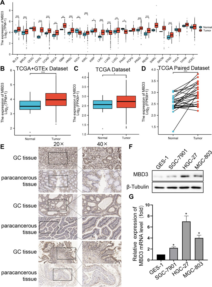
Background: Gastric cancer (GC) is a common malignancy and a leading cause of cancer-related death with high morbidity and mortality. Methyl-CpG binding domain protein 3 (MBD3), a key epigenetic regulator, is abnormally expressed in several cancers, participating in progression and metastasis. However, the role of MBD3 in GC remains unknown.

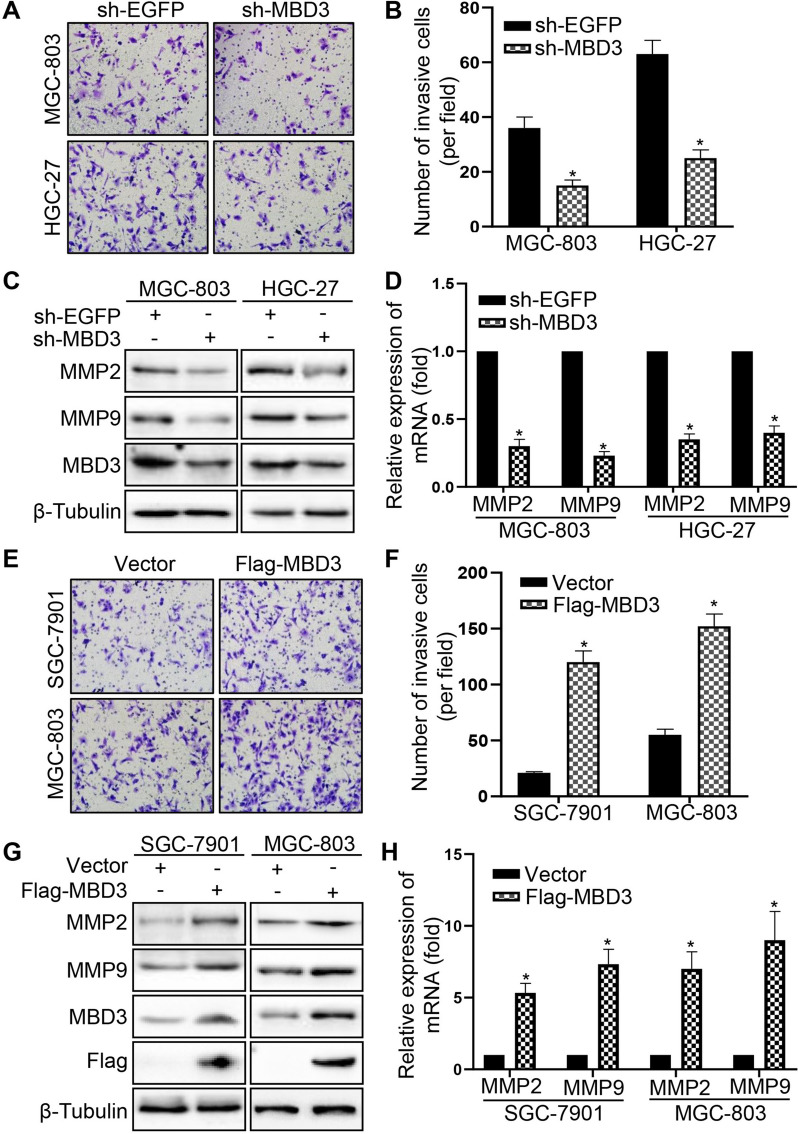
Methods: MBD3 expression was assessed via public databases and validated by western blotting and quantitative real-time polymerase chain reaction (qRT-PCR). The prognosis of MBD3 was analysed via bioinformatics based on the TCGA dataset. The migration, invasion and proliferation of GC cells were examined by transwell, wound healing, cell counting kit (CCK)-8, colony-formation and xenograft mouse models. Epithelial-mesenchymal transition (EMT) and phosphatidylinositide 3-kinases/ protein Kinase B (PI3K/AKT) pathway markers were evaluated by Western blotting. RNA sequencing was used to identify the target of MBD3.

Results: MBD3 expression was higher in GC tissues and cells than in normal tissues and cells. Additionally, high MBD3 levels were associated with poor prognosis in GC patients. Subsequently, we proved that MBD3 enhanced the migration, invasion and proliferation abilities of GC cells. Moreover, western blot results showed that MBD3 promoted EMT and activated the PI3K/AKT pathway. RNA sequencing analysis showed that MBD3 may increase actin γ1 (ACTG1) expression to promote migration and proliferation in GC cells.

Conclusion: MBD3 promoted migration, invasion, proliferation and EMT by upregulating ACTG1 via PI3K/AKT signaling activation in GC cells and may be a potential diagnostic and prognostic target.

Keywords: ACTG1; EMT; Gastric cancer; MBD3; PI3K/AKT.


Conflict of interest statement

The authors declare no competing interests.

Figures

Fig. 1
Fig. 1
The expression of MBD3 in GC tissues and cells. A MBD3 expression in different cancer types based on the TCGA database. B The expression of MBD3 was higher in GC than in normal gastric tissues from the GTEx database (normal 174 patients) and (C) TCGA database (normal 36 patients, tumor 414 patients). D MBD3 expression in TCGA-paired GC and normal tissues (normal 27 patients, tumor 27 patients). E IHC staining for MBD3 in GC and matched paracancerous tissues from three representative patients. Original magnifications 20 × and 40 × (inset panels). F, G Western blotting and qRT-PCR were used to measure the relative expression levels of MBD3 in different gastric cell lines (GES-1, SGC-7901, HGC-27 and MGC-803). (*P < 0.05, **P < 0.01, ***P < 0.001)
Fig. 2
Fig. 2
MBD3 enhanced the migration ability of GC cells. A, B Transwell assays were used to detect the migration ability of MGC-803 and HGC-27 cells transfected with sh-EGFP and sh-MBD3. C, D Transwell assays were used to examine the migration ability of SGC-7901 and MGC-803 cells transfected with vector and Flag-MBD3. E Wound healing assay showed the migration ability of MGC-803 and HGC-27 cells transfected with sh-EGFP and sh-MBD3. F Histograms represent the analysis of the wound healing rate in Fig. 2E. G Wound healing assay showed the migration ability of SGC-7901 and MGC-803 cells transfected with vector and Flag-MBD3. H Histograms represent the analysis of the wound healing rate in Fig. 2H. (*P < 0.05)
Fig. 3
Fig. 3
MBD3 enhanced the invasion ability of GC cells. A, B Transwell invasion assays showed the invasion ability of MGC-803 and HGC-27 cells transfected with sh-EGFP and sh-MBD3. C, D MMP2 and MMP9 expression were tested by western blot and qRT-PCR in MGC-803 and HGC-27 cells transfected with sh-EGFP and sh-MBD3. E, F Transwell invasion assays were used to examine the invasion ability of SGC-7901 and MGC-803 cells transfected with vector and Flag-MBD3. G, H MMP2 and MMP9 expression were tested by western blot and qRT-PCR in SGC-7901 and MGC-803 cells transfected with vector and Flag-MBD3. (*P < 0.05)
Fig. 4
Fig. 4
MBD3 promoted GC cell proliferation. A, B MBD3 downregulation inhibited the proliferation of MGC-803 and HGC-27 cells. C, D MBD3 upregulation promoted the proliferation of SGC-7901 and MGC-803 cells. All experiments were performed using at least three biological replicates. E, F MBD3 downregulation weakened colony formation in MGC-803 and HGC-27 cells. G, H MBD3 upregulation enhanced colony formation in SGC-7901 and MGC-803 cells. I, J MGC-803 cells with MBD3 upregulation were injected (2 × 10.6 cells/site) subcutaneously into a mouse, and the tumor volume was measured weekly (n = 5 mice). (*P < 0.05)
Fig. 5
Fig. 5
MBD3 promoted EMT in GC cells via activation of the PI3K/AKT pathway. A The expression of EMT proteins was examined in sh-MBD3-MGC-803 and HGC-27 cells by western blot. B The expression of EMT proteins was examined in Flag-MBD3-SGC-7901 and MGC-803 cells by western blot. C Western blotting was used to determine PI3K/AKT pathway protein expression in MGC-803 and HGC-27 cells after transfection with sh-EGFP or sh-MBD3. D Western blotting was used to test PI3K/AKT pathway protein expression in SGC-7901 and MGC-803 cells after transfection with vector or Flag-MBD3
Fig. 6
Fig. 6
Correlation between MBD3 and ACTG1 and the expression of ACTG1 in GC tissues and cells. A Volcano map analysis showed differentially expressed genes in response to sh-EGFP and sh-MBD3 treatment measured by RNA-seq. Green: low expression levels. Red: high expression levels. B The relationship between MBD3 and ACTG1 based on the TCGA dataset. C ACTG1 expression in different cancer types based on the TCGA database. D ACTG1 expression in TCGA-paired cancer and normal tissues. E The expression of ACTG1 was higher in GC than in normal gastric tissues from GTEx database (normal 174 patients) and TCGA database (normal 36 patients, tumor 414 patients). F ACTG1 expression in TCGA-paired GC and normal tissues (normal 27 patients, tumor 27 patients). G Western blotting was used to detect the relative expression levels of ACTG1 in different gastric cell lines (GES-1, SGC-7901, HGC-27 and MGC-803). (*P < 0.05, **P < 0.01, ***P < 0.001)
Fig. 7
Fig. 7
MBD3 promoted GC cell migration and growth by upregulating ACTG1 expression. A, B The migration ability of MGC-803 and HGC-27 cells transfected with plasmids was tested by transwell assays. C, D Transwell assays were used to examine the migration ability of SGC-7901 and MGC-803 cells after transfection. E, F Colony formation was detected in MGC-803 and HGC-27 cells transfected with plasmids. G, H Colony formation was detected in SGC-7901 and MGC-803 cells after transfection. (*P < 0.05)

References

    1. Siegel RL, Miller KD, Wagle NS, Jemal A. Cancer statistics, 2023. CA Cancer J Clin. 2023;73(1):17–48. doi: 10.3322/caac.21763. - DOI - PubMed
    1. Alsina M, Arrazubi V, Diez M, Tabernero J. Current developments in gastric cancer: from molecular profiling to treatment strategy. Nat Rev Gastroenterol Hepatol. 2023;20(3):155–170. doi: 10.1038/s41575-022-00703-w. - DOI - PubMed
    1. Grady WM, Yu M, Markowitz SD. Epigenetic Alterations in the Gastrointestinal Tract: Current and Emerging Use for Biomarkers of Cancer. Gastroenterology. 2021;160(3):690–709. doi: 10.1053/j.gastro.2020.09.058. - DOI - PMC - PubMed
    1. Sun L, Zhang H, Gao P. Metabolic reprogramming and epigenetic modifications on the path to cancer. Protein Cell. 2022;13(12):877–919. doi: 10.1007/s13238-021-00846-7. - DOI - PMC - PubMed
    1. Li C, Han T, Li Q, Zhang M, Guo R, Yang Y, et al. MKRN3-mediated ubiquitination of Poly(A)-binding proteins modulates the stability and translation of GNRH1 mRNA in mammalian puberty. Nucleic Acids Res. 2021;49(7):3796–3813. doi: 10.1093/nar/gkab155. - DOI - PMC - PubMed