

In-phasic cytosolic-nuclear Ca2+ rhythms in suprachiasmatic nucleus neurons
- PMID: 38178840
- PMCID: PMC10765503
- DOI: 10.3389/fnins.2023.1323565
In-phasic cytosolic-nuclear Ca2+ rhythms in suprachiasmatic nucleus neurons
Abstract
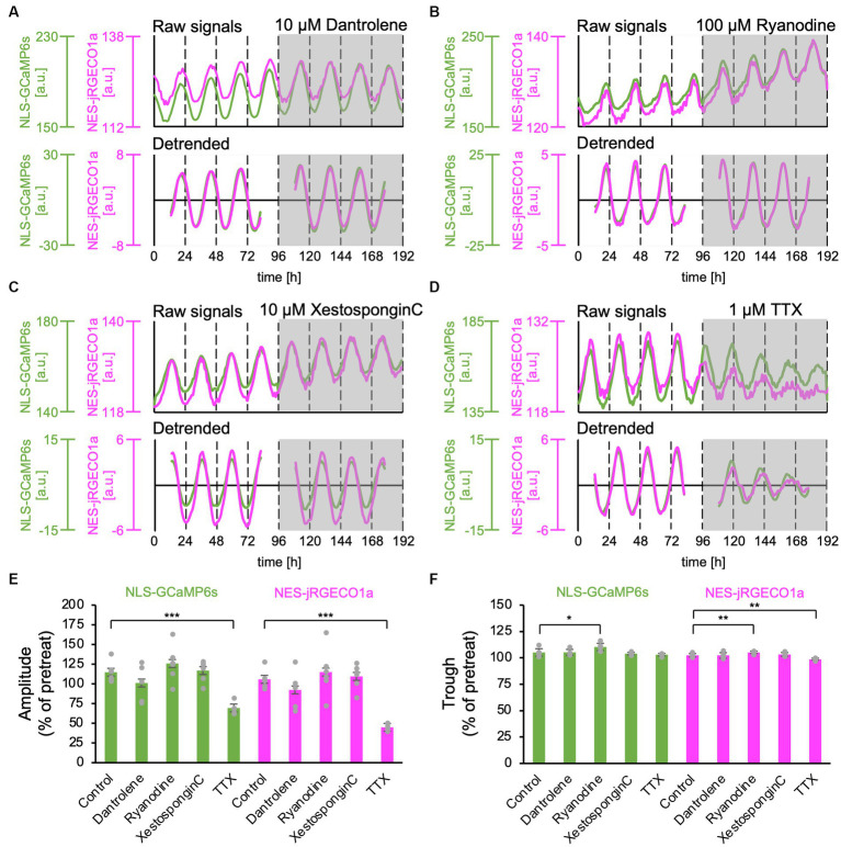
The suprachiasmatic nucleus (SCN) of the hypothalamus is the master circadian clock in mammals. SCN neurons exhibit circadian Ca2+ rhythms in the cytosol, which is thought to act as a messenger linking the transcriptional/translational feedback loop (TTFL) and physiological activities. Transcriptional regulation occurs in the nucleus in the TTFL model, and Ca2+-dependent kinase regulates the clock gene transcription. However, the Ca2+ regulatory mechanisms between cytosol and nucleus as well as the ionic origin of Ca2+ rhythms remain unclear. In the present study, we monitored circadian-timescale Ca2+ dynamics in the nucleus and cytosol of SCN neurons at the single-cell and network levels. We observed robust nuclear Ca2+ rhythm in the same phase as the cytosolic rhythm in single SCN neurons and entire regions. Neuronal firing inhibition reduced the amplitude of both nuclear and cytosolic Ca2+ rhythms, whereas blocking of Ca2+ release from the endoplasmic reticulum (ER) via ryanodine and inositol 1,4,5-trisphosphate (IP3) receptors had a minor effect on either Ca2+ rhythms. We conclude that the in-phasic circadian Ca2+ rhythms in the cytosol and nucleus are mainly driven by Ca2+ influx from the extracellular space, likely through the nuclear pore. It also raises the possibility that nuclear Ca2+ rhythms directly regulate transcription in situ.
Keywords: SCN; circadian clock; imaging; intracellular Ca2+; nucleus; organelle.
Copyright © 2023 Hiro, Kobayashi, Nemoto and Enoki.
Conflict of interest statement
The authors declare that the research was conducted in the absence of any commercial or financial relationships that could be construed as a potential conflict of interest.
Figures


Similar articles
-
Calcium Circadian Rhythmicity in the Suprachiasmatic Nucleus: Cell Autonomy and Network Modulation.eNeuro. 2017 Aug 18;4(4):ENEURO.0160-17.2017. doi: 10.1523/ENEURO.0160-17.2017. eCollection 2017 Jul-Aug. eNeuro. 2017. PMID: 28828400 Free PMC article.
-
Bmal1 is an essential regulator for circadian cytosolic Ca²⁺ rhythms in suprachiasmatic nucleus neurons.J Neurosci. 2014 Sep 3;34(36):12029-38. doi: 10.1523/JNEUROSCI.5158-13.2014. J Neurosci. 2014. PMID: 25186748 Free PMC article.
-
Calcium dynamics and circadian rhythms in suprachiasmatic nucleus neurons.Neuroscientist. 2004 Aug;10(4):315-24. doi: 10.1177/10738584031262149. Neuroscientist. 2004. PMID: 15271259 Review.
-
Circadian dynamics of cytosolic and nuclear Ca2+ in single suprachiasmatic nucleus neurons.Neuron. 2003 Apr 24;38(2):253-63. doi: 10.1016/s0896-6273(03)00164-8. Neuron. 2003. PMID: 12718859
-
Neural circuits in the central circadian clock and their regulation of sleep and wakefulness in mammals.Neurosci Res. 2022 Sep;182:1-6. doi: 10.1016/j.neures.2022.05.005. Epub 2022 May 18. Neurosci Res. 2022. PMID: 35597406 Review.
References
LinkOut - more resources
Full Text Sources
Miscellaneous