. 2023 Dec 18:10:1263913.
doi: 10.3389/fmolb.2023.1263913. eCollection 2023.

Transcriptional changes during isoproterenol-induced cardiac fibrosis in mice

Affiliations

Transcriptional changes during isoproterenol-induced cardiac fibrosis in mice

Disha Nanda et al. Front Mol Biosci. .

Abstract

Introduction: β-adrenergic stimulation using β-agonists such as isoproterenol has been routinely used to induce cardiac fibrosis in experimental animal models. Although transcriptome changes in surgical models of cardiac fibrosis such as transverse aortic constriction (TAC) and coronary artery ligation (CAL) are well-studied, transcriptional changes during isoproterenol-induced cardiac fibrosis are not well-explored. Methods: Cardiac fibrosis was induced in male C57BL6 mice by administration of isoproterenol for 4, 8, or 11 days at 50 mg/kg/day dose. Temporal changes in gene expression were studied by RNA sequencing. Results and discussion: We observed a significant alteration in the transcriptome profile across the different experimental groups compared to the saline group. Isoproterenol treatment caused upregulation of genes associated with ECM organization, cell-cell contact, three-dimensional structure, and cell growth, while genes associated with fatty acid oxidation, sarcoplasmic reticulum calcium ion transport, and cardiac muscle contraction are downregulated. A number of known long non-coding RNAs (lncRNAs) and putative novel lncRNAs exhibited differential regulation. In conclusion, our study shows that isoproterenol administration leads to the dysregulation of genes relevant to ECM deposition and cardiac contraction, and serves as an excellent alternate model to the surgical models of heart failure.

Keywords: ECM remodeling; cardiac fibrosis; isoproterenol; lncRNAs; transcriptomics.


Conflict of interest statement

The authors declare that the research was conducted in the absence of any commercial or financial relationships that could be construed as a potential conflict of interest. The authors declared that they were an editorial board member of Frontiers, at the time of submission. This had no impact on the peer review process and the final decision.

Figures

FIGURE 1
FIGURE 1
Induction of cardiac hypertrophy with a subcutaneous dose of isoproterenol. (A) Overview of the experimental setup where 12-week-old C57BL6 male mice were administered with 50 mg/kg/day isoproterenol for 4, 8, and 11 days, and the heart samples were collected for histology and RNA isolation. (B) Representative images of the hearts that were collected at different time points after isoproterenol treatment. (C) Graph showing heart weights normalized to body weight (BW) and tibia length (TL) at definitive timepoints. (D) Representative WGA staining and cardiomyocyte size analysis. (E) Picrosirius red staining indicative of ECM remodeling among the three groups and its quantification (n>=3) (marked in white arrow). (F) qRT PCR showing the levels of heart failure markers like ANP and β-MHC. * p value < 0.05, ** p value < 0.01, *** p value < 0.001, and **** p value < 0.0001. One way ANOVA followed by Dunnett’s test/Fishers test was performed to compute statistical differences between saline and isoproterenol groups.
FIGURE 2
FIGURE 2
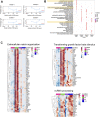
Transcriptomic analysis of the isoproterenol-treated hearts. RNA sequencing shows differential expression of common genes (DEGs) across the three groups. (A) Heatmap showing the DEGs across the three timepoints compared to control (LFC = 0, p < 0.05). (B) Gene Ontology showing the common pathways affected during isoproterenol-induced cardiac remodeling.
FIGURE 3
FIGURE 3
Time-course analysis of DEGS (A) Time-course analysis shows significant changes in gene expression over time. The gene expression profile was grouped into four clusters with distinct temporal profiles. (B) Gene Ontology (GO) of the genes obtained from the aforementioned four clusters. (C) Heatmap depicting the gene expression patterns of few pathways from each clusters.
FIGURE 4
FIGURE 4
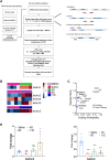
Expression profile of novel long non-coding RNA transcripts. (A) Schematic representation of the novel lncRNA prediction pipeline. (B) RNA sequencing shows differential expression of different novel lncRNAs across three groups. (C) Analysis of coding potential of Isolncs by CPAT compared to known coding and non-coding RNAs. (D) qRT PCR validation of Isolncs 2 and 4 in isoproterenol-treated animals. * p value < 0.05, ** p value < 0.01, and *** p value < 0.001. One-way ANOVA followed by Dunnett’s test was performed to compute statistical differences between saline and isoproterenol groups.
FIGURE 5
FIGURE 5
UCSC screenshots of ATAC-seq and qRT-PCR showing expression patterns of Isolncs 2 and 4 in different tissues and subcellular fractions. (A) UCSC Genome Browser (mouse mm10) screenshots using “open chromatin track” showing ATAC sequencing across the heart at E11 to E16 and P0 along with the liver and stomach. (B) Tissue profiling of Isolncs 2 and 4 in adult mice. (C) Expression of the novel Isolncs in a subcellular fraction of the heart (CM vs. CF).

References

    1. Ackers-Johnson M., Li P. Y., Holmes A. P., O’Brien S.-M., Pavlovic D., Foo R. S. (2016). A simplified, Langendorff-free method for concomitant isolation of viable cardiac myocytes and nonmyocytes from the adult mouse heart. Circulation Res. 119 (8), 909–920. 10.1161/CIRCRESAHA.116.309202 - DOI - PMC - PubMed
    1. Almehmadi F., Joncas S. X., Nevis I., Zahrani M., Bokhari M., Stirrat J., et al. (2014). Prevalence of myocardial fibrosis patterns in patients with systolic dysfunction: prognostic significance for the prediction of sudden cardiac arrest or appropriate implantable cardiac defibrillator therapy. Circ. Cardiovasc. Imaging 7 (4), 593–600. 10.1161/CIRCIMAGING.113.001768 - DOI - PubMed
    1. Azevedo P. S., Polegato B. F., Minicucci M. F., Paiva S. A., Zornoff L. A. (2015). Cardiac remodeling: concepts, clinical impact, pathophysiological mechanisms and pharmacologic treatment. Arq. Bras. Cardiol. 106, 62–69. 10.5935/abc.20160005 - DOI - PMC - PubMed
    1. Azlan A., Obeidat S. M., Yunus M. A., Azzam G. (2019). Systematic identification and characterization of Aedes aegypti long noncoding RNAs (lncRNAs). Sci. Rep. 9 (1), 12147. 10.1038/s41598-019-47506-9 - DOI - PMC - PubMed
    1. Bäck N., Luxmi R., Powers K. G., Mains R. E., Eipper B. A. (2020). Peptidylglycine α-amidating monooxygenase is required for atrial secretory granule formation. Proc. Natl. Acad. Sci. 117 (30), 17820–17831. 10.1073/pnas.2004410117 - DOI - PMC - PubMed