

Electroencephalographic Abnormalities in a Patient Suffering from Long-Term Neuropsychological Complications following SARS-CoV-2 Infection
- PMID: 38179211
- PMCID: PMC10764086
- DOI: 10.1159/000535241
Electroencephalographic Abnormalities in a Patient Suffering from Long-Term Neuropsychological Complications following SARS-CoV-2 Infection
Abstract
Introduction: Emotional apathy has recently been identified as a common symptom of long COVID. While recent meta-analyses have demonstrated generalized EEG slowing with the emergence of delta rhythms in patients hospitalized for severe SARS-CoV-2 infection, no EEG study or dopamine transporter scintigraphy (DaTSCAN) has been performed in patients with long COVID presenting with apathy. The objective of this case report was to explore the pathophysiology of neuropsychological symptoms in long COVID.
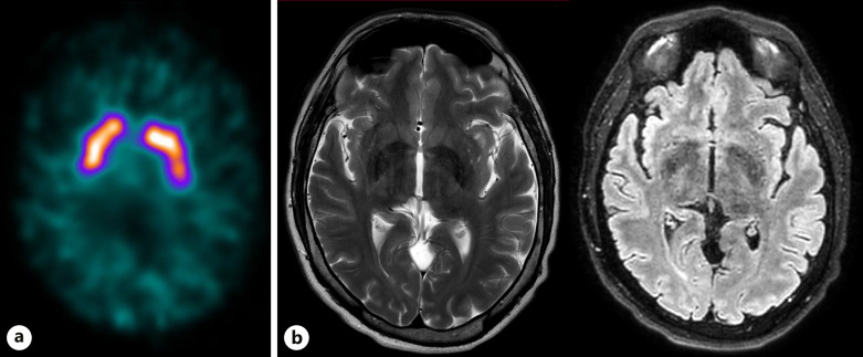
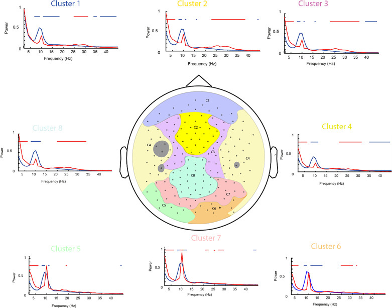
Case presentation: A 47-year-old patient who developed a long COVID with prominent apathy following an initially clinically mild SARS-CoV-2 infection underwent neuropsychological assessment, cerebral MRI, DaTSCAN, and resting-state high-density EEG 7 months after SARS-CoV-2 infection. The EEG data were compared to those of 21 healthy participants. The patient presented with apathy, cognitive difficulties with dysexecutive syndrome, moderate attentional and verbal episodic memory disturbances, and resolution of premorbid mild gaming disorder, mild mood disturbances, and sleep disturbances. His MRI and DaTSCAN were unremarkable. EEG revealed a complex pattern of oscillatory abnormalities compared to the control group, with a strong increase in whole-scalp delta and beta band activity, as well as a decrease in alpha band activity. Overall, these effects were more prominent in the frontal-central-temporal region.
Conclusion: These results suggest widespread changes in EEG oscillatory patterns in a patient with long COVID characterized by neuropsychological complications with prominent apathy. Despite the inherent limitations of a case report, these results suggest dysfunction in the cortical networks involved in motivation and emotion.
Keywords: Apathy; EEG; Long COVID; Neuropsychological disturbances.
© 2023 The Author(s). Published by S. Karger AG, Basel.
Conflict of interest statement
The authors declare that they have no conflicts of interest relevant to this work to declare.
Figures
Similar articles
-
Myoclonus status revealing COVID 19 infection.Seizure. 2023 Jan;104:12-14. doi: 10.1016/j.seizure.2022.11.010. Epub 2022 Nov 22. Seizure. 2023. PMID: 36446232 Free PMC article.
-
Letter to the Editor: Depression As The First Symptom Of Frontal Lobe Grade 2 Malignant Glioma.Turk Psikiyatri Derg. 2022 Summer;33(2):143-145. doi: 10.5080/u25957. Turk Psikiyatri Derg. 2022. PMID: 35730515 English, Turkish.
-
Persistence and emergence of new neuropsychological deficits following SARS-CoV-2 infection: A follow-up assessment of the Geneva COVID-COG cohort.J Glob Health. 2024 Mar 8;14:05008. doi: 10.7189/jogh.14.05008. J Glob Health. 2024. PMID: 38452292 Free PMC article.
-
What do we mean by long COVID? A scoping review of the cognitive sequelae of SARS-CoV-2 infection.Eur J Neurol. 2023 Dec;30(12):3968-3978. doi: 10.1111/ene.16027. Epub 2023 Aug 26. Eur J Neurol. 2023. PMID: 37540896
-
COVID-19-Associated Neurological Manifestations: An Emerging Electroencephalographic Literature.Front Physiol. 2021 Feb 19;11:622466. doi: 10.3389/fphys.2020.622466. eCollection 2020. Front Physiol. 2021. PMID: 33679425 Free PMC article. Review.
References
-
- Voruz P, Allali G, Benzakour L, Nuber-Champier A, Thomasson M, Jacot de Alcântara I, et al. . Long COVID neuropsychological deficits after severe, moderate, or mild infection. Clin Transl Neurosci. 2022;6(2):9.
Publication types
LinkOut - more resources
Full Text Sources
Miscellaneous
