. 2024 Feb 2;134(3):307-324.
doi: 10.1161/CIRCRESAHA.123.323365. Epub 2024 Jan 5.

Sox9 Accelerates Vascular Aging by Regulating Extracellular Matrix Composition and Stiffness

Affiliations

Sox9 Accelerates Vascular Aging by Regulating Extracellular Matrix Composition and Stiffness

Maria Faleeva et al. Circ Res. .

Abstract

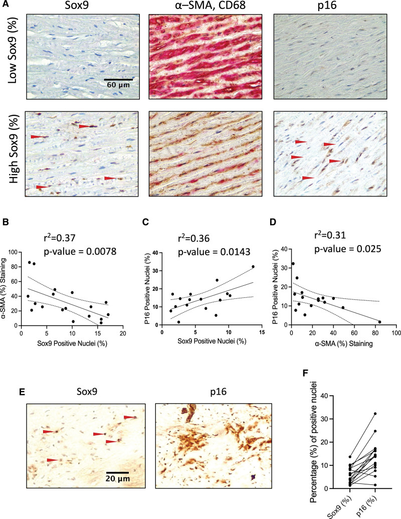
Background: Vascular calcification and increased extracellular matrix (ECM) stiffness are hallmarks of vascular aging. Sox9 (SRY-box transcription factor 9) has been implicated in vascular smooth muscle cell (VSMC) osteo/chondrogenic conversion; however, its relationship with aging and calcification has not been studied.

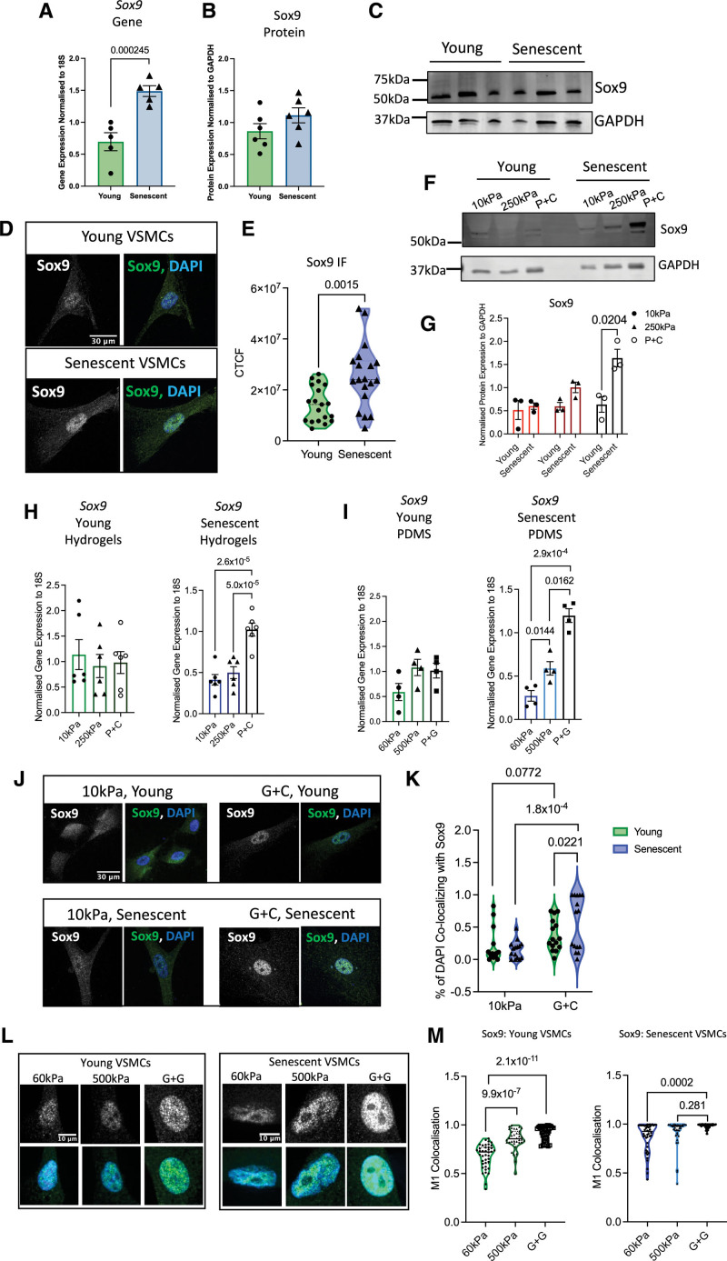
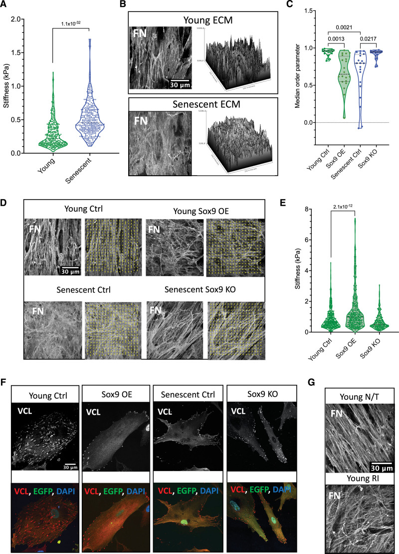
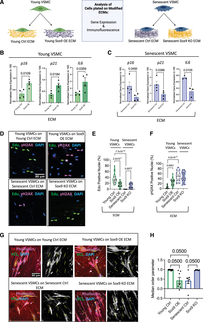
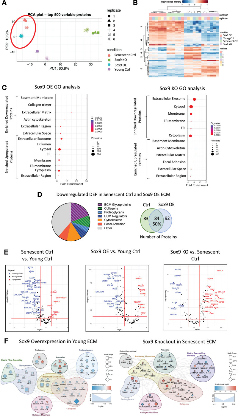
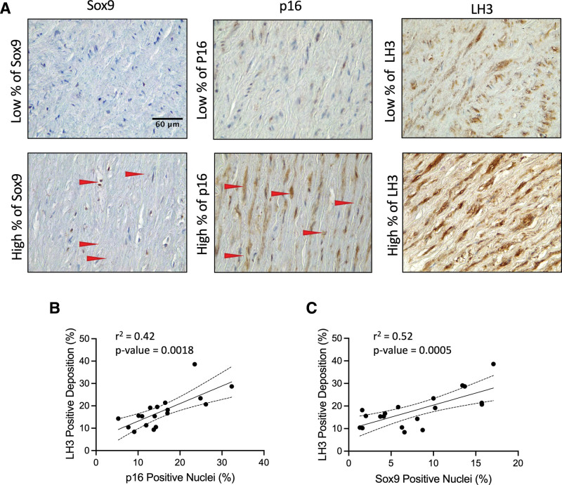
Methods: Immunohistochemistry was performed on human aortic samples from young and aged patients. Young and senescent primary human VSMCs were induced to produce ECM, and Sox9 expression was manipulated using adenoviral overexpression and depletion. ECM properties were characterized using atomic force microscopy and proteomics, and VSMC phenotype on hydrogels and the ECM were examined using confocal microscopy.

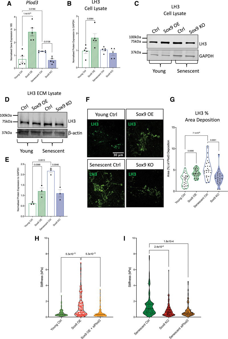
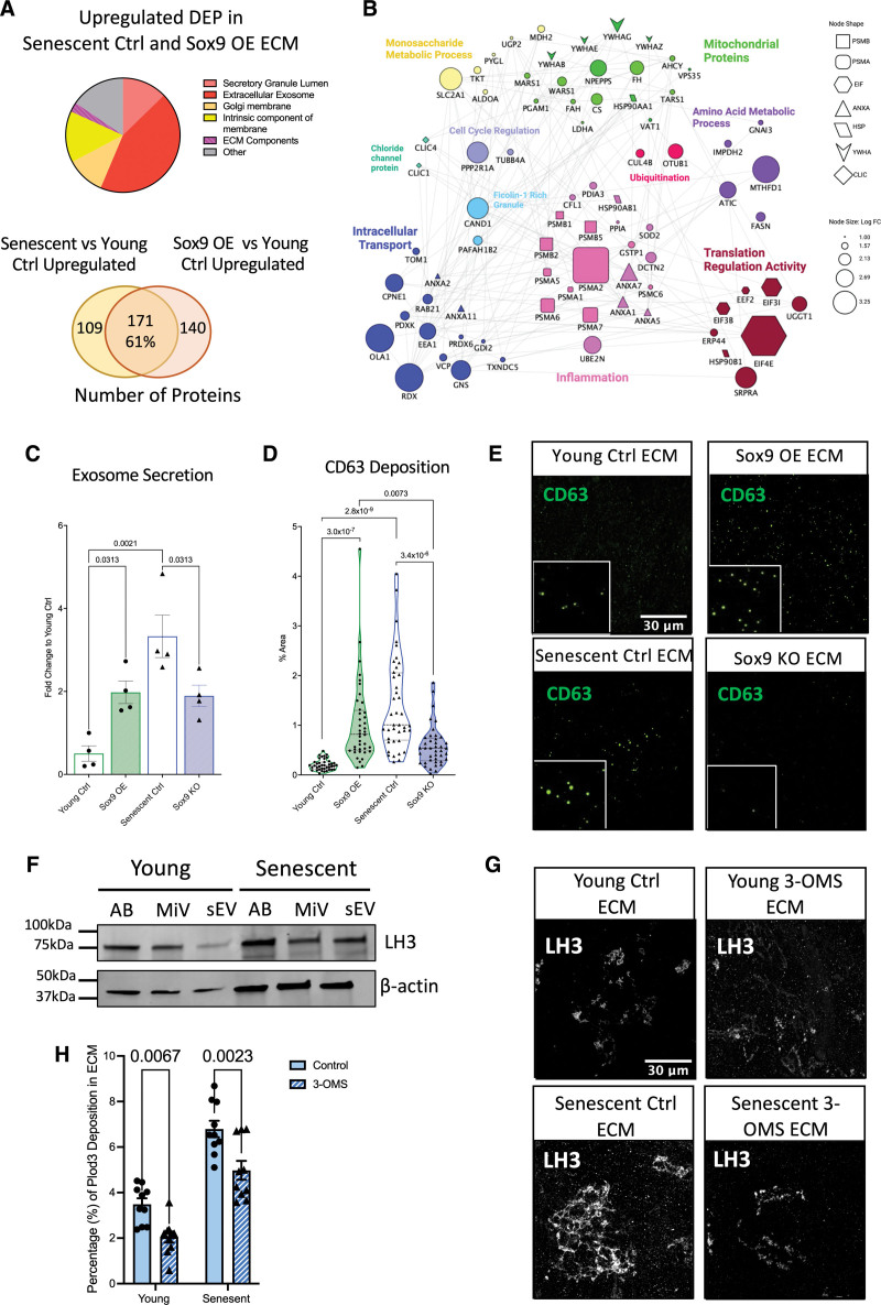
Results: In vivo, Sox9 was not spatially associated with vascular calcification but correlated with the senescence marker p16 (cyclin-dependent kinase inhibitor 2A). In vitro Sox9 showed mechanosensitive responses with increased expression and nuclear translocation in senescent cells and on stiff matrices. Sox9 was found to regulate ECM stiffness and organization by orchestrating changes in collagen (Col) expression and reducing VSMC contractility, leading to the formation of an ECM that mirrored that of senescent cells. These ECM changes promoted phenotypic modulation of VSMCs, whereby senescent cells plated on ECM synthesized from cells depleted of Sox9 returned to a proliferative state, while proliferating cells on a matrix produced by Sox9 expressing cells showed reduced proliferation and increased DNA damage, reiterating features of senescent cells. LH3 (procollagen-lysine, 2-oxoglutarate 5-dioxygenase 3) was identified as an Sox9 target and key regulator of ECM stiffness. LH3 is packaged into extracellular vesicles and Sox9 promotes extracellular vesicle secretion, leading to increased LH3 deposition within the ECM.

Conclusions: These findings highlight the crucial role of ECM structure and composition in regulating VSMC phenotype. We identify a positive feedback cycle, whereby cellular senescence and increased ECM stiffening promote Sox9 expression, which, in turn, drives further ECM modifications to further accelerate stiffening and senescence.

Keywords: atherosclerosis; calcium; extracellular matrix; extracellular vesicles; transcription factors.


Conflict of interest statement

Disclosures None.

Figures

Figure 1.
Figure 1.
Sox9 (SRY-box transcription factor 9) expression in vascular smooth muscle cells (VSMCs) correlates with increased cellular senescence and decreased expression of α-smooth muscle actin (α-SMA). A, Sox9, α-SMA, Cluster of Differentiation 68 (CD68), and p16 (cyclin-dependent kinase inhibitor 2A) staining in the medial layer of human aortic samples. B, Correlation of Sox9 positive nuclei with α-SMA (n=18), (C) p16 positive nuclei (n=16), and (D) α-SMA with p16 positive nuclei (n=16). Normality was validated via the Shapiro-Wilk test. Correlation was performed using the Pearson test. E, Immunohistochemistry of serial sections stained for Sox9 and p16. Red arrows highlight positively stained nuclei. F, Bar graph showing percentages of Sox9 and p16 nuclei in each sample.
Figure 2.
Figure 2.
Sox9 (SRY-box transcription factor 9) is mechanosensitive in senescent vascular smooth muscle cells (VSMCs). A through C, Sox9 gene and protein expression in young and senescent VSMCs (n=5, 6 from 3 isolates). Normality was confirmed with the Shapiro-Wilk test; 2-way ANOVA, false rate discovery (FDR) correction, and q-values are shown. D and E, Immunofluorescence (IF) in young and senescent VSMCs and quantification of Sox9 corrected total cell fluorescence (CTCF; n=18 from 3 isolates). Normality was confirmed with the Shapiro-Wilk test; 2-way ANOVA, FDR correction, and q-values are shown. F and G, Sox9 protein expression in young and senescent VSMCs on hydrogels and collagen-coated plastic (P+C; n=3). Normality was confirmed with the Shapiro-Wilk test; 2-way ANOVA, FDR correction, and q-values are shown. H, Sox9 gene expression in young and senescent VSMCs on different substrates (n=6 technical repeats from 3 isolates) and (I) 60- and 500-kPa Poly(dimethylsiloxane) (PDMS) gels and gelatin-coated plastic (P+G; n=4 technical repeats from 3 isolates). Normality was confirmed via the Shapiro-Wilk test; 2-way ANOVA, FDR correction, and q-values are shown. J through M, Quantification of Sox9 nuclear colocalization and IF in young and senescent VSMCs on different substrates (J and K) 10- and 250-kPa hydrogels and glass coated with collagen (G+C; n=15 technical repeats from 3 isolates) and (L and M) 60- and 500-kPa PDMS gels and glass coated with gelatin (G+G; n=45 technical repeats from 3 isolates). Sox9 is shown in gray and green, and nuclear 4’,6-diamidino-2-phenylindole (DAPI) staining is shown in blue. Normality was rejected via the Shapiro-Wilk test; mixed-effects analysis, FDR correction, and q-values are shown. GAPDH indicates glyceraldehyde-3-phosphate dehydrogenase.
Figure 3.
Figure 3.
Vascular smooth muscle cell (VSMC) senescence and Sox9 (SRY-box transcription factor 9) expression regulate the extracellular matrix (ECM). A, Atomic force microscopy (AFM) stiffness measurements of ECM synthesized from young (n=352 from 3 isolates) and senescent (n=354 from 3 isolates) VSMCs. Normality was rejected via the Shapiro-Wilk test; linear mixed-effect analysis and P value are shown. B, Representative fibronectin immunofluorescence (IF) staining and topology of young and senescent ECM. C, Median order parameter of analyzed fibronectin fiber alignment and (D) representative IF staining of ECM (n=20) control (Ctrl), overexpression (OE), and knockout (KO). Normality was rejected via the Shapiro-Wilk test; mixed-effect analysis, multiple testing correction via the false rate discovery (FDR) method of Benjamini and Hochberg, and q-values are presented. Fiber alignment is indicated by the yellow lines. E, AFM measurements of ECM synthesized from young VSMCs transfected with EGFP (enhanced green fluorescent protein; young Ctrl; n=532 from 3 isolates), Sox9 overexpression (Sox9 OE; n=448 from 3 repeats), and knockout (Sox9 KO; n=228 from 3 isolates) adenovirus. Normality was rejected via the Shapiro-Wilk test; mixed-effect analysis, multiple testing correction via the FDR method of Benjamini and Hochberg, and q-values are presented. F, Representative IF of VCL (vinculin) in young Ctrl, Sox9 OE, senescent Ctrl, and Sox9 KO. VCL is shown in grey/red, EGFP in green, and nuclear staining (4’,6-diamidino-2-phenylindole [DAPI]) in blue. G, Representative IF of fibronectin in ECM synthesized from young VSMCs with no treatment (young non-treated [N/T]) and young VSMCs treated with rock inhibitor (young Rho-associated protein kinase [ROCK] Inhibitor [RI]). FN indicates fibronectin.
Figure 4.
Figure 4.
Sox9 (SRY-box transcription factor 9)-modified extracellular matrix (ECM) regulates vascular smooth muscle cell (VSMC) phenotype. A, Schematic of experimental protocol. Reverse transcription-quantitative PCR (RT-qPCR) of p16 (cyclin-dependent kinase inhibitor 2A), p21 (cyclin-dependent kinase inhibitor 1), and IL6 (interleukin 6) from (B) young (n=4 from 3 isolates) and (C) senescent VSMCs (n=4/3 from 3 isolates) plated on transduced ECMs. Normality was validated via the Shapiro-Wilk test and the unpaired Student t test. D, Representative immunofluorescence (IF) and quantification (%) of (E) 5-ethynyl-2′-deoxyuridine (EdU) and (F) γH2AX (phosphorylation of the Ser-139 residue of the histone variant H2AX) from VSMCs plated on transduced ECMs. EdU, γH2AX, and nuclear 4’,6-diamidino-2-phenylindole (DAPI) staining are shown in green, magenta, and blue, respectively (n=20). Normality was validated via the Shapiro-Wilk test; 2-way ANOVA and q-values adjusted for multiple testing with Benjamini-Hochberg FDR correction are shown. G, Representative IF staining of young and senescent VSMCs plated on transduced ECMs with Fn (fibronectin) in red, phalloidin in magenta, VCL (vinculin) in green, and nuclear staining (DAPI) in blue. Adjacent black and white images of vinculin staining with yellow lines indicating cellular alignment within the frame. H, Quantified median order parameter of cellular alignment of young and senescent VSMCs plated on ECM synthesized from young Ctrl (n=7), Sox9 overexpression (OE; n=7), senescent Ctrl (n=7), and Sox9 knockout (KO; n=6) VSMCs (taken from 3 isolates). Normality was rejected via the Shapiro-Wilk test; mixed model analysis and q-values adjusted for multiple testing with Benjamini-Hochberg false rate discovery (FDR) correction are shown.
Figure 5.
Figure 5.
Sox9 (SRY-box transcription factor 9) drives extracellular matrix (ECM) composition toward a senescent-like phenotype (n=6 injections for each condition from a 35-year-old female and a 38-year-old female). A, Principle component analysis (PCA) plot of ECM samples synthesized from young vascular smooth muscle cells (VSMCs) treated with either EGFP (enhanced green fluorescent protein) control (young Ctrl) or Sox9 overexpression adenovirus (Sox9 overexpression [OE]) and senescent VSMCs treated with either short hairpin EGFP (shEGFP) control adenovirus (senescent Ctrl) or Sox9 knockout (KO) adenovirus. B, Heatmap of clustered differentially expressed proteins (DEPs), showing similarity between ECM synthesized from young Ctrl and Sox9 KO and between Sox9 OE and senescent Ctrl. C, Gene ontology (GO) analysis of DEPs in Sox9 OE ECM compared with young Ctrl and Sox9 KO compared with senescent Ctrl. D, Pie chart and Venn diagram of downregulated DEP in both Sox9 OE and senescent Ctrl ECM. E, Volcano plots of DEP comparing senescent Ctrl against young Ctrl, Sox9 OE against young Ctrl, and Sox9 KO against senescent Ctrl. F, Protein-protein interaction network showing interactions between significantly upregulated and downregulated proteins in Sox9 OE ECM compared with the young Ctrl ECM and in Sox9 KO ECM compared with the senescent Ctrl ECM. ER indicates endoplasmic reticulum.
Figure 6.
Figure 6.
Plod3/LH3 (procollagen-lysine, 2-oxoglutarate 5-dioxygenase 3) expression and deposition into the extracellular matrix (ECM) is regulated by Sox9 (SRY-box transcription factor 9). A, Reverse transcription-quantitative PCR (RT-qPCR) quantification of Plod3 expression in young vascular smooth muscle cells (VSMCs) transduced with EGFP (enhanced green fluorescent protein) control adenovirus (young Ctrl), Sox9 overexpression adenovirus (Sox9 overexpression [OE]), and senescent VSMCs transduced with short hairpin EGFP (shEGFP) control adenovirus (senescent Ctrl) and Sox9 knockout adenovirus (Sox9 knockout [KO]; n=5 from 3 isolates). Normality was validated via the Shapiro-Wilk test and 2-way ANOVA with q-values adjusted for multiple testing with Benjamini-Hochberg false discovery rate (FDR) correction. Quantification and representative Western blot of LH3 protein expression in young Ctrl, Sox9 OE, senescent Ctrl, and Sox9 KO VSMC (B and C) cell lysate (n=5 from 3 isolates) and (D and E) ECM (n=3 individual isolates). Normality was validated via the Shapiro-Wilk test and 2-way ANOVA with q-values adjusted for multiple testing with Benjamini-Hochberg FDR correction. F and G, Quantification and representative immunofluorescence of fluorescent LH3 signal in the ECM synthesized from young Ctrl, Sox9 OE, senescent Ctrl, and Sox9 KO VSMCs (n=25 from 3 isolates). Normality was validated via the Shapiro-Wilk test and 2-way ANOVA with q-values adjusted for multiple testing with Benjamini-Hochberg FDR correction. H, Atomic force microscopy (AFM) stiffness measurements (kPa) in decellularized ECM synthesized from (E) young Ctrl (n=151), Sox9 OE (n=183), and Sox9 OE with Plod3 knockout (siPlod3; n=197) and (I) senescent Ctrl (n=143), Sox9 KO (n=155), and Plod3 knockout (senescent siPlod3; n=142). Measurements were taken from ECM synthesized from 3 isolates. The Shapiro-Wilk test for normality distribution was rejected; mixed model analysis with q-values adjusted for multiple testing with Benjamini-Hochberg FDR correction is shown.
Figure 7.
Figure 7.
LH3 (procollagen-lysine, 2-oxoglutarate 5-dioxygenase 3) deposition increases in the medial aortic layer with age and Sox9 (SRY-box transcription factor 9) expression. A, Immunohistochemistry of Sox9, p16 (cyclin-dependent kinase inhibitor 2A), and LH3 staining in the aortic medial layer. Positive staining is shown in brown. Red arrows highlight the positive staining of Sox9 and p16. Correlation of LH3 positive staining (%) with (B) p16 and (C) Sox9 positive nuclei. Normality was validated via the Shapiro-Wilk test, and correlation was determined via the Pearson test (n=16).
Figure 8.
Figure 8.
Sox9 (SRY-box transcription factor 9) regulates LH3 (procollagen-lysine, 2-oxoglutarate 5-dioxygenase 3) deposition into the extracellular matrix (ECM) via increased extracellular vesicle (EV) secretion. A, Pie chart and Venn diagram depicting categories and percentages of upregulated differentially expressed proteins (DEPs) found in the ECM synthesized from both senescent vascular smooth muscle cells (VSMCs) transduced with EGFP (enhanced green fluorescent protein; senescent Ctrl) and young VSMCs transduced with Sox9 overexpression (OE) adenovirus. B, Protein-protein interaction of upregulated DEP found in Sox9 OE ECM related to EVs through gene ontology. C, Quantification of exosome secretion in young VSMCs transduced with EGFP control adenovirus (young Ctrl), Sox9 OE, senescent Ctrl, and Sox9 knockout (KO) adenovirus (n=4 from 3 isolates). Normality was validated via the Shapiro-Wilk test and 1-way ANOVA with Tukey post hoc. D and E, Quantification (n=20 from 3 isolates) and representative immunofluorescence (IF) images of CD63 molecule deposition in the young Ctrl, Sox9 OE, senescent Ctrl, and Sox9 KO ECM. Normality was rejected via the Shapiro-Wilk test; mixed model analysis with q-values adjusted for multiple testing with Benjamini-Hochberg FDR correction is shown. F, Western blot of LH3 protein expression in EVs, apoptotic bodies (ABs), micro-vesicles (miVs), and small EV (sEV). G and H, Representative IF images and quantification (n=10 from 3 isolates) of LH3 deposition into young and senescent ECM with or without the sEV secretion inhibitor 3-O-methyl-sphingomyelin. Normality was accepted via the Shapiro-Wilk test; 2-way ANOVA with q-values adjusted for multiple testing with Benjamini-Hochberg FDR correction is shown.

Comment in

References

    1. Lanzer P, Boehm M, Sorribas V, Thiriet M, Janzen J, Zeller T, St Hilaire C, Shanahan C. Medial vascular calcification revisited: review and perspectives. Eur Heart J. 2014;35:1515–1525. doi: 10.1093/eurheartj/ehu163 - PMC - PubMed
    1. Speer MY, Yang HY, Brabb T, Leaf E, Look A, Lin WL, Frutkin A, Dichek D, Giachelli CM. Smooth muscle cells give rise to osteochondrogenic precursors and chondrocytes in calcifying arteries. Circ Res. 2009;104:733–741. doi: 10.1161/CIRCRESAHA.108.183053 - PMC - PubMed
    1. Naik V, Leaf EM, Hu JH, Yang HY, Nguyen NB, Giachelli CM, Speer MY. Sources of cells that contribute to atherosclerotic intimal calcification: an in vivo genetic fate mapping study. Cardiovasc Res. 2012;94:545–554. doi: 10.1093/cvr/cvs126 - PMC - PubMed
    1. Bobryshev YV. Transdifferentiation of smooth muscle cells into chondrocytes in atherosclerotic arteries in situ: implications for diffuse intimal calcification. J Pathol. 2005;205:641–650. doi: 10.1002/path.1743 - PubMed
    1. Tyson KL, Reynolds JL, McNair R, Zhang Q, Weissberg PL, Shanahan CM. Osteo/chondrocytic transcription factors and their target genes exhibit distinct patterns of expression in human arterial calcification. Arterioscler Thromb Vasc Biol. 2003;23:489–494. doi: 10.1161/01.ATV.0000059406.92165.31 - PubMed

Publication types

Substances