. 2024 Feb 6;12(2):e0371723.
doi: 10.1128/spectrum.03717-23. Epub 2024 Jan 5.

Phylogenomics analysis of velvet regulators in the fungal kingdom

Affiliations

Phylogenomics analysis of velvet regulators in the fungal kingdom

Wanping Chen et al. Microbiol Spectr. .

Abstract

All life forms have evolved to respond appropriately to various environmental and internal cues. In the animal kingdom, the prototypical regulator class of such cellular responses is the Rel homology domain proteins including nuclear factor kappa-light-chain-enhancer of activated B cells (NF-κB). Fungi, the close relatives of animals, have also evolved with their own NF-κB-like regulators called velvet family proteins to govern cellular and chemical development. Here, we conducted a detailed investigation of the taxonomic broad presence of velvet proteins. We observed that velvet proteins are widely distributed in the fungal kingdom. Moreover, we have identified and characterized 21 major velvet clades in fungi. We have further revealed that the highly conserved velvet domain is composed of three distinct motifs and acts as an evolutionarily independent domain, which can be shuffled with various functional domains. Such rearrangements of the velvet domain have resulted in the functional and type diversity of the present velvet regulators. Importantly, our in-deep analyses of the primary and 3D structures of the various velvet domains showed that the fungal velvet domains can be divided into two major clans: the VelB and the VosA clans. The 3D structure comparisons revealed a close similarity of the velvet domain with many other eukaryotic DNA-binding proteins, including those of the Rel, Runt, and signal transducer and activator of transcription families, sharing a common β-sandwich fold. Altogether, this study improves our understanding of velvet regulators in the fungal kingdom.IMPORTANCEFungi are the relatives of animals in Opisthokonta and closely associated with human life by interactive ways such as pathogenicity, food, and secondary metabolites including beneficial ones like penicillin and harmful ones like the carcinogenic aflatoxins. Similar to animals, fungi have also evolved with NF-κB-like velvet family regulators. The velvet proteins constitute a large protein family of fungal transcription factors sharing a common velvet domain and play a key role in coordinating fungal secondary metabolism, developmental and differentiation processes. Our current understanding on velvet regulators is mostly from Ascomycota fungi; however, they remain largely unknown outside Ascomycota. Therefore, this study performed a taxonomic broad investigation of velvet proteins across the fungal kingdom and conducted a detailed analysis on velvet distribution, structure, diversity, and evolution. The results provide a holistic view of velvet regulatory system in the fungal kingdom.

Keywords: DNA-binding; Rel homology domain; fungal development; fungi; nuclear factor kappa-light-chain-enhancer of activated B cells (NF-κB); secondary metabolism; velvet regulators.


Conflict of interest statement

The authors declare no conflict of interest.

Figures

Fig 1
Fig 1
The domain architectures of the four velvet proteins in A. nidulans. AAs, amino acid residues.
Fig 2
Fig 2
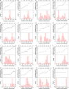
Distribution features of the velvet family in the fungal kingdom. (A) The genome numbers of each fungal group accessed in MycoCosm (38) are highlighted in green. (B) The percentage of genomes having velvet genes. (C) The mode of velvet gene numbers per genome in each fungal group. The detailed information is provided in Fig. 3. (D) The average length of velvet proteins (amino acid residues) in each fungal group. The detailed information is provided in Fig. 4. (E) The average length of velvet domains (amino acid residues) in each fungal group. (F) The percentage of N-terminal side located velvet domains in each fungal group. The detailed information is provided in Fig. 5. (G) The percentage of C-terminal side located velvet domains in each fungal group.
Fig 3
Fig 3
The frequency distribution of velvet gene number per genome in different fungal groups. The genomes without velvet genes were not counted. A, B, and C, respectively, correspond to the groups Agaricomycotina, Pucciniomycotina, and Ustilaginomycotina in the phylum Basidiomycota. D, E, and F, respectively, correspond to the groups Pezizomycotina, Saccharomycotina, and Taphrinomycotina in the phylum Ascomycota. G, H, and I, respectively, correspond to the groups Glomeromycotina, Mortierellomycotina, and Mucoromycotina in the phylum Mucoromycota. J, K, and L, respectively, correspond to the groups Entomophthoromycotina, Kickxellomycotina, and Zoopagomycotina in the phylum Zoopagomycota. M corresponds to the phylum Blastocladiomycota. N and O, respectively, correspond to the groups Chytridiomycetes and Neocallimastigomycetes in the phylum Chytridiomycota. P corresponds to the phylum Cryptomycota.
Fig 4
Fig 4
Length box charts of velvet proteins and domains in different fungal groups. The length was calculated as the number of amino acid residues. Normal distribution was used to fit the length distribution. A and B, respectively, correspond to the charts of velvet proteins and domains.
Fig 5
Fig 5
The distribution of velvet domain position in different fungal groups. The position of velvet domain in a protein was calculated as the midpoint of velvet domain divided by the protein length (the number of amino acid residues). The position of velvet domain locating before 40% was defined as N-terminal side of the protein, while that locating after 60% was defined as C-terminal side of the protein and others are middle part of the protein. A, B, and C, respectively, correspond to the groups Agaricomycotina, Pucciniomycotina, and Ustilaginomycotina in the phylum Basidiomycota. D, E, and F, respectively, correspond to the groups Pezizomycotina, Saccharomycotina, and Taphrinomycotina in the phylum Ascomycota. G, H, and I, respectively, correspond to the groups Glomeromycotina, Mortierellomycotina, and Mucoromycotina in the phylum Mucoromycota. J, K, and L, respectively, correspond to the groups Entomophthoromycotina, Kickxellomycotina, and Zoopagomycotina in the phylum Zoopagomycota. M corresponds to the phylum Blastocladiomycota. N and O, respectively, correspond to the groups Chytridiomycetes and Neocallimastigomycetes in the phylum Chytridiomycota. P corresponds to the phylum Cryptomycota.
Fig 6
Fig 6
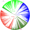
Phylogenetic relationship of the Pezizomycotina velvet proteins. The branch length of the tree is indicated by the scale bar in the upper left corner. The clades of these velvet proteins are indicated by their colors. The figure in high definition is provided as Fig. S1.
Fig 7
Fig 7
Length distribution of clades Pez-VeA, Pez-VelB, Pez-VelC, and Pez-VosA in Pezizomycotina shown as box plots. The clades were based on the phylogenetic relationship shown in Fig. 6. The length was calculated as the number of amino acid residues of velvet proteins. The normal distribution was used to fit the length distribution. Two-group comparisons were performed using the t-test.
Fig 8
Fig 8
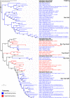
Phylogenetic relationship of Saccharomycotina and Taphrinomycotina velvet proteins. The Capsaspora velvet protein was used as an outgroup. Aspergillus flavus VelD and A. nidulans VeA, VelB, VelC, and VosA were used as references. Bootstrap values for each node are presented. The branch length of each tree is indicated by the scale bar in the lower left corner. The taxonomic groups of these velvet proteins are indicated by their colors paraphrasing in the lower left corner. The clades are marked on the right.
Fig 9
Fig 9
Phylogenetic relationship of the Basidiomycota velvet proteins. The position of the references Capsaspora velvet protein, A. nidulans VeA, VelB, VelC, and VosA is indicated on the outer. The tree branch length is indicated by the scale bar in the lower left corner. The taxonomic groups of these velvet proteins are indicated by their colors paraphrasing in the lower left corner. The clades are marked on the outer. The figure in high definition is provided as Fig. S2 to S7.
Fig 10
Fig 10
Length distribution of clades Bas-Velvet1, Bas-Velvet2, and Bas-Velvet3 in Basidiomycota shown as box plots. The clades were based on the phylogenetic relationship shown in Fig. 9. The length was calculated as the number of amino acid residues of velvet proteins. The normal distribution was used to fit the length distribution. Two-group comparisons were performed using the t-test.
Fig 11
Fig 11
Phylogenetic relationship of the Mucoromycota velvet proteins. The Capsaspora velvet protein was used as the outgroup. A. nidulans VeA, VelB, VelC, and VosA were used as references. The branch length of each tree is indicated by the scale bar in the lower left corner. The taxonomic groups of these velvet proteins are indicated by their colors paraphrasing in the lower left corner. The clades are marked on the outer. The figure in high definition is provided as Fig. S3.
Fig 12
Fig 12
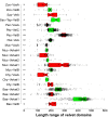
Length distribution of the clades Muc-Velvet1, Muc-Velvet2, Muc-Velvet3, Muc-Velvet4, Muc-VelB, and Muc-VosA in Mucoromycota shown as box plots. The clades were based on the phylogenetic relationship shown in Fig. 11. The length was calculated as the number of amino acid residues of velvet proteins. The normal distribution was used to fit the length distribution. Two-group comparisons were performed using the t-test.
Fig 13
Fig 13
Phylogenetic relationship of Chytridiomycota velvet proteins. The Capsaspora velvet protein was used as the outgroup. A. nidulans VeA, VelB, VelC, and VosA were used as references. They are highlighted in bold. The branch length of each tree is indicated by the scale bar in the lower left corner. The taxonomic groups of these velvet proteins are indicated by their colors paraphrasing in the lower left corner. The clades are marked on the right.
Fig 14
Fig 14
Phylogenetic relationship of Zoopagomycota velvet proteins. The Capsaspora velvet protein was used as the outgroup. A. nidulans VeA, VelB, VelC, and VosA were used as references. They are highlighted in bold. Bootstrap values for each node are presented. The branch length of each tree is indicated by the scale bar in the lower left corner. The taxonomic groups of these velvet proteins are indicated by their colors paraphrasing in the lower middle. The clades are marked on the right of protein IDs. The VelB clade is collapsed in the tree, and expands on the right.
Fig 15
Fig 15
Phylogenetic relationship of Blastocladiomycota and Cryptomycota velvet proteins. The Capsaspora velvet protein was used as the outgroup. A. nidulans VeA, VelB, VelC, and VosA were used as references. Bootstrap values for each node are presented. The branch length of each tree is indicated by the scale bar in the lower left corner. The taxonomic groups of these velvet proteins are indicated by their colors paraphrasing in the lower middle. The clades are marked on the right of protein IDs.
Fig 16
Fig 16
Length distribution of velvet domains from the 21 major clades shown as box plots. The clades were based on the aforementioned phylogenetic analysis. The length was calculated as the number of amino acid residues of velvet domains. The figure was generated by BoxPlotR (41). Data points are shown in a jittered mode with the Tukey whisker extent. The notches were added to the boxes in the presence of medians, and the symbol + indicates the mean value.
Fig 17
Fig 17
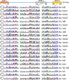
Comparison of the three characteristic motifs of velvet domains among the 21 major clades. The alignment of velvet domains was performed against the profile hidden Markov model of velvet domain PF11754 with 243 residues (https://www.ebi.ac.uk/interpro/entry/pfam/PF11754/) and then subjected to WebLogo (https://weblogo.threeplusone.com/) to generate sequence logos. In the logo, the total stack height represents the information content of residues at that position. The relative height of each residue in the stack is proportional to its frequency at the position, and the residues were sorted so that the most common one was on the top of the stack. The full sequence logos of velvet domains are provided in Fig. S4. The residues are colored according to their chemical properties, of which polar ones G, S, T, Y, and C are in green; neutral ones Q and N are in purple; basic ones K, R, and H are in blue; acidic ones D and E are in red; and hydrophobic ones A, V, L, I, P, W, F, and M are in black. The black balls at the bottom indicate the consensus dominant residues in the 21 clades, and the red balls indicate the other conserved residues revealed by the ConSurf analysis.
Fig 18
Fig 18
Phylogenetic relationship of the 21 velvet domains based on their consensus sequences. Bootstrap values for each node are highlighted in red.
Fig 19
Fig 19
Alignment of the 21 velvet domains based on their secondary structures. The predicted 3D structures of the 21 velvet domains (Fig. S5) modeled by AlphaFold 2 with their consensus sequences were submitted to PROMALS3D for structure alignment. Consensus secondary structure (SS) symbols: alpha-helix: h; beta-strand: e. Consensus AA symbols: conserved amino acids are in bold and uppercase letters; aliphatic (I, V, L): l; aromatic (Y, H, W, F): @; hydrophobic (W, F, Y, M, L, I, V, A, C, T, H): h; alcohol (S, T): o; polar residues (D, E, H, K, N, Q, R, S, T): p; tiny (A, G, C, S): t; small (A, G, C, S, V, N, D, T, P): s; bulky residues (E, F, I, K, L, M, Q, R, W, Y): b; positively charged (K, R, H): +; negatively charged (D, E): -; charged (D, E, K, R, H): c.
Fig 20
Fig 20
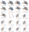
3D structure comparison of the 21 velvet domains. (A) The structural similarity dendrogram of the 21 velvet domains. The 3D structures of the 21 velvet domains were submitted to the Dali server with all against all structure comparison for generating their structural similarity dendrogram. (B) The pairwise structure alignment summary of velvet domains with Pez-VelB and Pez-VosA as references. The detailed comparison was given in Fig. 21. For measuring the alignments, the lower the root mean square deviation (RMSD), the better the structure alignment between the pair of structures. TM-score ranges between 0 and 1, and scores >0.5 generally indicate that the proteins have the same fold (42).
Fig 21
Fig 21
The pairwise structure alignment of velvet domains with Pez-VelB and Pez-VosA as references. The 3D structures of velvet domains were submitted to the Protein Data Bank (PDB) server (https://www.rcsb.org/alignment) for pairwise structure alignment with the jFATCT (rigid) method. The comparison was summarized in Fig. 20B.
Fig 22
Fig 22
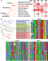
Features of velvet proteins outside the fungal kingdom. (A) A cladogram of species beyond the fungi harboring velvet proteins and their distribution. The taxonomic relationship was based on the NCBI taxonomy database (45), and A. nidulans was used as a representative fungus. (B) Phylogenetic relationship of velvet proteins outside the fungal kingdom and their three velvet characteristic motifs. A. nidulans VeA, VelB, VelC, and VosA were used as references and highlighted in bold on the tree. The velvet proteins in the same clade are highlighted with the same background color. The residues are colored with the Clustal X default coloring scheme.
Fig 23
Fig 23
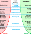
Two evolutionary clans of velvet clades in the fungal kingdom. The phylogenetic relationship of fungal velvet clades was based on their velvet domains.
Fig 24
Fig 24
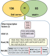
Comparison of structurally similar proteins of the VosA velvet domain by the Dali server and VAST+. The detailed lists of structurally similar proteins by the Dali server and the VAST+ analysis are provided in Table S2 and S3. The upper part is a Venn diagram between the Dali server analysis and the VAST+ analysis. The lower part is a detailed list of the 49 shared proteins by the Dali server and the VAST+ analysis. The PDB IDs are listed inside the box.

Similar articles

Cited by

References

    1. Mafessoni F, Lachmann M, Gokhale CS. 2021. On the fitness of informative cues in complex environments. J Theor Biol 527:110819. doi:10.1016/j.jtbi.2021.110819 - DOI - PubMed
    1. Wingfield JC, Patrick Kelley J, Angelier F, Chastel O, Lei F, Lynn SE, Miner B, Davis JE, Li D, Wang G. 2011. Organism–environment interactions in a changing world: a mechanistic approach. J Ornithol 152:279–288. doi:10.1007/s10336-011-0668-3 - DOI
    1. Leger MM, Ros-Rocher N, Najle SR, Ruiz-Trillo I, Baldauf S. 2022. Rel/NF-κB transcription factors emerged at the onset of opisthokonts. Genome Biol Evol 14:evab289. doi:10.1093/gbe/evab289 - DOI - PMC - PubMed
    1. Ahmed YL, Gerke J, Park H-S, Bayram Ö, Neumann P, Ni M, Dickmanns A, Kim SC, Yu J-H, Braus GH, Ficner R, Stock AM. 2013. The velvet family of fungal regulators contains a DNA-binding domain structurally similar to NF-κB. PLoS Biol 11:e1001750. doi:10.1371/journal.pbio.1001750 - DOI - PMC - PubMed
    1. Ghosh S, Hayden MS. 2012. Celebrating 25 years of NF-κB research. Immunol Rev 246:5–13. doi:10.1111/j.1600-065X.2012.01111.x - DOI - PMC - PubMed

LinkOut - more resources