

Polysaccharide utilization loci from Bacteroidota encode CE15 enzymes with possible roles in cleaving pectin-lignin bonds
- PMID: 38179933
- PMCID: PMC10807430
- DOI: 10.1128/aem.01768-23
Polysaccharide utilization loci from Bacteroidota encode CE15 enzymes with possible roles in cleaving pectin-lignin bonds
Abstract
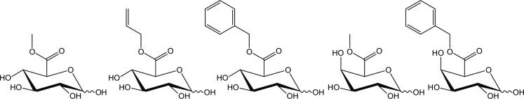
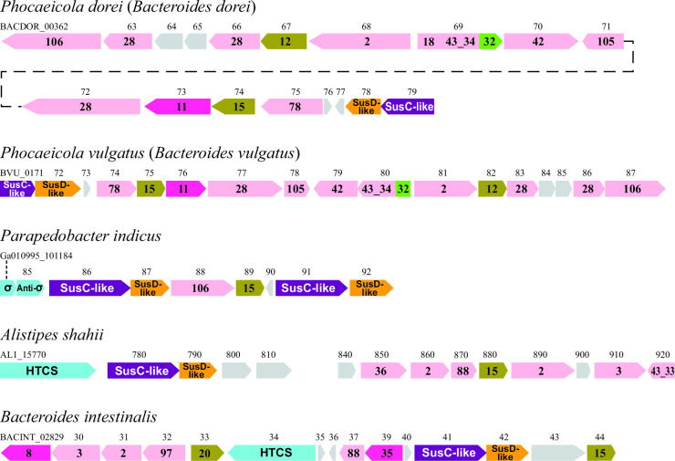
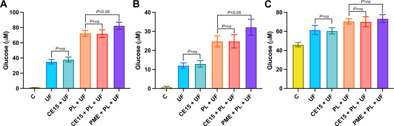
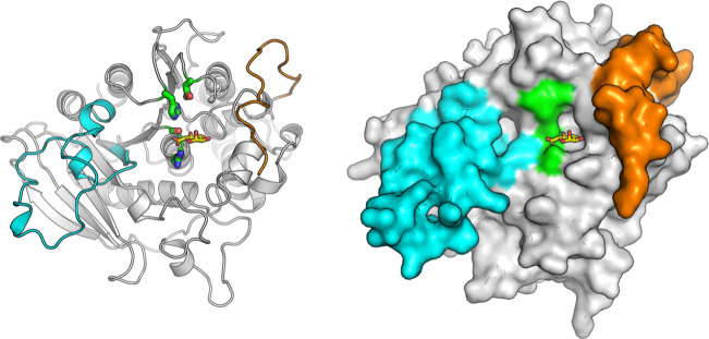
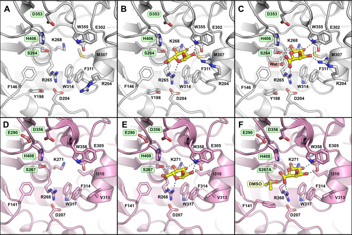
Lignocellulose is a renewable but complex material exhibiting high recalcitrance to enzymatic hydrolysis, which is attributed, in part, to the presence of covalent linkages between lignin and polysaccharides in the plant cell wall. Glucuronoyl esterases from carbohydrate esterase family 15 (CE15) have been proposed as an aid in reducing this recalcitrance by cleaving ester bonds found between lignin and glucuronoxylan. In the Bacteroidota phylum, some species organize genes related to carbohydrate metabolism in polysaccharide utilization loci (PULs) which encode all necessary proteins to bind, deconstruct, and respond to a target glycan. Bioinformatic analyses identified CE15 members in some PULs that appear to not target the expected glucuronoxylan. Here, five CE15 members from such PULs were investigated with the aim of gaining insights on their biological roles. The selected targets were characterized using glucuronoyl esterase model substrates and with a new synthetic molecule mimicking a putative ester linkage between pectin and lignin. The CE15 enzyme from Phocaeicola vulgatus was structurally determined by X-ray crystallography both with and without carbohydrate ligands with galacturonate binding in a distinct conformation than that of glucuronate. We further explored whether these CE15 enzymes could act akin to pectin methylesterases on pectin-rich biomass but did not find evidence to support the proposed activity. Based on the evidence gathered, the CE15 enzymes in the PULs expected to degrade pectin could be involved in cleavage of uronic acid esters in rhamnogalacturonans.IMPORTANCEThe plant cell wall is a highly complex matrix, and while most of its polymers interact non-covalently, there are also covalent bonds between lignin and carbohydrates. Bonds between xylan and lignin are known, such as the glucuronoyl ester bonds that are cleavable by CE15 enzymes. Our work here indicates that enzymes from CE15 may also have other activities, as we have discovered enzymes in PULs proposed to target other polysaccharides, including pectin. Our study represents the first investigation of such enzymes. Our first hypothesis that the enzymes would act as pectin methylesterases was shown to be false, and we instead propose that they may cleave other esters on complex pectins such as rhamnogalacturonan II. The work presents both the characterization of five novel enzymes and can also provide indirect information about the components of the cell wall itself, which is a highly challenging material to chemically analyze in fine detail.
Keywords: carbohydrate esterase; carbohydrate esterase family 15; glucuronoyl esterase; lignocellulose; pectin; protein structure.
Conflict of interest statement
The authors declare no conflict of interest.
Figures
Similar articles
-
Biochemical and structural features of diverse bacterial glucuronoyl esterases facilitating recalcitrant biomass conversion.Biotechnol Biofuels. 2018 Aug 1;11:213. doi: 10.1186/s13068-018-1213-x. eCollection 2018. Biotechnol Biofuels. 2018. PMID: 30083226 Free PMC article.
-
A New Functional Classification of Glucuronoyl Esterases by Peptide Pattern Recognition.Front Microbiol. 2017 Feb 28;8:309. doi: 10.3389/fmicb.2017.00309. eCollection 2017. Front Microbiol. 2017. PMID: 28293230 Free PMC article.
-
Glucuronoyl esterases - enzymes to decouple lignin and carbohydrates and enable better utilization of renewable plant biomass.Essays Biochem. 2023 Apr 18;67(3):493-503. doi: 10.1042/EBC20220155. Essays Biochem. 2023. PMID: 36651189 Free PMC article. Review.
-
Structural and functional investigation of a fungal member of carbohydrate esterase family 15 with potential specificity for rare xylans.Acta Crystallogr D Struct Biol. 2023 Jun 1;79(Pt 6):545-555. doi: 10.1107/S205979832300325X. Epub 2023 May 25. Acta Crystallogr D Struct Biol. 2023. PMID: 37227091 Free PMC article.
-
Glucuronoyl esterases: diversity, properties and biotechnological potential. A review.Crit Rev Biotechnol. 2018 Nov;38(7):1121-1136. doi: 10.1080/07388551.2018.1468316. Epub 2018 May 8. Crit Rev Biotechnol. 2018. PMID: 29739247 Review.
Cited by
-
Structural, Biochemical, and Phylogenetic Analysis of Bacterial and Fungal Carbohydrate Esterase Family 15 Glucuronoyl Esterases in the Rumen.Protein J. 2024 Aug;43(4):910-922. doi: 10.1007/s10930-024-10221-0. Epub 2024 Aug 17. Protein J. 2024. PMID: 39153129 Free PMC article.
-
Short-Term Application of Alfalfa Green Manure Increases Maize Yield and Soil Fertility While Altering Microbial Communities in Karst Yellow Clay Soil.Microorganisms. 2025 Jun 21;13(7):1445. doi: 10.3390/microorganisms13071445. Microorganisms. 2025. PMID: 40731955 Free PMC article.
-
Exploration of three Dyadobacter fermentans enzymes uncovers molecular activity determinants in CE15.Appl Microbiol Biotechnol. 2024 May 15;108(1):335. doi: 10.1007/s00253-024-13175-6. Appl Microbiol Biotechnol. 2024. PMID: 38747981 Free PMC article.
-
Amplicon Sequencing Analysis of Submerged Plant Microbiome Diversity and Screening for ACC Deaminase Production by Microbes.Int J Mol Sci. 2024 Dec 12;25(24):13330. doi: 10.3390/ijms252413330. Int J Mol Sci. 2024. PMID: 39769095 Free PMC article.
-
Stem lodging Resistance-1 controls stem strength by positively regulating the biosynthesis of cell wall components in Capsicum annuum L.Hortic Res. 2024 Jun 20;11(8):uhae169. doi: 10.1093/hr/uhae169. eCollection 2024 Aug. Hortic Res. 2024. PMID: 39135730 Free PMC article.
References
-
- Oh YH, Eom IY, Joo JC, Yu JH, Song BK, Lee SH, Hong SH, Park SJ. 2015. Recent advances in development of biomass pretreatment technologies used in biorefinery for the production of bio-based fuels, chemicals and polymers. Korean J Chem Eng 32:1945–1959. doi:10.1007/s11814-015-0191-y - DOI
-
- Du X, Pérez-Boada M, Fernández C, Rencoret J, del Río JC, Jiménez-Barbero J, Li J, Gutiérrez A, Martínez AT. 2014. Analysis of lignin-carbohydrate and lignin-lignin linkages after hydrolase treatment of xylan-lignin, glucomannan-lignin and glucan-lignin complexes from spruce wood. Planta 239:1079–1090. doi:10.1007/s00425-014-2037-y - DOI - PubMed
Publication types
MeSH terms
Substances
Grants and funding
LinkOut - more resources
Full Text Sources
Molecular Biology Databases
