Review
. 2024 Jul;34(7):547-565.
doi: 10.1016/j.tcb.2023.09.006. Epub 2023 Oct 26.

Astrocyte morphology

Affiliations
Review

Astrocyte morphology

Katherine T Baldwin et al. Trends Cell Biol. 2024 Jul.

Abstract

Astrocytes are predominant glial cells that tile the central nervous system (CNS). A cardinal feature of astrocytes is their complex and visually enchanting morphology, referred to as bushy, spongy, and star-like. A central precept of this review is that such complex morphological shapes evolved to allow astrocytes to contact and signal with diverse cells at a range of distances in order to sample, regulate, and contribute to the extracellular milieu, and thus participate widely in cell-cell signaling during physiology and disease. The recent use of improved imaging methods and cell-specific molecular evaluations has revealed new information on the structural organization and molecular underpinnings of astrocyte morphology, the mechanisms of astrocyte morphogenesis, and the contributions to disease states of reduced morphology. These insights have reignited interest in astrocyte morphological complexity as a cornerstone of fundamental glial biology and as a critical substrate for multicellular spatial and physiological interactions in the CNS.

Keywords: Sholl; electron microscopy; glia; imaging; morphology; neuropil; territory.


Conflict of interest statement

Declaration of interests The authors have no interests to declare.

Figures

Figure 1:
Figure 1:. Views of astrocytes at the microscopic scale.
A. Sagittal brain section of an adult mouse expressing GFP and tdTomato from two separate AAV 2/5 constructs using the astrocyte specific GfaABC1D promoter, illustrating astrocyte specificity and tiling of the CNS by astrocytes. B. Higher magnification view of the striatum from panel A. C. Higher magnification view of a region from panel B, showing astrocyte tiling and elaborate morphology. D. A single striatal astrocyte loaded with Lucifer Yellow with intracellular iontophoresis. Panels A-C were captured by Baljit Khakh and Ling Wu. Panel D is reproduced from a published study [29].
Figure 2:
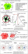
Figure 2:. Metrics used to assess astrocyte morphology using light microscopy.
A. Cartoon illustrates various morphological parameters that can be measured from confocal images of astrocytes expressing a reporter protein such as GFP. These numbers provide metrics by which to assess astrocyte morphology and its changes. The panel is adapted from a published study [32]. A stated in the text, these 2D measurements do not accurately reflect 3D morphology, which is impossible to image with light microscopy. However, they do quantify astrocyte territories. B. Illustrates the method of Sholl analysis that is used to assess cellular complexity. From such Sholl plots, the process maximum (Nm) represents the highest number of intersections an astrocyte makes, the critical value (rc) is the distance from the soma at which Nm occurs, and the maximum radius (rm) is the maximum width of the Sholl plot. In addition, the number of primary branches emanating directly from the soma of each astrocyte (Np) can be used to calculate the Schoenen ramification index, which quantifies overall branching. These numbers provide metrics by which to assess astrocyte morphology and its changes, as shown by Sholl [114] and Shoenen [115]. Standard imaging programs can be used and the mentioned analysis methods are available or easily implemented in ImageJ (https://imagej.nih.gov/ij/). However, we note that while methods exist to preform Sholl analysis on 3D images, this analysis is subject to the caveat of z-axis “stretching” and, like 2D analysis, lacks the resolution to resolve astrocyte leaflets. C. Schematic illustrating the neuropil infiltration volume and how the neuropil infiltration volume fraction can be measured using small voxels placed within astrocyte territories. In this measurement too, the absolute volume of leaflets is not possible to measure with light microscopy. D. Schematic illustrating astrocyte territory volume, and how this method can be used to examine astrocyte tiling behavior.
Figure 3:
Figure 3:. Astrocytes at the nanoscale.
Complexity of astrocytic nanoarchitecture revealed by ultra-high resolution focused ion beam scanning electron microscopy (FIB-SEM). A. FIB-SEM instrumentation (left) used to image a portion of an astrocyte (schematized in the center) using scanning electron microscopy and reconstruction in three dimensions (right; portion of mouse Layer 2/3 cortical astrocyte shown). B. Three specific nanostructural building blocks can be identified in astrocytes including cores (cyan), expansions (purple), and constrictions (orange). Most perisynaptic astrocytic processes (PAPs) are formed by constrictions. C-D. Astrocytic building blocks can be used to examine the hierarchical branching (C) and connectivity (D) of astrocytic nanoarchitecture using directed graphs. E. Astrocytic nanoarchitecture can be described as having a wide, but shallow hierarchical branching organization with major connectivity hubs (asterisks, left), signature distances between astrocytic processes, mitochondria, and synapses (magenta lines; center), and distributed sets of astrocyte-defined synaptic clusters (ADSCs; multi-color synaptic clusters; right). Scale cube in A, 1μm3. Figure reproduced and adapted from Ref [49]. Thank you to Dr. David Polcari (Systems for Research) and Dr. Chris Salmon (McGill University) for the image of the FIB-SEM instrument and the schematic of astrocyte in Panel A, respectively.
Figure 4:
Figure 4:. Molecular regulators of astrocyte morphogenesis.
A number of cell surface receptors on astrocytes play key roles in astrocyte morphogenesis via their interaction with neuron-secreted factors, neuronal membrane proteins, and astrocytic membrane proteins. Astrocyte-neuron interaction: Several members of the FGFR family, including heartless [77] in Drosophila and fgfr3 and fgfr4 [78] in Zebrafish, promote astrocyte outgrowth and neuropil infiltration. Glutamate promotes astrocyte arborization and synapse ensheathment via mGluR5 [79], GABA promotes astrocyte morphogenesis via GABAB receptors [82], while BDNF induces morphogenesis via the TrkB isoform, TrkB.T1 [80]. Expression of Shh by layer V cortical neurons in enhances astrocyte morphological complexity in a layer-specific manner [81]. Astrocytic Neuroligins promote astrocyte territory size and neuropil infiltration via interaction with neuronal neurexins during early postnatal development [84] (see text for discussion of recent findings by Golf and colleagues at later time points [85]). δ-catenin regulates surface expression of N-cadherin in both astrocytes and neurons to regulate cortical layer-specific astrocyte morphogenesis [90]. Homophilic NRCAM interaction restricts astrocyte neuropil infiltration. Both NRCAM [87] and Necl2 [89] promote astrocyte-synapse association, while Cx30 [88] restricts the extent of astrocyte process infiltration into the synaptic cleft. Astrocyte-astrocyte interaction: During the first three postnatal weeks, astrocytes grow in size and complexity and establish non-overlapping territories with neighboring astrocytes. Transcellular hepaCAM interaction plays a key role in astrocyte territory establishment and gap junction coupling through regulation of Cx43 [94]. This figure was created with Biorender.com.

References

    1. Khakh BS and Sofroniew MV (2015) Diversity of astrocyte functions and phenotypes in neural circuits. Nat Neurosci 18. - PMC - PubMed
    1. Allen NJ and Lyons DA (2018) Glia as architects of central nervous system formation and function. Science 362 (6411), 181–185. - PMC - PubMed
    1. Attwell D et al. (2010) Glial and neuronal control of brain blood flow. Nature 468 (7321). - PMC - PubMed
    1. Verkhratsky A and Nedergaard M (2018) Physiology of astroglia. Physiol Rev 98 (1), 239–389. - PMC - PubMed
    1. Nagai J et al. (2021) Behaviorally consequential astrocytic regulation of neural circuits. Neuron 109 (4), 576–596. - PMC - PubMed