. 2024 Mar 4;223(3):e202211048.
doi: 10.1083/jcb.202211048. Epub 2024 Jan 5.

Fission-independent compartmentalization of mitochondria during budding yeast cell division

Affiliations

Fission-independent compartmentalization of mitochondria during budding yeast cell division

Saori R Yoshii et al. J Cell Biol. .

Erratum in

Abstract

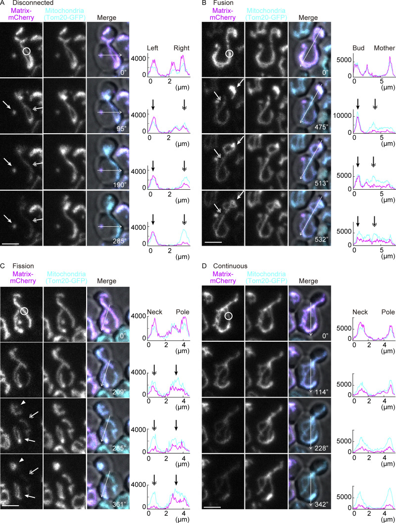
Lateral diffusion barriers compartmentalize membranes to generate polarity or asymmetrically partition membrane-associated macromolecules. Budding yeasts assemble such barriers in the endoplasmic reticulum (ER) and the outer nuclear envelope at the bud neck to retain aging factors in the mother cell and generate naïve and rejuvenated daughter cells. However, little is known about whether other organelles are similarly compartmentalized. Here, we show that the membranes of mitochondria are laterally compartmentalized at the bud neck and near the cell poles. The barriers in the inner mitochondrial membrane are constitutive, whereas those in the outer membrane form in response to stresses. The strength of mitochondrial diffusion barriers is regulated positively by spatial cues from the septin axis and negatively by retrograde (RTG) signaling. These data indicate that mitochondria are compartmentalized in a fission-independent manner. We propose that these diffusion barriers promote mitochondrial polarity and contribute to mitochondrial quality control.


Conflict of interest statement

Disclosures: The authors declare no competing interests exist.

Figures

Figure 1.
Figure 1.
Monitoring of mitochondrial continuity. (A–D) mCherry FLIP in wild-type cells expressing Tom20-GFP (mitochondrial marker; without photobleach) and matrix-mCherry (photobleach target). The line graphs are the intensity profiles along the lines in the respective images. (A) Loss of matrix-mCherry serves as a continuity marker; a physically separate mitochondrion, indicated by an arrow, retained mCherry signals while a photobleached mitochondrion, indicated by a double arrow, lost the fluorescence. (B) An example of a fusion event. The photobleached mitochondrion in the mother (indicated by a double arrow) lost mCherry fluorescence while a physically separate mitochondrion in the bud (indicated by an arrow) retained the fluorescence, until these two mitochondria fused with each other, leading to equilibration of the mCherry fluorescence between the structures. The fusion event took place between 475 and 513 s. (C) An example of a fission event. A continuous mitochondrion was photobleached and lost mCherry fluorescence from the entire structure until it underwent fission forming three separate mitochondria. One mitochondrion at the bleaching area (indicated by a double arrow) further lost mCherry fluorescence whereas two separated mitochondria (indicated by an arrow and arrowhead) retained the fluorescence after fission. The fission event took place between 209 and 285 s. (D) An example of a continuous mitochondrion between the mother and bud throughout the imaging period. The mCherry fluorescence was lost from the entire structure. Photobleach was applied in the mCherry channel as indicated by white circles. Images are a sum projection of five z-stacks taken at 0.5-μm intervals. Scale bar: 3 µm.
Figure 2.
Figure 2.
Lateral compartmentalization of continuous mitochondria. (A–C) Dual-color FLIP in wild-type cells expressing GFP-tagged mitochondrial proteins and matrix-mCherry. Representative images, pooled quantification data of GFP and mCherry FLIP, and tX (time to reduce to X% of the total fluorescence) of GFP FLIP from three independent experiments are shown. (A) Wild-type cells expressing Hem1-GFP and matrix-mCherry (n = 30 cells, n = 10 cells for each experiment). (B) Wild-type cells expressing Tom20-GFP and matrix-mCherry (n = 32 cells, n ≥ 10 cells for each experiment). (C) Wild-type cells expressing Atp1-GFP and matrix-mCherry (n = 25 cells, n ≥ 8 cells for each experiment). Photobleach was applied in the GFP and mCherry channels as indicated with white circles. Images are a sum projection of five z-stacks taken at 0.5-μm intervals. Scale bar: 3 μm. Data from three independent clones were pooled to obtain the bleaching curves. Shadows represent mean ± SD. Error bar: mean ± SE. Welch’s two-tailed t test was applied to compare the tX in the mother and bud.
Figure S1.
Figure S1.
Matrix-mCherry FLIP. (A–C) t20 (time to reduce to 20% of the total fluorescence) of mCherry FLIP from three independent experiments are shown (mCherry data from Fig. 2). Note that tX for mCherry cannot be directly compared with tX for GFP due to the different bleaching conditions and different susceptibility to photobleaching. (A) Wild-type cells expressing Hem1-GFP and matrix-mCherry (n = 30 cells, n = 10 cells for each experiment). (B) Wild-type cells expressing Tom20-GFP and matrix-mCherry (n = 32 cells, n ≥ 10 cells for each experiment). (C) Wild-type cells expressing Atp1-GFP and matrix-mCherry (n = 25 cells, n ≥ 8 cells for each experiment). (D–G) Pooled quantification data of mCherry FLIP and t20 from three independent experiments are shown (mCherry data from Fig. 3, A–D). (D) fis1∆ cells expressing Tom20-GFP and matrix-mCherry (n = 32 cells, n ≥ 10 cells for each experiment). (E) dnm1∆ cells expressing Tom20-GFP and matrix-mCherry (n = 28 cells, n ≥ 7 cells for each experiment). (F) fis1∆ cells expressing Hem1-GFP and matrix-mCherry (n = 21 cells, n = 7 cells for each experiment). (G) fis1∆ cells expressing Atp1-GFP and matrix-mCherry (n = 25 cells, n ≥ 8 cells for each experiment). Data from three independent clones were pooled to obtain the bleaching curves. Shadows represent mean ± SD. Error bar: mean ± SE. Welch’s two-tailed t test was applied to compare the t20 in the mother and bud.
Figure 3.
Figure 3.
Compartmentalization of mitochondria is independent of the fission machinery. (A–D) Dual-color FLIP in fis1∆ cells expressing GFP-tagged mitochondrial proteins and matrix-mCherry. Representative images, pooled quantification data of GFP FLIP, and tX of GFP FLIP from three independent experiments are shown. Photobleach was applied in the GFP and mCherry channels as indicated with white circles. (Note here that the bleaching and imaging conditions were changed and the graphs cannot be directly compared with Fig. 2). (A) fis1∆ cells expressing Tom20-GFP and matrix-mCherry (n = 32 cells, n ≥ 10 cells for each experiment). (B) dnm1∆ cells expressing Tom20-GFP and matrix-mCherry (n = 28 cells, n ≥ 7 cells for each experiment). (C) fis1∆ cells expressing Hem1-GFP and matrix-mCherry (n = 21 cells, n = 7 cells for each experiment). (D) fis1∆ cells expressing Atp1-GFP and matrix-mCherry (n = 25 cells, n ≥ 8 cells for each experiment). Images are a sum projection of five z-stacks taken at 0.5 μm intervals. Scale bar: 3 μm. Data from three independent clones were pooled to obtain the bleaching curves. Shadows represent mean ± SD. Error bar: mean ± SE. Welch’s two-tailed t test was applied to compare the tX in the mother and bud.
Figure 4.
Figure 4.
Visualization of protein diffusion in mitochondria. (A and B) Representative images from photoconversion experiments in fis1∆ cells expressing Tom20-2×Kaede in the absence of matrix-targeted fluorescence proteins (A) and fis1∆ cells expressing matrix-2×Kaede (B). (C) Quantification of converted-to-nonconverted Kaede fluorescence ratios (red/green) in the mother compartment compared with the bud compartment using Tom20-2×Kaede (n = 17 cells) or matrix-2×Kaede (n = 17 cells). Photoconversion was applied as indicated with white circles with 15 imaging-photoconversion cycles (10 s/cycle). The line graphs are the intensity profiles along the lines in the respective images after photoconversion. Images are a sum projection of five z-stacks taken at 0.5 μm intervals. Scale bar: 3 μm. Error bar: mean ± SE. Welch’s two-tailed t test was applied to compare the conversion ratios between two groups.
Figure 5.
Figure 5.
A mitochondrial diffusion barrier exists at the bud neck. (A) Two possible scenarios upon change of photobleaching areas from the mother (upper images) to bud (lower images). In the presence of a diffusion barrier at the bud neck, indicated by the red disk, bleaching in the bud would result in reversed bleaching curves (left). Slower diffusion in the bud compartment (absence of a diffusion barrier) would result in both compartments losing fluorescence in a similar manner upon bleaching in the bud (right). (B) Example images, pooled quantification of GFP FLIP, and t70 of GFP FLIP in fis1∆ cells expressing Tom20-GFP and matrix-mCherry (n = 34 cells, n ≥ 10 cells for each experiment). Photobleach was applied in the GFP and mCherry channels in the bud as indicated by white circles. Images are a sum projection of five z-stacks taken at 0.5-μm intervals. Scale bar: 3 μm. Shadows represent mean ± SD. Error bar: mean ± SE. Welch’s two-tailed t test was applied to compare the tX in the mother and bud.
Figure 6.
Figure 6.
Mitochondrial masses tethered at cell poles are compartmentalized. (A–H) Dual-color FLIP in fis1∆ cells expressing GFP-tagged mitochondrial proteins and matrix-mCherry, categorized according to their morphologies. Example images (A–D), pooled quantification of GFP FLIP, and t70 of GFP FLIP from three or four independent experiments (E–H). Shadows represent mean ± SD. (A and E) Cells with mitochondrial accumulation both at the mother and bud poles (E; n = 40 cells, n = 10 cells for each experiment). (B and F) Cells with mitochondrial accumulation only at the bud pole (F; n = 28 cells, n ≥ 9 cells for each experiment). (C and G) Cells with mitochondrial accumulation only at the mother pole (G; n = 39 cells, n ≥ 10 cells for each experiment). (D and H) Cells without mitochondrial accumulation at the poles (H; n = 29 cells, n ≥ 9 cells for each experiment). Arrows indicate mitochondrial accumulation at cell poles. Welch’s two-tailed t test was applied to compare the tX in the mother and bud. (I) t70 (bud) from E-H are compared among the morphology groups. t70 (bud) values in each category were compared by one-way ANOVA followed by Tukey’s method. (J) FLIP was applied in the mitochondrial tubules at 10 pxs away from the mitochondrial masses as illustrated in the model (left). Representative images of FLIP experiments in cells with mitochondrial masses without (left, indicated by an arrow) or with (right, indicated by a double arrow) attachment at the mother cell poles. (K) The pooled mother bleaching curves in cells that have mitochondrial masses with (n = 23 cells) or without (n = 24 cells) attachment at the mother cell poles. Light shadows represent mean ± SD and dark shadows represent mean ± SE. Data were fitted to the nonlinear regression curves and analyzed based on a null hypothesis “one curve for all data sets” and an alternative hypothesis of “different curve for each data set” using the extra sum-of-squares F-test. (L) FRAP was performed within the mitochondrial masses with (n = 35 cells) or without (n = 36 cells) attachment at the mother cell poles. Tom20-GFP fluorescence within the bleached area was obtained and normalized to the average of the final 200 data points (frame numbers 401–600; 100%). Shadows represent mean ± SD. Photobleaching was applied in GFP and mCherry channels as indicated with white circles. Images are a sum projection of five z-stacks taken at 0.5-μm intervals. Scale bar: 3 μm. Error bar: mean ± SE.
Figure S2.
Figure S2.
Compartmentalization in mitochondria tethered to the cell poles. (A–F) Dual-color FLIP in fis1∆ cells expressing GFP-tagged mitochondrial proteins and matrix-mCherry. (A and B) Representative images of fis1∆ cells expressing Atp1-GFP and matrix-targeted mCherry with mitochondrial accumulation both at the mother and bud poles (A) or only at the bud pole (B). Atp1-GFP shows delayed bleaching in the tethered mitochondrial masses whereas matrix-mCherry does not. (C and D) Representative images of fis1∆ cells expressing Hem1-GFP and matrix-targeted mCherry with mitochondrial accumulation both at the mother and bud poles (C) or only at the bud pole (D). Soluble matrix proteins do not show delayed bleaching in the tethered mitochondrial masses. (E and F) Representative images (E) and GFP quantification (F) of fis1∆ cells expressing Tom20-GFP and matrix-targeted mCherry after cytokinesis. Mitochondrial masses that are tethered to the former bud pole-side of daughter cells (Pole; n = 15 cells) show a delayed diffusion pattern of Tom20-GFP whereas those located at the former bud neck side (Neck; n = 15 cells) do not. Light shadows represent mean ± SD and dark shadows represent mean ± SE. Photobleach was applied in the GFP and mCherry channels as indicated by white circles. Images are a sum projection of five z-stacks taken at 0.5-μm intervals. Scale bar: 3 μm. Data were fitted to the nonlinear regression curves and analyzed based on a null hypothesis “one curve for all data sets” and an alternative hypothesis of “different curve for each data set” using the extra sum-of-squares F-test.
Figure 7.
Figure 7.
IMM diffusion barriers exist constitutively and OMM diffusion barriers are formed in response to stresses. (A and B) Dual-color FLIP in fis1∆ cells expressing Yta12-GFP (IMM protein) and matrix-mCherry. Representative images and pooled quantification of Yta12-GFP FLIP and t70 of Yta12-GFP are shown. Cells grown in YPD medium (A; n = 23 cells; n ≥ 6 cells for each experiment). Cells grown in YPE (B; n = 25 cells; n ≥ 5 cells for each experiment). (C) Compartmentalization indexes (tX) are calculated as tX (time to reduce to X% of the total fluorescence) in the bud (tX [B]) compared with tX in the mother (tX [M]) (upper panel). Compartmentalization indexes (t70) of Yta12-GFP FLIP data from A and B (lower panel). Welch’s two-tailed t test was applied to compare the tX in the mother and bud. (D) GFP FLIP in fis1∆ cells expressing Alo1-GFP (OMM protein) in the absence of matrix-mCherry. Representative images and pooled quantification of Alo1-GFP FLIP are shown (n = 23 cells). (E and F) Dual-color FLIP in fis1∆ cells expressing Tom20-GFP and mCherry fused to the transmembrane region of Tom20 (Tom20TM-mCherry). Representative images and line graphs of the intensity profiles along the indicated lines (E). Quantification of Tom20-GFP and Tom20TM-mCherry (F; n = 17 cells). (G) Dual-color FLIP in fis1∆ cells expressing Alo1-GFP (OMM) and matrix-mCherry grown in YPD medium (n = 30 cells). (H) Compartmentalization indexes (t50) were calculated from Alo1-GFP (OMM protein) FLIP in fis1∆ cells grown on YPD, YPG, YPE, or YPDE in the presence or absence of matrix-mCherry. Data from at least three independent experiments are shown. n ≥ 5 cells for each clone, and a total of 42 cells (YPD without mCherry), 34 cells (YPG without mCherry), 54 cells (YPD with mCherry), 49 cells (YPE with mCherry), or 25 cells (YPDE with mCherry) were analyzed. Compartmentalization indexes (t50) were compared by one-way ANOVA followed by the Tukey’s method. (I) Single-color FLIP in fis1∆ cells expressing Alo1-GFP (OMM) in the absence of matrix-mCherry grown in YPG medium (n = 34 cells). (J) Dual-color FLIP in fis1∆ cells expressing Alo1-GFP (OMM) and matrix-mCherry grown in YPE medium (n = 49 cells). Photobleach was applied as indicated by white circles. Images are a sum projection of five z-stacks taken at 0.5-μm intervals. Scale bar: 3 μm. Shadows represent mean ± SE (hereafter, shadowed error bars in the FLIP experiments are changed to SE to compare the means among groups instead of showing distributions within a group). Error bar: mean ± SE.
Figure S3.
Figure S3.
Other IMM and OMM proteins. (A–E) Single-color FLIP in fis1∆ cells expressing GFP-tagged mitochondrial IMM (A–C) or OMM (D and E) proteins in the absence of matrix-mCherry. Representative images and quantification of Oxa1-GFP (A; IMM, n = 10 cells), Yme1-GFP (B; IMM, n = 12 cells), Atm1-GFP (C; IMM, n = 10 cells), Om45-GFP (D; OMM, n = 12 cells), and Por1-GFP (E; OMM, n = 11 cells). Photobleach was applied in the GFP channel as indicated by white circles. Shadows represent mean ± SD. Images are a sum projection of five z-stacks taken at 0.5-μm intervals. Scale bar: 3 μm.
Figure 8.
Figure 8.
Regulation of IMM and OMM diffusion barriers. (A–D) Dual-color FLIP cells expressing Yta12-GFP (IMM) and matrix-mCherry. Representative images and quantification of Yta12-GFP FLIP in fis1∆ (A; n = 37 cells), fis1bud6∆ (B; n = 34 cells), fis1shs1∆ (C; n = 35 cells), and fis1mks1∆ (D; n = 36 cells) cells. Yta12-GFP bleaching curves in fis1∆ cells are overlayed as gray lines (the same set of fis1∆ data from A are overlayed in B–D and H). (E) Compartmentalization indexes (t70) were calculated from three independent clones (the same set of data as D and H). n ≥ 10 cells for each experiment. Shadows represent mean ± SE. (F) t70 values for the bud curves were calculated from three independent experiments using the same set of data as in A–E and H. (G) FRAP was performed within the mitochondrial masses that are attached at mother cell poles as exemplified (right) in fis1∆ cells (n = 45 cells) or in fis1mks1∆ cells (n = 45 cells). Yta12-GFP fluorescence within the bleached area was obtained and normalized to the average of the final 200 data points (frame numbers 401–600; 100%). Shadows represent mean ± SD. (H) Representative images and quantification of Yta12-GFP FLIP in the presence of matrix-mCherry in fis1rtg2∆ cells (n = 33 cells). Bleaching curves of fis1∆ cells are overlayed as in B–D. (I–K) Dual-color FLIP in cells expressing Alo1-GFP (OMM) and matrix-mCherry grown on agar plates containing SD + 2% glucose and 2% ethanol. Representative images and quantification of Alo1-GFP FLIP in fis1∆ (n = 30 cells), fis1rtg2∆ (n = 30 cells), and fis1mks1∆ cells (n = 30 cells). Alo1-GFP bleaching curves in fis1∆ cells are overlayed as gray lines (the same set of fis1∆ data from I are overlayed in J and K). Shadows represent mean ± SE. (L) Compartmentalization indexes (t50) were calculated from three independent clones with 10 cells per clone (the same data as I–K). Error bar: mean ± SE. Photobleach was applied in the GFP and mCherry channels as indicated by white circles. Images are a sum projection of five z-stacks taken at 0.5 μm intervals. Scale bar: 3 μm. Statistical analyses were performed by one-way ANOVA followed by Dunnett’s method.
Figure S4.
Figure S4.
Regulation of IMM diffusion barriers. (A–C) Dual-color FLIP in fis1∆ cells expressing Tom20-GFP and matrix-mCherry. Quantification of Tom20-GFP FLIP in fis1∆ (A; n = 30 cells), fis1∆ bud6∆ (B; n = 23 cells), fis1shs1∆ (C; n = 30 cells) cells. Tom20-GFP bleaching curves from A are overlayed in B and C as gray lines. (D–G) Dual-color FLIP in fis1∆ cells expressing Yta12-GFP and matrix-mCherry. Quantification of Yta12-GFP FLIP in fis1∆ (D; n = 36 cells), fis1sur2∆ (E; n = 21 cells), fis1∆ crd1∆ (F; n = 20 cells), and fis1∆ mic60∆ (G; n = 25 cells). Yta12-GFP bleaching curves from D are overlayed in E–G as gray lines. Shadows represent mean ± SE.

References

    1. Altmann, K., Frank M., Neumann D., Jakobs S., and Westermann B.. 2008. The class V myosin motor protein, Myo2, plays a major role in mitochondrial motility in Saccharomyces cerevisiae. J. Cell Biol. 181:119–130. 10.1083/jcb.200709099 - DOI - PMC - PubMed
    1. Arlt, H., Tauer R., Feldmann H., Neupert W., and Langer T.. 1996. The YTA10-12 complex, an AAA protease with chaperone-like activity in the inner membrane of mitochondria. Cell. 85:875–885. 10.1016/S0092-8674(00)81271-4 - DOI - PubMed
    1. Baldi, S., Bolognesi A., Meinema A.C., and Barral Y.. 2017. Heat stress promotes longevity in budding yeast by relaxing the confinement of age-promoting factors in the mother cell. Elife. 6:e28329. 10.7554/eLife.28329 - DOI - PMC - PubMed
    1. Barral, Y., Mermall V., Mooseker M.S., and Snyder M.. 2000. Compartmentalization of the cell cortex by septins is required for maintenance of cell polarity in yeast. Mol. Cell. 5:841–851. 10.1016/S1097-2765(00)80324-X - DOI - PubMed
    1. bin Imtiaz, M.K., Royall L.N., Gonzalez-Bohorquez D., and Jessberger S.. 2022. Human neural progenitors establish a diffusion barrier in the endoplasmic reticulum membrane during cell division. Development. 149:dev200613. 10.1242/dev.200613 - DOI - PMC - PubMed

Publication types