. 2024 Mar;229(2):359-385.
doi: 10.1007/s00429-023-02725-9. Epub 2024 Jan 5.

Immunohistochemical field parcellation of the human hippocampus along its antero-posterior axis

Affiliations

Immunohistochemical field parcellation of the human hippocampus along its antero-posterior axis

Emilio González-Arnay et al. Brain Struct Funct. 2024 Mar.

Abstract

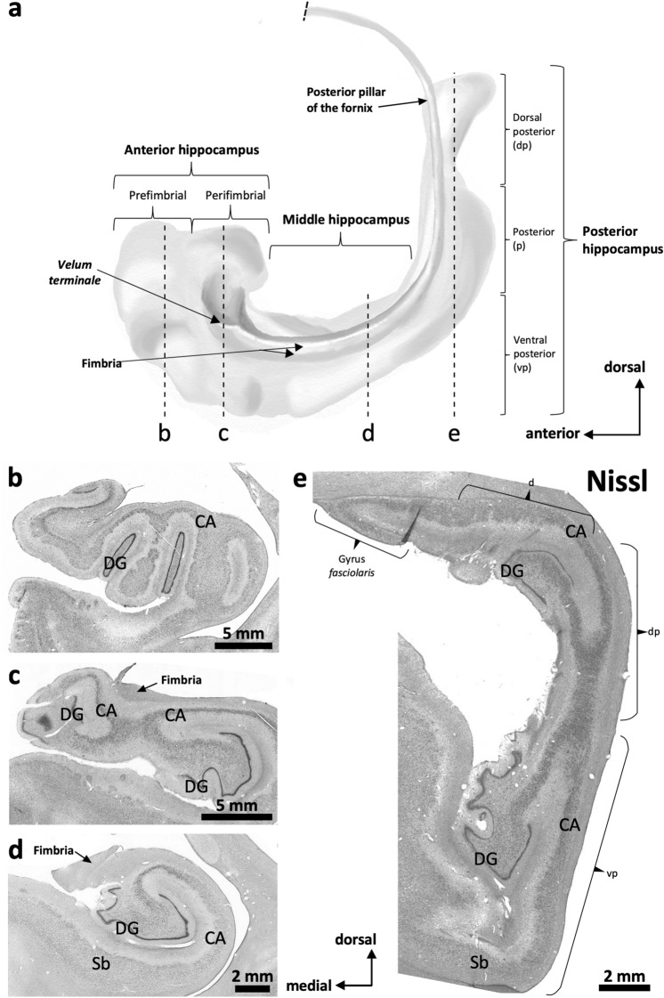
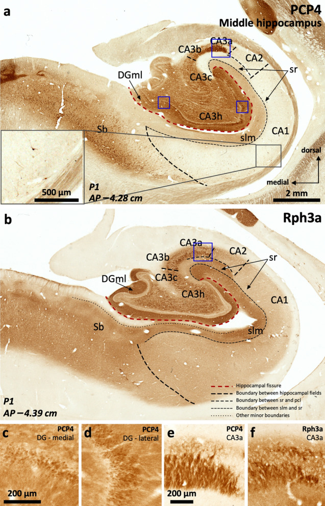
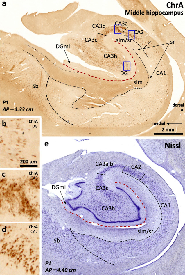
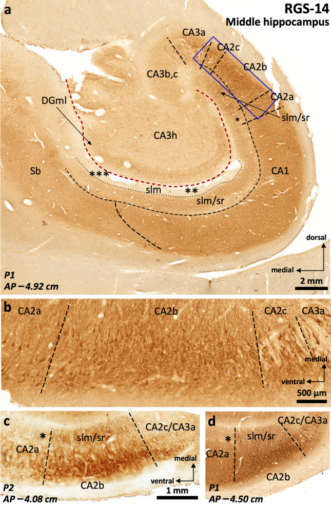
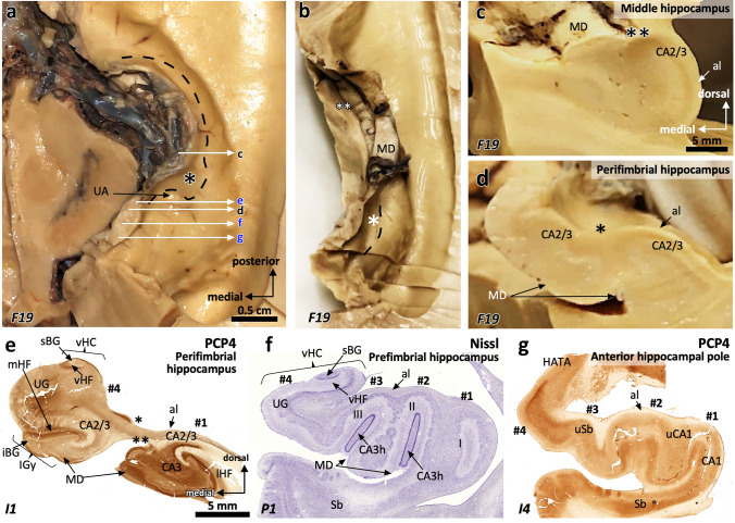
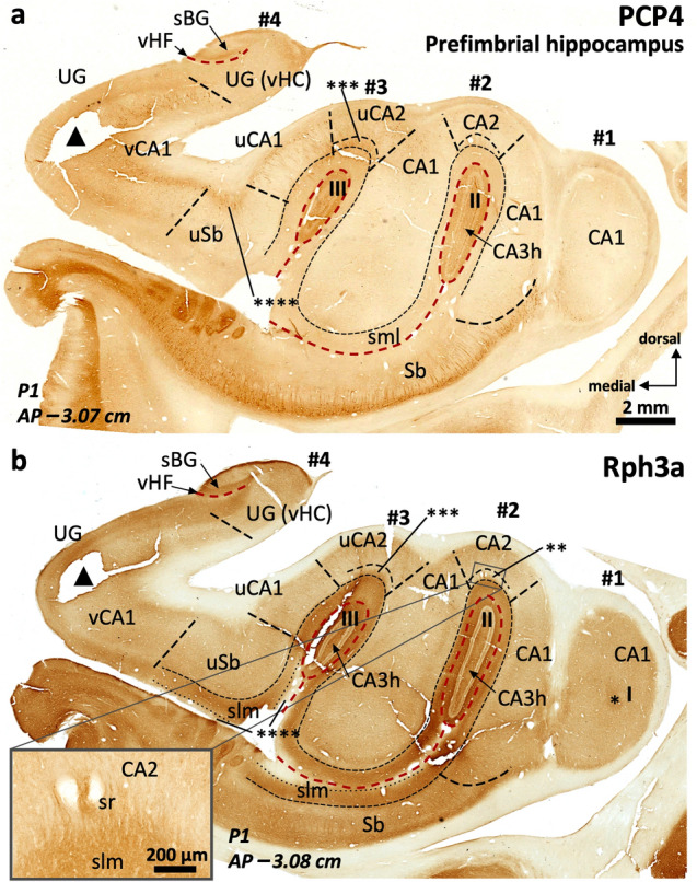
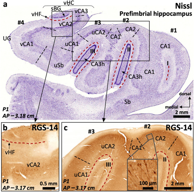
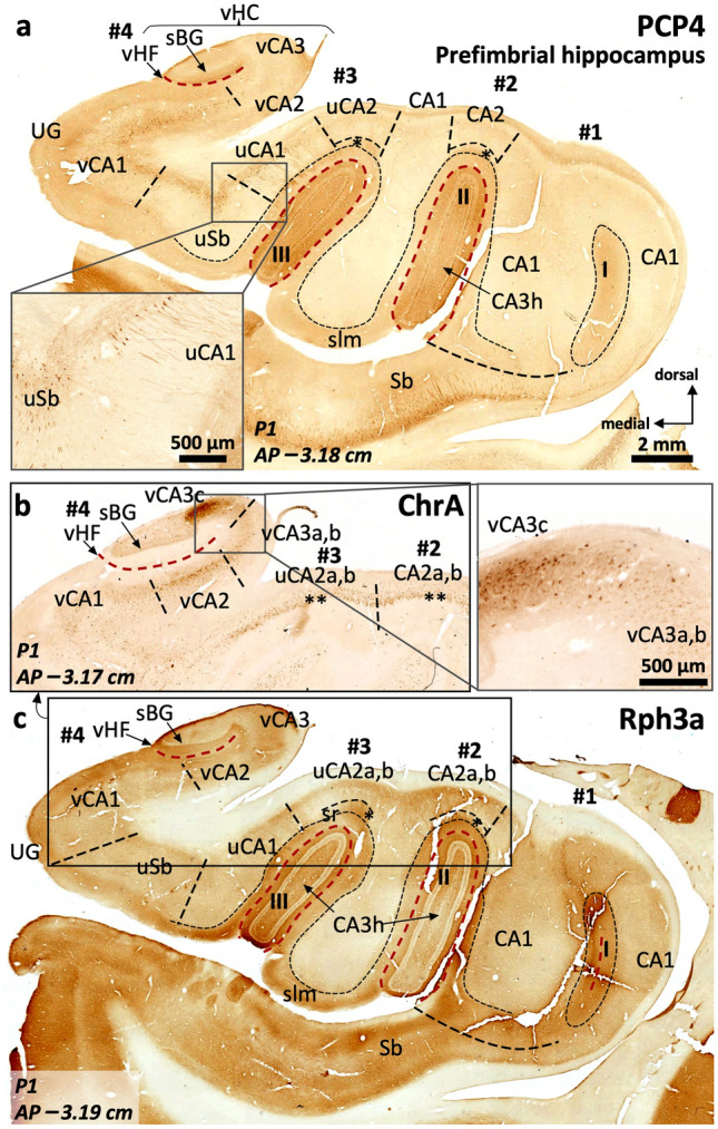
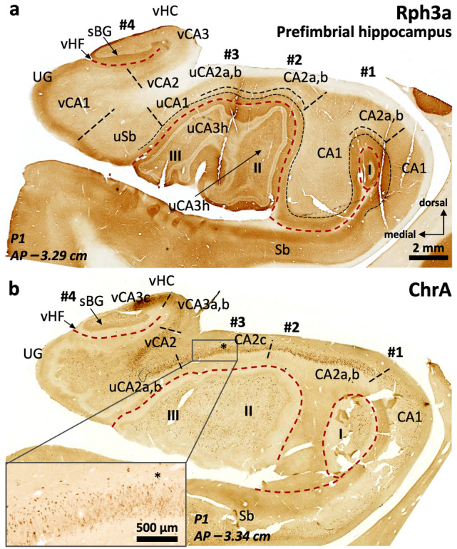
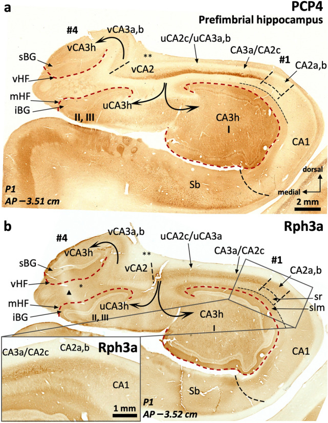
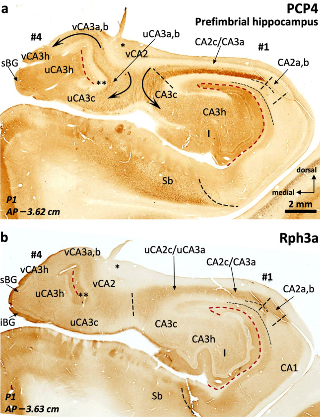
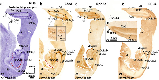
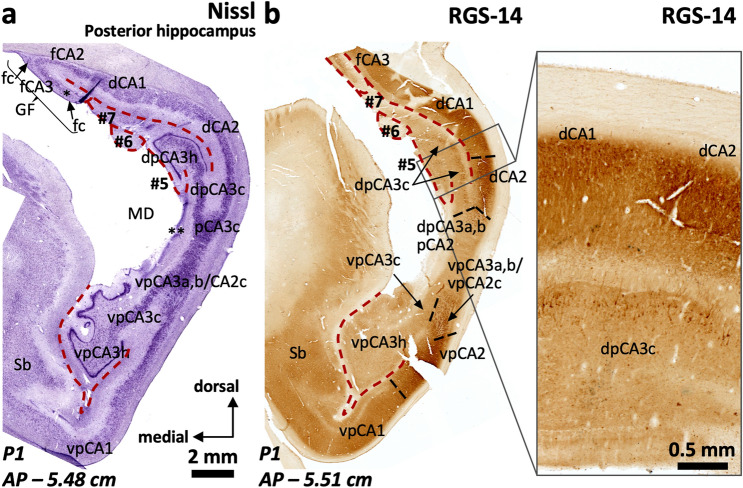
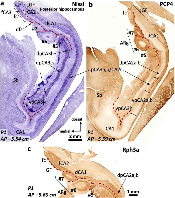
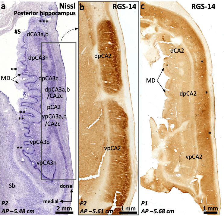
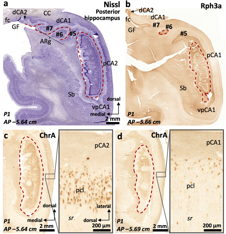
The primate hippocampus includes the dentate gyrus, cornu ammonis (CA), and subiculum. CA is subdivided into four fields (CA1-CA3, plus CA3h/hilus of the dentate gyrus) with specific pyramidal cell morphology and connections. Work in non-human mammals has shown that hippocampal connectivity is precisely patterned both in the laminar and longitudinal axes. One of the main handicaps in the study of neuropathological semiology in the human hippocampus is the lack of clear laminar and longitudinal borders. The aim of this study was to explore a histochemical segmentation of the adult human hippocampus, integrating field (medio-lateral), laminar, and anteroposterior longitudinal patterning. We provide criteria for head-body-tail field and subfield parcellation of the human hippocampus based on immunodetection of Rabphilin3a (Rph3a), Purkinje-cell protein 4 (PCP4), Chromogranin A and Regulation of G protein signaling-14 (RGS-14). Notably, Rph3a and PCP4 allow to identify the border between CA3 and CA2, while Chromogranin A and RGS-14 give specific staining of CA2. We also provide novel histological data about the composition of human-specific regions of the anterior and posterior hippocampus. The data are given with stereotaxic coordinates along the longitudinal axis. This study provides novel insights for a detailed region-specific parcellation of the human hippocampus useful for human brain imaging and neuropathology.

Keywords: Fasciola cinerea; Uncus; Andreas Retzius gyri; Band of Giacomini; Gyrus fasciolaris; Hippocampal body; Hippocampal head; Hippocampal tail; Hippocampus; Intralimbic gyrus; Parcellation; Posterior hippocampus; Segmentation; Vertical hippocampus.


Conflict of interest statement

The authors have no relevant financial or non-financial interests to disclose.

Figures

Fig. 1
Fig. 1
Macroscopic regionalization of the human hippocampus (a). There are three main regions from anterior to posterior. The most anterior one, anterior hippocampus or hippocampal head (b and c), bends back on itself surrounding anteriorly the anterior end of the fimbria defining a prefimbrial (b) and a perifimbrial region (c) that is continued posteriorly with the middle hippocampus or hippocampal body (d). Once the fimbria acquires a vertical orientation and becomes the posterior pillar or crus of the fornix, a C-shaped concave posterior hippocampus appears (e). This posterior hippocampus is divided into a ventral posterior region (vp; ventral posterior hippocampus), a dorsal posterior region (dp, dorsal posterior hippocampus), and the posterior region (p, posterior hippocampus ‘proper’) which forms the posterior end of the hippocampus. A dorsal hippocampal region (d, dorsal hippocampus) links the dorsal posterior hippocampus and the posterior hippocampus proper –not shown in e– with the gyrus fasciolaris (e), which lies ventral to the posterior end of the corpus callosum. Abbreviations: CA, cornu ammonis; DG, dentate gyrus; Sb, subiculum
Fig. 2
Fig. 2
Distribution of immunohistochemical markers PCP4 (a) and Rph3a (b) in the human middle hippocampus. Blue squares in (a) and (b) identify fields enlarged in cf. The dentate gyrus molecular layer (DGml) shows neuropil immunoreactivity for PCP4 and Rph3a. In the hilar region (CA3h), Rph3a is present in neuropil and interneurons (b, see also Supplementary Fig. 1). A dense plexus of PCP4 + , Rph3a + mossy fibers reaches lateral CA3 (CA3a, CA3b) thus highlighting the medial boundary of CA2. The boundary between CA2 and CA1 is defined by a PCP4 + , Rph3a + stratum radiatum (sr) in CA2, in contrast to the PCP4–, Rph3a– stratum radiatum (sr) of CA1. Meanwhile, the boundary between CA1 and subiculum is defined by the lack of immunostaining of PCP4 in pyramidal cells of CA1 in contrast to PCP4 + pyramidal cells in subiculum; The inset in (a) shows some scattered non-pyramidal neurons in the stratum oriens of CA1. c, d, Enlarged regions from the dentate gyrus -DG- in (a) showing cell somata immunoreactive for PCP4 in the granule cell layer. Note the less marked staining of cell somata in the medial DG (c) compared to the lateral DG (d). e, f, high magnification (from a and b, respectively) of the PCP4 + (e), Rph3a + (f) mossy fibers reaching lateral CA3
Fig. 3
Fig. 3
Parcellation of the human middle hippocampus using ChrA immunohistochemistry (a) and Nissl staining (e). Blue squares in (a) identify fields enlarged in b–d. ChrA immunostaining shows immunoreactive somata in the granule cell layer of the dentate gyrus (DG) (a, high magnification in b). ChrA immunostaining reveals boundaries between CA fields: CA3 presents mostly neuropil staining, together with some ChrA somata (c), CA2 presents pyramidal cells showing ChrA + cytoplasmic staining (d), and CA1 shows ChrA + neuropil in stratum lacunosum moleculare (slm), ChrA– neuropil in stratum radiatum (sr), and ChrA + cytoplasmic staining in some scattered non-pyramidal cells in the deep pyramidal layer and stratum oriens; a similar pattern is present in the subiculum (Sb). e, Nissl staining of a histological section in a close anteroposterior level to that of (a). Abbreviation: DGml, dentate gyrus molecular layer. The code for the broken lines as in Fig. 2b
Fig. 4
Fig. 4
RGS-14 immunostaining in human middle hippocampus (a) and RGS-14-based compartmentalization of field CA2 (b-d). The blue square in (a) refers to the field shown at higher magnification in (b). RGS-14 is diffusely and sparsely present in hilar neuropil (h), as well as throughout the whole depth of CA3b, CA3c (CA3b,c), and CA3h pyramidal cell layer. The presence of deep RGS-14 + pyramidal cells allows the demarcation of CA3a. The main medio-lateral dimension of CA2 (CA2b) features an intense RGS-14 + pyramidal cell layer, RGS-14 + dendrites are also found in the stratum radiatum (sr) and in stratum lacunosum moleculare (slm). Note that an internal boundary between both strata is not distinguishable with this staining. A subset of RGS-14 + pyramidal cells extends further laterally, progressively intermingling with deep CA1 pyramidal cells and progressively losing the sr/slm immunoreactivity (* in a, c, d), this short subfield is termed CA2a. RGS-14 neuropil immunoreactivity in the slm tends to disappear in ventromedial regions of CA1 (** in a) and is not present in subicular slm (*** in a). Note the inter-individual differences in RGS-14 neuropil staining (bd). Abbreviation: DGml, dentate gyrus molecular layer. The code for the broken lines as in Fig. 2b
Fig. 5
Fig. 5
Macro (a-d) microscopic (eg) correlation in the human anterior hippocampus (hippocampal head). Dorsal view of a left human hippocampus (a, prior to dissection, b, partially dissected). White arrows in (a) depict approximate levels of cutting to obtain the macroscopic (black characters) and microscopic (blue characters) sections shown in this figure. The hippocampal head is organized around the anterior end of the fimbria (* in a, b, d, e). Therefore, any region showing a macroscopic recognizable medial fimbriodentate junction (** in b,c) should be considered as middle hippocampus or hippocampal body. Note that the fimbriodentate junction is also present in e (**), but it is not easily recognizable macroscopically. Anterior to the middle hippocampus, three gross anatomical regions are present: 1. Perifimbrial hippocampus (e) including the most lateral (#1) and medial (#4) hippocampal digitations, separated by the anterior fimbria (*) which is the main gross feature of this region along with the presence of a margo denticulatus (MD) laying ventrally (d, e) and not medially (c) to the fimbriodentate junction (**). 2. Prefimbrial hippocampus (f): There is no fimbriodentate junction, but a minute margo denticulatus (MD, the surface of the dentate gyrus) is still present in the ventral surface of the hippocampal head. All classical hippocampal fields are represented. Intermediate hippocampal digitations (#2, #3) are present, including their dentate gyrus (II, III). 3. Anterior hippocampal pole (g), which is the most anterior area, and where only subicular modified subfields and CA1 fields are present (uSb, uCA1). Abbreviations: al, alveus; -h as a suffix, hilar; HATA, hippocampo-amygdaloid transitional area; iBG, inferior Band of Giacomini; IGy, intralimbic gyrus; lHF, lateral hippocampal fissure; MD, margo denticulatus; mHF, medial hippocampal fissure; sBG, superior Band of Giacomini; Sb, subiculum; u- as a prefix, uncal; UA, uncal apex; UG, uncinate gyrus; vHC, vertical hippocampus; vHF vertical hippocampal fissure
Fig. 6
Fig. 6
Field delimitation in the human prefimbrial hippocampus. Fields in the lateral digitations (#1, #2) follow a middle hippocampus-like pattern. Level –3,08 (b) is remarkable as it shows the most anterior histological evidence of field CA2 (a Rph3a + stratum radiatum (sr) ** in b, inset in b) and the most anterior stratum lacunosum moleculare (slm) of CA1 corresponding to digitation #1. Single * in b is at the most anterior end of digitation #1 dentate gyrus. Fields in the medial digitations (#3, #4) are strongly modified. Uncal CA1 (uCA1) shows a deep, discontinuous PCP4 + pyramidal cell layer (a) with strongly positive ascending projections. This deep PCP4 + layer is also present in uncal CA2 (uCA2), which still shows a lightly Rph3a + , PCP4 + stratum radiatum (*** in a, b). The uncal subiculum (uSb) shows rather typical features presenting both PCP4 immunoreactive cells at the pyramidal cell layer (a) and weaker Rph3a neuropil staining in the superficial part of slm (****) as compared to deeper part of this stratum (b, compare with Fig. 2). Note the density of PCP4 + cells (a) in the vertical CA1 (vCA1) deep pyramidal layer and how it narrows and bends dorsally upon converging with the remaining vertical hippocampus proper (vHC), in the uncinate gyrus (UG), where a vertical hippocampal fissure (vHF) is also identifiable. Triangles are within a tissue loss zone. Abbreviations: -h as a suffix; hilar; sBG, superior Band of Giacomini; Sb, subiculum; u- as a prefix, uncal; I, II, III, dentate gyrus of digitations #1, #2, #3, respectively. The code for the broken lines as in Fig. 2b
Fig. 7
Fig. 7
Field delimitation in the human prefimbrial hippocampus combining cytoarchitecture and CA2 immunomarker RGS-14. Molecularly defined CA2 (i.e., RGS-14 + in our context) in the prefimbrial hippocampus is represented in three different medio-lateral levels (a). Vertical CA2 (vCA2) corresponding to the vertical hippocampus in the uncinate gyrus (UG) shows dense packing of pyramidal cells (a), some of them RGS-14 + (b). Laterally (c), uncal CA2 (uCA2), corresponding to the third digitation (#3) and its dentate gyrus (III), and a ‘standard’ CA2 (corresponding to the second digitation #2 and its dentate gyrus: II) display dense cytoplasmic immunoreactivity for RGS-14, as well as RGS-14 + apical dendrites (inset in c).). Abbreviations: -h as a suffix, hilar; sBG, superior Band of Giacomini; Sb, subiculum; u- as a prefix, uncal; UG, uncinate gyrus; vHC, vertical hippocampus; vHF vertical hippocampal fissure; v- as a prefix, vertical; I, II, III, dentate gyrus of digitations #1, #2, #3, respectively. The code for the broken lines as in Fig. 2b
Fig. 8
Fig. 8
CA2 delimitation and uncinate gyrus – vertical hippocampus (UG, vHC) parcellation in the human prefimbrial hippocampus. The vCA1-vCA2 boundary is characterized by the loss of pyramidal cell PCP4 immunostaining (a) and the appearance of a strongly ChrA + pyramidal cell layer in vCA2 (b). A lateral vCA3a,b shows weak ChrA immunoreactivity, while the most medial region (CA3c) shows both strong neuropil and cytoplasmic staining in pyramidal cells (inset in b). Laterally, a PCP4 + , Rph3a + stratum radiatum (* in a, c) coincides with an elongated CA2 showing ChrA cytoplasmic staining in superficial pyramidal cells (** in b). Note that this CA2 is the posterior juncture of uCA2 and CA2 (b). In between uCA2 and vCA1, two additional subfields are found: the uncal subiculum (uSb) and a modified subfield with intermediate features between subiculum and CA1; termed uCA1 (a, c). Inset in (a) shows PCP4 + deep pyramidal neurons in uCA1 and uSb, with their apical dendrites stained only in uCA1. Abbreviations: -h as a suffix, hilar; sBG, superior Band of Giacomini; Sb, subiculum; slm, stratum lacunosum moleculare; u- as a prefix, uncal; UG, uncinate gyrus; vHC, vertical hippocampus; vHF vertical hippocampal fissure; v- as a prefix, vertical; I, II, III, dentate gyrus of digitations #1, #2, #3, respectively. The code for the broken lines as in Fig. 2b
Fig. 9
Fig. 9
Continuity of anterior CA2 fields. Immediately anterior to the anterior end of the fimbria, CA2 typical and modified fields, including vertical CA2, start to fuse (a) forming a continuous band, as visible in (b). Note the ChrA + neuronal bodies in pyramidal layer of CA2 (b). A zone in CA2c shows lower cytoplasmatic ChrA staining and moderate neuropil staining (inset in b: CA2c), which are features similar to the boundary between CA2 and CA3 in the middle hippocampus, and represent the convergence between modified (uCA2, vCA2) and typical CA2. Note the horse hoof-like appearance of the combined hilus of digitations #2 and #3 (II, III). A hair-like artifact in (b) has been toned down. Abbreviations: -h as a suffix, hilar; sBG, superior Band of Giacomini; Sb, subiculum; u- as a prefix, uncal; UG, uncinate gyrus; vHC, vertical hippocampus; vHF, vertical hippocampal fissure; v- as a prefix, vertical; I, II, III, dentate gyrus of digitations #1, #2, #3, respectively. The code for the broken lines as in Fig. 2b
Fig. 10
Fig. 10
The perifimbrial hippocampus. The hippocampal head is organized around the anterior insertion of the fimbria (**). Beneath the anterior insertion, the pyramidal cell layer bends ventrally to reach the hilar region (curved arrows), where it splits between the hilar region of digitation #1 laterally (I), and the hilar region of the remaining digitations anteriorly and medially (II, III) (digitations #2 and #3 extend well beyond the anterior limit of the fimbria, see Figs. 5, 6, 7, 8, 9). Stratum lacunosum moleculare (slm) in the medial end of vCA2 appears expanded (* in b) as consequence of tangential sectioning. In this region, CA2 is almost horizontal. Note the vicinity between the vertical hippocampal fissure (vHF) and the medial hippocampal fissure (mHF). Inset in (b) shows Rph3a + stratum radiatum in CA2, as shown in previous figures. Triangle in (b) indicates an artifact: linear-shaped lower Rph3a staining in the uncus due to a folding in the tissue while processing. Abbreviations: -h as a suffix, hilar; iBG, inferior Band of Giacomini; mHF, medial hippocampal fissure; sBG, superior Band of Giacomini; Sb, subiculum; u- as a prefix, uncal; UG, uncinate gyrus; vHC, vertical hippocampus; vHF vertical hippocampal fissure; v- as a prefix, vertical; I, II, III, dentate gyrus of digitations #1, #2, #3, respectively. The code for the broken lines as in Fig. 2b
Fig. 11
Fig. 11
The perifimbrial hippocampus near the uncal apex. The anterior part of the fimbria is indicated by *. The uncal portion of CA3 reaches, from lateral to medial, the hilar region of the vertical hippocampus (left two arrows in a). The superior and inferior portions of the Band of Giacomini (i.e., the vertical dentate gyrus; sBG, superior Band of Giacomini; iBG, inferior Band of Giacomini,) are seen as a single, superficial, and non-lobulated dentate gyrus. Note that the medial hippocampal fissure is fused with the vertical hippocampal fissure (**), but there is no communication between this fissure and the non-fissural pial surface. Also, the density of PCP4 + , Rph3a + mossy fibers in the uncal CA3 is quite reduced compared to typical CA3. Abbreviations: -h as a suffix, hilar; Sb, subiculum; u- as a prefix, uncal; v- as a prefix, vertical; I, dentate gyrus of digitation #1. The code for the broken lines as in Fig. 2b
Fig. 12
Fig. 12
General organization and histochemical features of the posterior hippocampus, the dorsal hippocampus and the gyrus fasciolaris. The posterior hippocampus or hippocampal tail is the hippocampal region posterior to the transition between the fimbria and the posterior pillar (crus, *) of the fornix. In a coronal section, it is composed by inferior (ventral posterior, vp), superior (dorsal posterior, dp, not present in this figure; see Figs. 13, 14, 15) and posterior proper (posterior, p, not present in this figure; see Fig. 16) fields. The gyrus fasciolaris (GF) and the dorsal hippocampus are ventral to the posterior part of the corpus callosum (CC); GF is and separated from CC by the subcallosal fissure (scf). While cytoarchitectonic organization (a) of the ventral posterior hippocampus is largely equivalent to the cytoarchitectonic organization of the middle hippocampus (body), there is a lack of correlation between classic cytoarchitectonic landmarks and the distribution of immunohistochemical markers in the gyrus fasciolaris, particularly those that define boundaries in the CA2/CA3 region like Chromogranin A (b). However, this latter marker allows identification of CA2 and CA3 subfields both in ventral posterior hippocampus and in the gyrus fasciolaris (b). Note the different patterns of Chromogranin A immunoreactivity in fCA2 and fCA3 (inset in b). Neither Rph3a (c) nor PCP4 (d) are useful to properly demarcate CA2 boundaries, because the CA2 stratum radiatum in these regions is not as evident as in anterior regions. Mossy fibers retain their immunohistochemical features (Rph3a + , PCP4 +) in both ventral posterior and dorsal areas, allowing the delimitation of CA3 (vpCA3, dCA3). Dorsal CA3 (dCA3) is continuous with a CA3-like area in the gyrus fasciolaris that also shows a deep layer of RGS-14 + pyramidal cells (e); this modified fasciolar CA3 field (fCA3) lays underneath a narrow band of dentate gyrus-like tissue corresponding with the fasciola cinerea (fc in b, c, e; see Sect. "Field parcellation of the human dorsal hippocampus and the gyrus fasciolaris"). Fasciolar CA2 (fCA2) shows an arrangement of RGS-14 + cells and neuropil that matches the one found in more anterior hippocampal regions (e, compare with Fig. 4). Triangle in (c) indicates a staining artifact: lower Rph3a immunostaining due to a folding in the tissue while processing. Abbreviations: CC, corpus callosum, d- as a prefix, dorsal; f- as a prefix, fasciolar; GF, gyrus fasciolaris; Sb, subiculum; scf, subcallosal fissure; vp- as a prefix, ventral posterior. The code for the broken lines as in Fig. 2b
Fig. 13
Fig. 13
Continuity of posterior hippocampal fields. Immediately lateral and posterior to the posterior attachment of the crus of the fornix to the dentate gyrus (**), the posterior hippocampus unfolds its full complexity. An X-shaped structure (a) composed by densely packed pyramidal cells organized as two dorsal (dp) and two ventral (vp) blades corresponds to regions with immunohistochemical features resembling typical CA3. This resemblance is more marked in the ventral posterior hippocampus (b), where the continuity CA2-CA3a,b-CA3c-CA3h follows a standard staining pattern for RGS-14 (see also Fig. 4). However, dorsal posterior CA3 (dpCA3) shows a deep RGS-14 + pyramidal cell layer (dpCA3c) which is unique of this subfield (inset in b). Dorsal CA1 (dCA1) is continuous with a fasciolar CA2/CA3 complex (fCA2, fCA3) that forms a narrow, vestigial, hilus (*) with the fasciola cinerea (fc in a). Laterally, the most anterior apex of two posterior-to-anterior bulges appear in the medial hippocampal surface, between the fasciola cinerea and the dorsal, smooth, component of the margo denticulatus (termed #5). In this plane, these bulges are formed mainly by an undefined stratum moleculare and are termed #6 and 7# from lateral to medial in keeping with the nomenclature used in the anterior hippocampus. Both correspond to the short gyri of Andreas Retzius (Retzius ; Ziogas and Triarhou ; ten Donkelaar et al. 2018). Abbreviations: d- as a prefix, dorsal; dp- as a prefix, dorsal posterior; f- as a prefix, fasciolar; GF, gyrus fasciolaris; p- as a prefix, posterior; Sb, subiculum; vp- as a prefix, ventral posterior. The code for the broken lines as in Fig. 2b
Fig. 14
Fig. 14
Posterior hippocampus posterior to the crus, dorsal hippocampus and gyrus fasciolaris (GF). The fasciola cinerea (fc) progressively acquires the shape of a minute ‘standard’ dentate gyrus (a). It is separated from the medial gyrus of Andreas Retzius (ARg #7) and the gyrus fasciolaris (GF) by the dentatofasciolar sulcus (dfs). A remnant from the X-shaped (pCA3a,b) is present in the lateral surface of the posterior hippocampus, displaying PCP4 + mossy fibers (b). Rph3a immunostaining (c) shows the composition of the Andreas Retzius gyri (ARg): levels anterior to AP –5,5 show no dentate gyrus axis in the ARg; these coronal sections involve only an anterior cul-de-sac which is composed of a transversally sliced molecular layer; this molecular layer shows CA1-like features in medial ARg (#7) and subicular-like features in lateral ARg (#6). Abbreviations: d- as a prefix, dorsal; dp- as a prefix, dorsal posterior; f- as a prefix, fasciolar; GF, gyrus fasciolaris; p- as a prefix, posterior; Sb, subiculum; vp- as a prefix, ventral posterior. The code for the broken lines as in Fig. 2b
Fig. 15
Fig. 15
Continuity of posterior CA2 subfields. Posterior to the attachment of the crus of the fornix the margo denticulatus (MD) spans a long dorso-ventral distance, acquiring in some individuals a remarkably dentate shape (**). Lateral to the X-shaped CA3 complex, a single CA2 field runs parallel to the surface of the MD. This CA2 field is composed by the posterior convergence of vpCA2, dpCA2 and dCA2. Ventral posterior CA2 (vpCA2) shows cytoarchitectonic features (a) akin to typical CA2 while dorsal posterior CA2 (dpCA2) and dorsal CA2 (dCA2) show an intermediate architecture between a ‘canonical’ CA1 and a typical CA2. However, all three CA2 fields show a deep layer of RGS-14 + pyramidal cells (b, * in c), therefore matching the definition of molecularly defined CA2. A presumptive fasciola cinerea appears attached to the dorsal surface of the dorsal posterior hippocampus (*** in a). Abbreviations: d- as a prefix, dorsal; dp- as a prefix, dorsal posterior; p- as a prefix, posterior; Sb, subiculum; vp- as a prefix, ventral posterior
Fig. 16
Fig. 16
Cytoarchitectonic organization around the posterior end of the dentate gyrus. A single pCA2 evolves into a subcallosal dorsal CA1 (dCA1) that is connected to the fasciola cinerea (fc) through a very narrow dCA2/CA3 complex. From dCA1 (a) two short-based convolutions emerge, bulging in the ventromedial surface of the dorsal hippocampus (Andreas Retzius gyri, ARg, #6 and #7). The most lateral (#6 in b) shows an internal core composed of dentate gyrus-like molecular layer. This is highlighted with Rph3a in b, with the red dashed line representing the limit of the molecular layer surface of the dentate gryus. Note in c and d the abrupt transition between a posterior CA2 and a posterior CA1, highlighted by the loss of ChrA + pyramidal cell bodies in the pyramidal cell layer (pcl). Abbreviations: CC, corpus callosum; d- as a prefix, dorsal; GF, gyrus fasciolaris; p- as a prefix, posterior; Sb, subiculum; sr, stratum radiatum; vp- as a prefix, ventral posterior. The code for the broken line as in Fig. 2b

Similar articles

Cited by

References

    1. Adickes ED, Folkerth RD, Sims KL. Use of perfusion fixation for improved neuropathologic examination. Arch Pathol Lab Med. 1997;121:1199–1206. - PubMed
    1. Adler DH, Pluta J, Kadivar S, Craige C, Gee JC, Avants BB, Yushkevich PA. Histology-derived volumetric annotation of the human hippocampal subfields in postmortem MRI. Neuroimage. 2014;84:505–523. doi: 10.1016/j.neuroimage.2013.08.067. - DOI - PMC - PubMed
    1. Adler DH, Wisse LEM, Ittyerah R, Pluta JB, Ding SL, Xie L, Wang J, Kadivar S, Robinson JL, Schuck T, Trojanowski JQ, Grossman M, Detre JA, Elliott MA, Toledo JB, Liu W, Pickup S, Miller MI, Das SR, Wolk DA, Yushkevich PA. Characterizing the human hippocampus in aging and Alzheimer's disease using a computational atlas derived from ex vivo MRI and histology. Proc Natl Acad Sci USA. 2018;115:4252–4257. doi: 10.1073/pnas.1801093115. - DOI - PMC - PubMed
    1. Andrey P, Maurin Y. Free-D: an integrated environment for three-dimensional reconstruction from serial sections. J Neurosci Meth. 2005;145:233–244. doi: 10.1016/j.jneumeth.2005.01.006. - DOI - PubMed
    1. Annese J, Schenker-Ahmed NM, Bartsch H, Maechler P, Sheh C, Thomas N, Kayano J, Ghatan A, Bresler N, Frosch MP, Klaming R, Corkin S. Postmortem examination of patient H M 's brain based on histological sectioning and digital 3D reconstruction. Nat Commun. 2014;5:3122. doi: 10.1038/ncomms4122. - DOI - PMC - PubMed