. 2024 Feb 9;52(3):1498-1511.
doi: 10.1093/nar/gkad1218.

Rapid human genomic DNA cloning into mouse artificial chromosome via direct chromosome transfer from human iPSC and CRISPR/Cas9-mediated translocation

Affiliations

Rapid human genomic DNA cloning into mouse artificial chromosome via direct chromosome transfer from human iPSC and CRISPR/Cas9-mediated translocation

Hitomaru Miyamoto et al. Nucleic Acids Res. .

Abstract

A 'genomically' humanized animal stably maintains and functionally expresses the genes on human chromosome fragment (hCF; <24 Mb) loaded onto mouse artificial chromosome (MAC); however, cloning of hCF onto the MAC (hCF-MAC) requires a complex process that involves multiple steps of chromosome engineering through various cells via chromosome transfer and Cre-loxP chromosome translocation. Here, we aimed to develop a strategy to rapidly construct the hCF-MAC by employing three alternative techniques: (i) application of human induced pluripotent stem cells (hiPSCs) as chromosome donors for microcell-mediated chromosome transfer (MMCT), (ii) combination of paclitaxel (PTX) and reversine (Rev) as micronucleation inducers and (iii) CRISPR/Cas9 genome editing for site-specific translocations. We achieved a direct transfer of human chromosome 6 or 21 as a model from hiPSCs as alternative human chromosome donors into CHO cells containing MAC. MMCT was performed with less toxicity through induction of micronucleation by PTX and Rev. Furthermore, chromosome translocation was induced by simultaneous cleavage between human chromosome and MAC by using CRISPR/Cas9, resulting in the generation of hCF-MAC containing CHO clones without Cre-loxP recombination and drug selection. Our strategy facilitates rapid chromosome cloning and also contributes to the functional genomic analyses of human chromosomes.


Figures

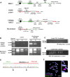
Graphical Abstract
Graphical Abstract
Figure 1.
Figure 1.
Comparative schematic diagram of the previous and optimized hCF-MAC construction. (A) Schematic diagram represents the previous method, simplified to show a minimum of seven steps, including chromosome labeling of human cell (step 1), whole-cell fusion of human cell and mouse A9 cell (step 2), MMCT into another A9 cell (step 3), MMCT into chicken DT40 cell (step 4), loxP insertion by homologous recombination in DT40 cells (step 5), chromosome transfer into CHO cell containing MAC (step 6), Cre-loxP recombination in CHO cell containing human chromosome and MAC (step 7). (B) Schematic diagram represents the optimized hCF-MAC construction method in this study, efficiently refined to include chromosome labeling of hiPSC (step 1), direct chromosome transfer from hiPSC into CHO cell containing MAC (step 2), CRISPR/Cas9-induced translocation between human chromosome and MAC in CHO cell (step 3). The diagrams visually highlight the considerable decrease in steps.
Figure 2.
Figure 2.
Chromosome labeling of hiPSC. (A) Scheme for insertion of selection marker (HS4-CAG-mCherry-HS4-PGK-HygroR) by co-transfection of the specific targeting vector and single RNP induced homology-dependent recombination on HSA6. The specific targeting vector also contains homologous arms flanked by gRNA-6T3 recognition sequence, and when introduced into the cell, cleavage occurs simultaneously with the same sequence on HSA6. (B) Representative brightfield and fluorescent images of hygromycin resistant HFL1-L6 clone. Scale bars represent 200 μm. (C) FISH on metaphase spreads of HFL1-L6 clone with HLA-A specific BAC probe (CH501-309N1, red signal) and targeting vector (green signal). Yellow arrowhead denotes the labelled HSA6 with a magnified view. Scale bars represent 10 μm. (D) Scheme for insertion of selection marker (HS4-CAG-EGFP-HS4-PGK-NeoR) by co-transfection of targeting vector and two Cas9-RNPs induced non-homologous end joining on HSA21. Targeting vector with pUC backbone and HSA21 are simultaneously cleaved by gRNA-VIKING and gRNA-21qteloT9, respectively. (E) Representative brightfield and fluorescent images of G418 resistant HFL1-L21 clone. Scale bars represent 200 μm. (F) FISH on metaphase spreads of HFL1-L21 clone with HSA21 α-satellite specific probe (p11-4, red signal) and targeting vector (green signal). Yellow arrowhead denotes the labeled and non-labeled HSA21 with a magnified view. Scale bars represent 10 μm.
Figure 3.
Figure 3.
Direct chromosome transfer from hiPSCs to CHO-MAC cells. (A, D) Representative brightfield and fluorescent images of the hygromycin and G418 co-resistant CHO colony. Scale bars represent 200 μm. (B, C) FISH on metaphase spreads of CHO MAC1/HSA6 #07-2 clone with human DNA specific probe (human Cot-1, red signal) and mouse DNA probe (mouse Cot-1, green signal), or HLA-A specific probe (CH501-309N1, red signal) and targeting vector (green signal). Yellow arrow heads denote the MAC1 and HSA6 with a magnified view. Scale bars represent 10 μm. (E, F) FISH on metaphase spreads of CHO MAC2/HSA21 #06-1 clone with human DNA specific probe (human Cot-1, red signal) and mouse DNA probe (mouse Cot-1, green signal), or HSA21 α-satellite specific probe (p11-4, red signal) and targeting vector (green signal). Yellow arrowheads denote the MAC2 and HSA21 with a magnified view. Scale bars represent 10 μm.
Figure 4.
Figure 4.
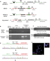
HSA6p-MAC1 construction via CRISPR/Cas9-mediated chromosome translocation. (A) Schematic representation of MAC1 and HSA6 with translocational regions highlighted. The subsequent panels depict the resulting HSA6p-MAC1 and by-product post-translocation. Red lines indicate human chromosome and green lines indicate mouse chromosome. Arrows indicate primer for detection of translocated DNA, and colors correspond to recognition chromosomes. (B) Agarose gel electrophoresis images of PCR-amplified products from bulk genomic DNA of CHO MAC1/HSA6 post-simultaneous cleavage induction of MAC1 and HSA6, or non-RNP introduced (Control). The top row shows the detection of translocated DNA (<700 bp) in HSA6p-MAC1 and the bottom row shows the by-product DNA (<450 bp). (C) Agarose gel electrophoresis image of crude PCR products derived from samples seeded at 10 cells/well. PCR product from each well is represented in two lanes: the left lane targets the by-product, while the right lane targets HSA6p-MAC1. (D) Agarose gel electrophoresis image of crude PCR products derived from 1 cell/well colonies. PCR product from each well is represented in two lanes: the left lane targets the HSA6p-MAC1, while the right lane targets by-product. (E) Sanger sequencing electropherogram excerpt, highlighting the translocation junction of genomic DNA samples derived from a pool of 10 cells. Black arrowheads show the breakpoints. (F) FISH on metaphase spreads of CHO HSA6p-MAC1 clone with human DNA specific probe (human Cot-1, red signal) and mouse DNA probe (mouse Cot-1, green signal). Yellow arrowheads denote the HSA6p-MAC1 and HSA6 with p-arm deletion with a magnified view. Scale bars represent 10 μm.
Figure 5.
Figure 5.
HSA21q-MAC2 construction via CRISPR/Cas9-mediated chromosome translocation. (A) Schematic representation of MAC2 and HSA21 with translocational regions highlighted. The subsequent panels depict the resulting HSA21q-MAC2 and by-product post-translocation. Red lines indicate human chromosome and green lines indicate mouse chromosome. Arrows indicate primer for detection of translocated DNA, and colors correspond to recognition chromosomes. MAC2-derived MI-MAC was used in this study. (B) Agarose gel electrophoresis images of PCR-amplified products from bulk genomic DNA of CHO MAC2/HSA21q post-simultaneous cleavage induction of MAC2 and HSA21 or non-RNP introduced (Control). The top row shows the detection of translocated DNA (<400 bp) in HSA21q-MAC2 and the bottom row shows the by-product DNA (<1500 bp). (C) Agarose gel electrophoresis image of crude PCR products derived from samples seeded at 100 cells/well. PCR product from each well is represented in two lanes: the left lane targets the by-product, while the right lane targets. (D) Sanger sequencing electropherogram excerpt highlighting the translocation junction of two independent 100-cell pools genomic DNA samples after enrichment by 10-cell pools. Black arrowheads show the breakpoints. (E) FISH on metaphase spreads of CHO HSA21q-MAC2 clone with human DNA specific probe (human Cot-1, red signal) and mouse DNA probe (mouse Cot-1, green signal). Yellow arrow heads denote the HSA21q-MAC2 and HSA21 with q-arm deletion with a magnified view. Scale bars represent 10 μm.

References

    1. Kuroiwa Y., Yoshida H., Ohshima T., Shinohara T., Ohguma A., Kazuki Y., Oshimura M., Ishida I., Tomizuka K.. The use of chromosome-based vectors for animal transgenesis. Gene Ther. 2002; 9:708–712. - PubMed
    1. Tomizuka K., Yoshida H., Uejima H., Kugoh H., Sato K., Ohguma A., Hayasaka M., Hanaoka K., Oshimura M., Ishida I.. Functional expression and germline transmission of a human chromosome fragment in chimaeric mice. Nat. Genet. 1997; 16:133–143. - PubMed
    1. Moriwaki T., Abe S., Oshimura M., Kazuki Y.. Transchromosomic technology for genomically humanized animals. Exp. Cell. Res. 2020; 390:111914. - PubMed
    1. Ma X., Shah Y., Cheung C., Guo G.L., Feigenbaum L., Krausz K.W., Idle J.R., Gonzalez F.J.. The pregnane X receptor gene-humanized mouse: a model for investigating drug-drug interactions mediated by cytochromes P450 3A. Drug Metab. Dispos. 2007; 35:194–200. - PubMed
    1. Yang Q., Nagano T., Shah Y., Cheung C., Ito S., Gonzalez F.J.. The PPAR alpha-humanized mouse: a model to investigate species differences in liver toxicity mediated by PPAR alpha. Toxicol. Sci. 2008; 101:132–139. - PMC - PubMed