. 2024 Mar 21;52(5):2340-2354.
doi: 10.1093/nar/gkad1237.

USP1-dependent nucleolytic expansion of PRIMPOL-generated nascent DNA strand discontinuities during replication stress

Affiliations

USP1-dependent nucleolytic expansion of PRIMPOL-generated nascent DNA strand discontinuities during replication stress

Alexandra Nusawardhana et al. Nucleic Acids Res. .

Abstract

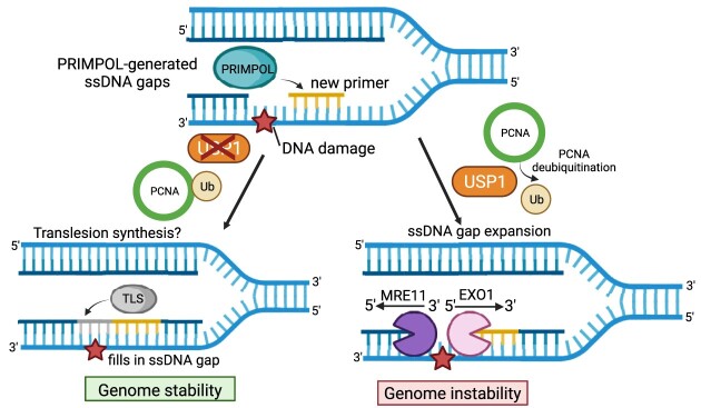
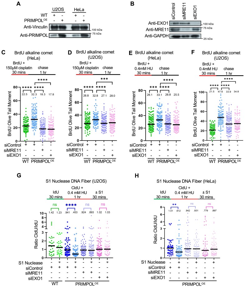
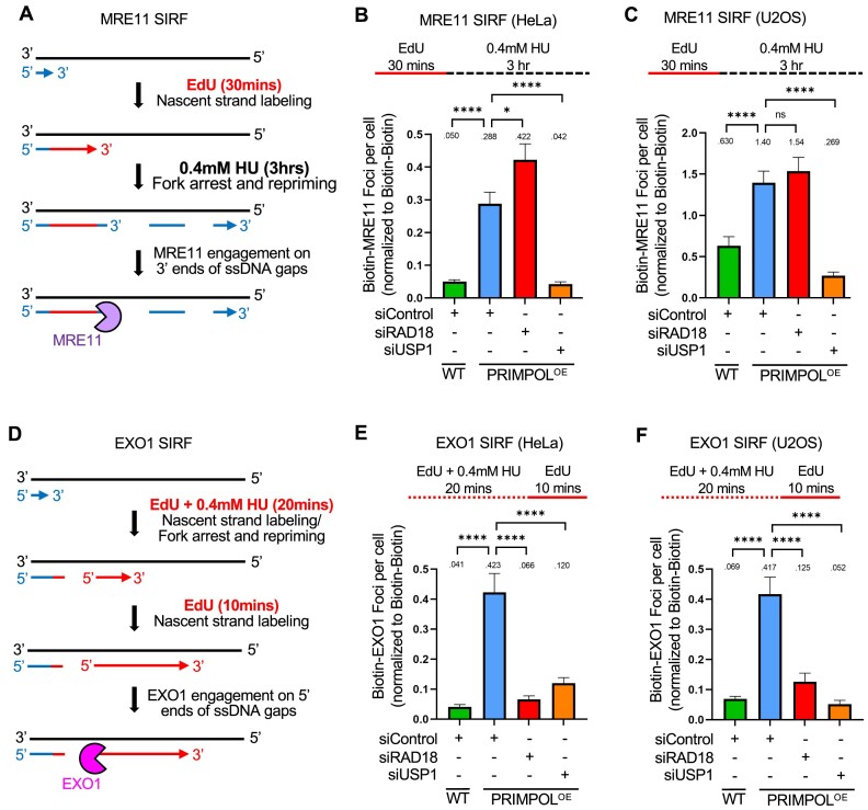
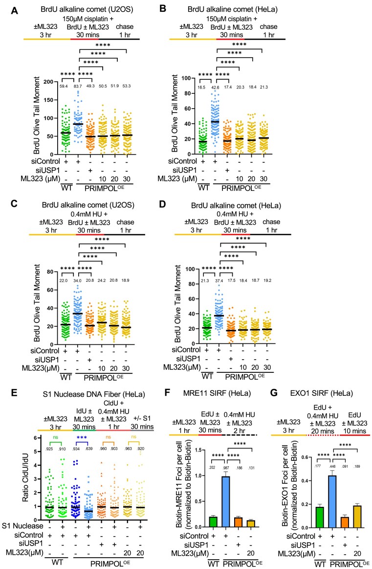
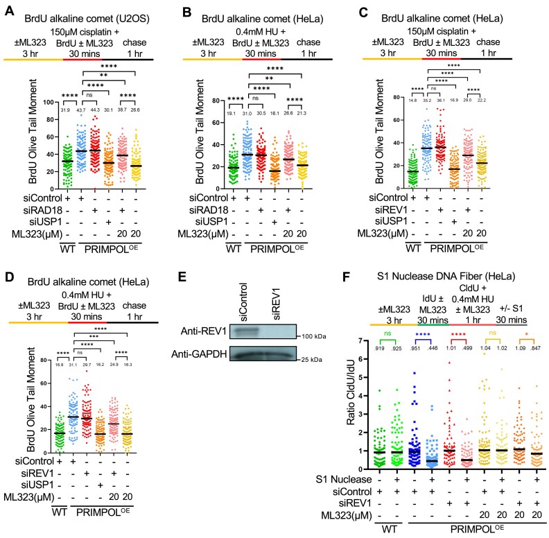
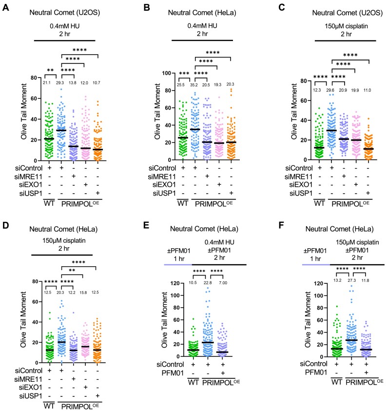
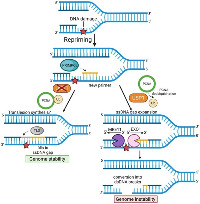
DNA replication stress-induced fork arrest represents a significant threat to genomic integrity. One major mechanism of replication restart involves repriming downstream of the arrested fork by PRIMPOL, leaving behind a single-stranded DNA (ssDNA) gap. Accumulation of nascent strand ssDNA gaps has emerged as a possible determinant of the cellular hypersensitivity to genotoxic agents in certain genetic backgrounds such as BRCA deficiency, but how gaps are converted into cytotoxic structures is still unclear. Here, we investigate the processing of PRIMPOL-dependent ssDNA gaps upon replication stress induced by hydroxyurea and cisplatin. We show that gaps generated in PRIMPOL-overexpressing cells are expanded in the 3'-5' direction by the MRE11 exonuclease, and in the 5'-3' direction by the EXO1 exonuclease. This bidirectional exonucleolytic gap expansion ultimately promotes their conversion into DSBs. We moreover identify the de-ubiquitinating enzyme USP1 as a critical regulator of PRIMPOL-generated ssDNA gaps. USP1 promotes gap accumulation during S-phase, and their expansion by the MRE11 and EXO1 nucleases. This activity of USP1 is linked to its role in de-ubiquitinating PCNA, suggesting that PCNA ubiquitination prevents gap accumulation during replication. Finally, we show that USP1 depletion suppresses DSB formation in PRIMPOL-overexpressing cells, highlighting an unexpected role for USP1 in promoting genomic instability under these conditions.


Figures

Graphical Abstract
Graphical Abstract
Figure 1.
Figure 1.
Loss of EXO1 suppresses the accumulation of nascent strand ssDNA gaps induced by HU and cisplatin in PRIMPOL-overexpressing cells. (A) Western blots showing the expression of PRIMPOL in control and PRIMPOL-overexpressing HeLa and U2OS cells. (B) Western blots showing siRNA-mediated knockdown of EXO1 and MRE11 in HeLa-PRIMPOLOE cells. (C–F) BrdU alkaline comet assay showing that knockdown of EXO1 suppresses the accumulation of replication-associated ssDNA gaps induced by treatment with 150μM cisplatin (C, D) or 0.4mM HU (E, F) in HeLa (C, E) and U2OS (D, F) PRIMPOL-overexpressing cells, similar to MRE11 depletion. At least 100 nuclei were quantified for each condition. The median values are marked on the graph and listed at the top. Asterisks indicate statistical significance (Mann-Whitney, two-tailed). Schematic representations of the assay conditions are shown at the top. (G, H) S1 nuclease DNA fiber combing assays showing that knockdown of EXO1 suppresses the accumulation of nascent strand ssDNA gaps induced by treatment with 0.4mM HU in U2OS (G) and HeLa (H) PRIMPOL-overexpressing cells, similar to MRE11 depletion. The ratio of CldU to IdU tract lengths is presented, with the median values marked on the graphs and listed at the top. At least 35 tracts were quantified for each sample. Asterisks indicate statistical significance (Mann-Whitney, two-tailed). Schematic representations of the assay conditions are shown at the top.
Figure 2.
Figure 2.
Loss of USP1 suppresses the accumulation of nascent strand ssDNA gaps induced by HU and cisplatin in PRIMPOL-overexpressing cells. (A) Western blots showing siRNA-mediated knockdown of RAD18 and USP1 in HeLa-PRIMPOLOE cells, and the impact on PCNA ubiquitination, using an antibody that specifically recognizes mono-ubiquitinated PCNA. (B–E) BrdU alkaline comet assay showing that knockdown of USP1 suppresses the accumulation of replication-associated ssDNA gaps induced by treatment with 0.4 mM HU (B, C) or 150 μM cisplatin (D, E) in HeLa (B, D) and U2OS (C, E) PRIMPOL-overexpressing cells. At least 100 nuclei were quantified for each condition. The median values are marked on the graph and listed at the top. Asterisks indicate statistical significance (Mann–Whitney, two-tailed). Schematic representations of the assay conditions are shown at the top. (F, G) S1 nuclease DNA fiber combing assays showing that knockdown of USP1 suppresses the accumulation of nascent strand ssDNA gaps induced by treatment with 0.4 mM HU in HeLa (F) and U2OS (G) PRIMPOL-overexpressing cells. The ratio of CldU to IdU tract lengths is presented, with the median values marked on the graphs and listed at the top. At least 30 tracts were quantified for each sample. Asterisks indicate statistical significance (Mann-Whitney, two-tailed). Schematic representations of the assay conditions are shown at the top.
Figure 3.
Figure 3.
USP1 depletion suppresses the binding of MRE11 and EXO1 to replication stress-induced nascent strand ssDNA gaps in PRIMPOL-overexpressing cells. (A–C) SIRF experiments showing that treatment with 0.4mM HU induces binding of MRE11 to nascent DNA in HeLa (B) and U2OS (C) PRIMPOL-overexpressing cells, and this binding is suppressed by USP1 depletion. The labeling scheme (A) is designed to capture MRE11 binding to the 3′ end of the gap (for simplicity, only one strand, e.g. the leading strand, is shown in the schematic representation; EdU-labeled nascent DNA is indicated in red). At least 100 cells were quantified for each condition. Bars indicate the mean values, error bars represent standard errors of the mean, and asterisks indicate statistical significance (t-test, two-tailed, unpaired). Schematic representations of the assay conditions are shown at the top. (D–F) SIRF experiments showing that treatment with 0.4mM HU induces binding of EXO1 to nascent DNA in HeLa (E) and U2OS (F) PRIMPOL-overexpressing cells, and this binding is suppressed by USP1 depletion. The labeling scheme (D) is designed to capture EXO1 binding to the 5′ end of the gap (for simplicity, only one strand, e.g. the leading strand, is shown in the schematic representation; EdU-labeled nascent DNA is indicated in red). At least 100 cells were quantified for each condition. Bars indicate the mean values, error bars represent standard errors of the mean, and asterisks indicate statistical significance (t-test, two-tailed, unpaired). Schematic representations of the assay conditions are shown at the top.
Figure 4.
Figure 4.
Loss of USP1 suppresses the accumulation of nascent strand ssDNA gaps induced by HU and cisplatin in PRIMPOL-overexpressing cells. (A-D) BrdU alkaline comet assay showing that inhibition of USP1 by treatment with ML323 as indicated, suppresses the accumulation of replication-associated ssDNA gaps induced by exposure to 150μM cisplatin (A, B) or 0.4mM HU (C, D) in U2OS (A, C) and HeLa (B, D) PRIMPOL-overexpressing cells. At least 70 nuclei were quantified for each condition. The median values are marked on the graph and listed at the top. Asterisks indicate statistical significance (Mann–Whitney, two-tailed). Schematic representations of the assay conditions are shown at the top. (E) S1 nuclease DNA fiber combing assay showing that inhibition of USP1 by treatment with ML323 as indicated suppresses the accumulation of replication-associated ssDNA gaps induced by exposure to 0.4 mM HU in HeLa PRIMPOL-overexpressing cells. The ratio of CldU to IdU tract lengths is presented, with the median values marked on the graphs and listed at the top. At least 50 tracts were quantified for each sample. Asterisks indicate statistical significance (Mann–Whitney, two-tailed). A schematic representation of the assay conditions is shown at the top. (F, G) SIRF experiments showing that USP1 inhibition by ML323 treatment as indicated suppresses the binding of MRE11 (F) and EXO1 (G) to nascent DNA in HeLa PRIMPOL-overexpressing cells, similar to USP1 depletion. At least 100 cells were quantified for each condition. Bars indicate the mean values, error bars represent standard errors of the mean, and asterisks indicate statistical significance (t-test, two-tailed, unpaired). Schematic representations of the assay conditions are shown at the top.
Figure 5.
Figure 5.
Inhibition of USP1 de-ubiquitination activity promotes TLS-mediated gap filling in PRIMPOL-overexpressing cells. (A, B) BrdU alkaline comet assays showing that RAD18 depletion reverses the suppression of ssDNA gap accumulation caused by inhibition of USP1 in U2OS (A) and HeLa (B) PRIMPOL-overexpressing cells upon treatment with cisplatin or HU as indicated. At least 100 nuclei were quantified for each condition. The median values are marked on the graph and listed at the top. Asterisks indicate statistical significance (Mann-Whitney, two-tailed). Schematic representations of the assay conditions are shown at the top. (C, D) BrdU alkaline comet assays showing that REV1 depletion reverses the suppression of ssDNA gap accumulation caused by inhibition of USP1 in HeLa PRIMPOL-overexpressing cells upon treatment with cisplatin (C) or HU (D) as indicated. At least 100 nuclei were quantified for each condition. The median values are marked on the graph and listed at the top. Asterisks indicate statistical significance (Mann-Whitney, two-tailed). Schematic representations of the assay conditions are shown at the top. (E) Western blots showing siRNA-mediated knockdown of REV1 in HeLa-PRIMPOLOE cells. (F) S1 nuclease DNA fiber combing assay showing that depletion of REV1 partially restores HU-induced ssDNA gap accumulation in HeLa PRIMPOL-overexpressing cells treated with the USP1 inhibitor ML323. The ratio of CldU to IdU tract lengths is presented, with the median values marked on the graphs and listed at the top. At least 70 tracts were quantified for each sample. Asterisks indicate statistical significance (Mann-Whitney, two-tailed). A schematic representation of the assay conditions is shown at the top.
Figure 6.
Figure 6.
Processing of PRIMPOL-derived ssDNA gaps by MRE11, EXO1 and USP1 leads to DSB formation. (A–D) Neutral comet assays showing that treatment with 0.4 mM HU (A, B) or 150μM cisplatin (C, D) for 2 h causes accumulation of DSBs in U2OS (A, C) and HeLa (B, D) PRIMPOL-overexpressing cells, which are suppressed by depletion of MRE11, EXO1 or USP1. At least 100 comets were quantified for each sample. The median values are marked on the graph, and asterisks indicate statistical significance (Mann–Whitney, two-tailed). (E, F) Neutral comet assays showing that inhibition of MRE11 endonuclease activity using the specific inhibitor PFM01 (100μM) suppresses DSB accumulation in PRIMPOL-overexpressing HeLa cells upon treatment with 0.4mM HU (E) or 150μM cisplatin (F) for 2 h. At least 100 comets were quantified for each sample. The median values are marked on the graph, and asterisks indicate statistical significance (Mann–Whitney, two-tailed). Schematic representations of the assay conditions are shown at the top.
Figure 7.
Figure 7.
Schematic representation of the proposed model. PRIMPOL-generated gaps are engaged by MRE11 and EXO1, even in BRCA-proficient cells, and expanded bidirectionally. USP1, through de-ubiquitination of PCNA, promotes gap expansion by MRE11 and EXO1, presumably by suppressing TLS-mediated gap filling. Under these conditions, upon extensive processing by MRE11 and EXO1, PRIMPOL-generated gaps can be converted into DSBs. Created with BioRender.com.

Similar articles

Cited by

References

    1. Zeman M.K., Cimprich K.A. Causes and consequences of replication stress. Nat. Cell Biol. 2014; 16:2–9. - PMC - PubMed
    1. Berti M., Vindigni A. Replication stress: getting back on track. Nat. Struct. Mol. Biol. 2016; 23:103–109. - PMC - PubMed
    1. Barbari S.R., Shcherbakova P.V. Replicative DNA polymerase defects in human cancers: consequences, mechanisms, and implications for therapy. DNA Repair (Amst.). 2017; 56:16–25. - PMC - PubMed
    1. Thakar T., Moldovan G.L. The emerging determinants of replication fork stability. Nucleic Acids Res. 2021; 49:7224–7238. - PMC - PubMed
    1. Schleicher E.M., Dhoonmoon A., Jackson L.M., Clements K.E., Stump C.L., Nicolae C.M., Moldovan G.L. Dual genome-wide CRISPR knockout and CRISPR activation screens identify mechanisms that regulate the resistance to multiple ATR inhibitors. PLos Genet. 2020; 16:e1009176. - PMC - PubMed

MeSH terms