. 2024 Jan 5;10(1):eadj7067.
doi: 10.1126/sciadv.adj7067. Epub 2024 Jan 5.

Pain-free oral delivery of biologic drugs using intestinal peristalsis-actuated microneedle robots

Affiliations

Pain-free oral delivery of biologic drugs using intestinal peristalsis-actuated microneedle robots

Xize Gao et al. Sci Adv. .

Abstract

Biologic drugs hold immense promise for medical treatments, but their oral delivery remains a daunting challenge due to the harsh digestive environment and restricted gastrointestinal absorption. Here, inspired by the porcupinefish's ability to inflate itself and deploy its spines for defense, we proposed an intestinal microneedle robot designed to absorb intestinal fluids for rapid inflation and inject drug-loaded microneedles into the insensate intestinal wall for drug delivery. Upon reaching the equilibrium volume, the microneedle robot leverages rhythmic peristaltic contraction for mucosa penetration. The robot's barbed microneedles can then detach from its body during peristaltic relaxation and retain in the mucosa for drug releasing. Extensive in vivo experiments involving 14 minipigs confirmed the effectiveness of the intestinal peristalsis for microrobot actuation and demonstrated comparable insulin delivery efficacy to subcutaneous injection. The ingestible peristalsis-actuated microneedle robots may transform the oral administration of biologic drugs that primary relies on parenteral injection currently.


Figures

Fig. 1.
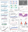
Fig. 1.. Intestinal peristalsis–actuated porcupinefish-inspired microneedle robots for oral delivery of biologic drugs.
(A and B) Photographs of a porcupine fish (Diodon holocanthus) before (A) and after (B) inflation. The fish inflates its own body and erects spines to defend itself when threatened. (A) is reprinted with permission from John White. Copyright 2005 John White. (B) is reprinted from Prescriber, 31: 28–32. Copyright 2020, with permission from Wiley Interface Ltd. (C) A fluorescent image showing the stretchable membrane and barb-equipped microneedles. Scale bar, 1 mm. (D) Photograph showing the stretched microneedle patch. Scale bar, 5 mm. (E) Photograph of the superabsorbent hydrogel before (top) and after swelling (bottom). Scale bar, 15 mm. (F) Schematic of the bioinspired microneedle robots containing drug-loaded microneedles, stretchable membrane, and superabsorbent hydrogel, which mimics the spines, skin, and water in belly of the porcupinefish, respectively. (G) Photographs of the microneedle robot before and after swelling. Scale bar, 10 mm. (H) Photographs of a swollen microneedle robot in ex vivo minipig intestine. Scale bar, 10 mm. (I) Schematic of the delivery route of the microneedle robots through a digestive tract. The microneedle robot swells and uses inherent intestinal peristalsis to achieve microneedle penetration, mucosa retention, and drug release. Upon prolonged peristaltic contraction, the microneedle robot reaches its fatigue limit, disintegrates into pieces, and is excreted through the gastrointestinal tracts.
Fig. 2.
Fig. 2.. Preparation and characterization of the microneedle robots.
(A) Workflow to fabricate the swellable microneedle robots. (B) Schematic of the rigid-flexible coupling between the microneedles and membrane through covalent cross-linking and physical entanglement. PAAm forms double network with polyvinyl alcohol (PVA). PEG doping leads to lower cross-linking degree inside the microneedle network and more PEGDA residual double bonds left on the microneedle surface as PAAm clicking sites, thereby improving drug release and enhancing the interfacial strength. (C) Representative cryo-SEM images of the PEGDA network with (right) and without (left) PEG doping. Scale bar, 2.5 μm. (D) In vitro accumulated release of rhodamine-labeled mPEG-5000 in simulated intestinal fluids at 37°C. n = 3 technical replicates. (E) Compressive (black) and penetration (red) force-displacement curves of the microneedles confirming the robustness of the microneedles and the force required to penetrate into a freshly dissected ex vivo minipig intestine. n = 3 technical replicates. The purple band represents the potential range of peristaltic forces acting on the microneedles calculated based on the data from (25). (F) Representative histological image of the penetrated ex vivo small intestine tissue. Scale bar, 0.5 mm. Red dotted line denoted locations of microneedle penetration. (G) Representative force-displacement curve showing the adhesive force between the microneedle and membrane with various PEG molecular weights and doping percentages tested by 90° peeling experiments. n = 5 technical replicates. (H) Calculated adhesive force per microneedle at the needle-membrane interface, ND, nondetectable, two-tailed Student’s t test; PEG percentage, ***P = 0.0004, **P = 0.0012; PEG molecular weight, ****P < 0.0001, **P = 0.0019. (I) Tensile stress-strain curves of the membranes with various PVA/AAm ratios. n = 3 technical replicates. (J) Swelling ratio versus time of hydrogel particles with different cross-linking densities. n = 3 technical replicates. (K) Swelling pressure versus swelling ratio of hydrogel particles with different cross-linking densities.
Fig. 3.
Fig. 3.. Microneedle penetration through peristaltic contraction.
(A) Schematic of in vivo measurement of intestinal peristaltic contraction force of the minipigs using a pressure sensor attached to a gastroscope. (B) Schematic of the peristaltic pressure (Pintestine) acting on the microneedles. (C) Representative excerpted 5-min peristaltic pressure profile of the three minipigs measured by pressure sensors with diameters of 10, 12, and 14 mm. n = 3 biological replicates. (D) Calculated peristaltic force acting on the microneedles of the microrobot with diameters of 10, 12, and 14 mm. Two-tailed Student’s t test, 12 versus 14, *P = 0.0405; 10 versus 14, *P = 0.0236; 10 versus 12; ns, nonsignificant. (E) Comparison of the experimental and theoretical expansion volume values of the microneedle robots with various parameter values. n = 3 technical replicates. (F) Dimensional change of the microneedle robots of various hydrogel loading ratios and membrane modulus before and after the swelling. (G and H) Gastroscopic images showing a microneedle robot at the swelling equilibrium state (G) contracted by the intestinal lumen (H). Scale bar, 5 mm. (I) Histology staining sections showing efficiency of the in vivo penetration. Scale bar, 0.5 mm. Red dotted line denotes locations of microneedle penetration. (J) Representative compressive curves of the microneedle robots at the equilibrated state of different hydrogel loading ratios and membrane modulus. n = 3 technical replicates. (K) Compressive tests of 1000 compression cycles of the equilibrated microneedle robots under peristaltic pressure.
Fig. 4.
Fig. 4.. Microneedle retention through peristaltic contraction and relaxation.
(A) Schematic of peristaltic contraction and relaxation, and the interactions between luminal wall and the microneedle robot during the contraction duration (TC) and relaxation duration (TR). (B) Analysis of the alternations of contraction and relaxation phases of intestinal peristalsis on three minipigs. (C) Statistical comparisons between the durations of contraction and relaxation phases of intestinal peristalsis. n = 3 technical replicates each for three biological replicates totaling n = 9. Two-tailed Student’s t test, ****P < 0.0001. (D) Schematic of the barbed microneedle retention through peristaltic contraction and relaxation (left). The barbed microneedles are constructed with different layers (nbarb) and side length ratios (Lbarb) to increase the retraction force for microneedle separation and tissue retention (middle). SEM images of the barbed microneedles (right). Scale bar, 400 μm. (E) Photographs of the barbed microneedle patches. Scale bars, 2 mm (top) and 1 mm (bottom). (F) Representative force-displacement curves of the barbless microneedles and barbed microneedles during the penetration and retraction cycles. Fout is used to evaluate the retraction force. (G) The retraction force of the barbed microneedles with various side length ratio (left) and barb layer numbers (right). Mean ± SD, n = 3 technical replicates. Two-way ANOVA test; *P = 0.0247, **P = 0.0067, ***P = 0.0001, ****P < 0.0001. (H) Merged microscopic images show the retention of barbs in the agarose gel. Scale bar, 200 μm. (I) Comparisons of penetration, retention, and drug delivery efficiency of the barbless and barbed microneedles after penetration-retraction cycles. Mean ± SD, n = 3 technical replicates. Two-way ANOVA test, ****P < 0.0001.
Fig. 5.
Fig. 5.. In vivo drug delivery in minipigs.
(A) X-ray images showing the oral delivery and excretion routes in vivo. The capsule-coated microneedle robots keep intact in the esophagus and stomach (a) and then pass through the pylorus and into the intestinal lumen (b). Once being exposed to the gut fluids, the microneedle robots begin to swell and interacted with the intestinal lumen (c). Last, the microneedle robots are contracted to crush under continuous peristaltic contraction and excreted from the gastrointestinal tracts (d). (B and D) Blood glucose (mean ± SD, n = 3 biological replicates) and plasma human insulin levels (mean ± SD, n = 5 biological replicates) tested in minipig after endoscopy-aided intestinal gavage (1.0 mg), subcutaneously injection (0.2 mg), and delivery via robotic capsules with (0.8 mg, barbed) or without (0.8 mg, barbless) barbed microneedles. (C) Blood glucose changes after delivered by various delivery methods for 3 hours, two-tailed Student’s t test; intestinal gavage, *P = 0.0117; robot, *P = 0.0142. (E) Normalized human insulin delivered into the blood plasma within 4 hours calculated by the area under the plasma concentration-time curve (AUC) divided by dose, two-tailed Student’s t test; *P = 0.0226, **P = 0.0099.

References

    1. Brown T. D., Whitehead K. A., Mitragotri S., Materials for oral delivery of proteins and peptides. Nat. Rev. Mater. 5, 127–148 (2020).
    1. Brayden D. J., Alonso M. J., Oral delivery of peptides: Opportunities and issues for translation. Adv. Drug Deliver. Rev. 106, 193–195 (2016). - PubMed
    1. Drucker D. J., Advances in oral peptide therapeutics. Nat. Rev. Drug Discov. 19, 277–289 (2020). - PubMed
    1. Wallace J. L., Granger D. N., The cellular and molecular basis of gastric mucosal defense. FASEB J. 10, 731–740 (1996). - PubMed
    1. Melo-Gonzalez F., Fenton T. M., Forss C., Smedley C., Goenka A., MacDonald A. S., Thornton D. J., Travis M. A., Intestinal mucin activates human dendritic cells and IL-8 production in a glycan-specific manner. J. Biol. Chem. 293, 8543–8553 (2018). - PMC - PubMed

Publication types

Substances