. 2024 Apr 30;120(5):506-518.
doi: 10.1093/cvr/cvae003.

The role of cellular senescence in profibrillatory atrial remodelling associated with cardiac pathology

Affiliations

The role of cellular senescence in profibrillatory atrial remodelling associated with cardiac pathology

Mozhdeh Mehdizadeh et al. Cardiovasc Res. .

Abstract

Aims: Cellular senescence is a stress-related or aging response believed to contribute to many cardiac conditions; however, its role in atrial fibrillation (AF) is unknown. Age is the single most important determinant of the risk of AF. The present study was designed to (i) evaluate AF susceptibility and senescence marker expression in rat models of aging and myocardial infarction (MI), (ii) study the effect of reducing senescent-cell burden with senolytic therapy on the atrial substrate in MI rats, and (iii) assess senescence markers in human atrial tissue as a function of age and the presence of AF.

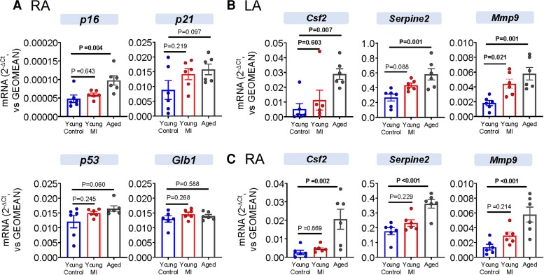
Methods and results: AF susceptibility was studied with programmed electrical stimulation. Gene and protein expression was evaluated by immunoblot or immunofluorescence (protein) and digital polymerase chain reaction (PCR) or reverse transcriptase quantitative PCR (messenger RNA). A previously validated senolytic combination, dasatinib and quercetin, (D+Q; or corresponding vehicle) was administered from the time of sham or MI surgery through 28 days later. Experiments were performed blinded to treatment assignment. Burst pacing-induced AF was seen in 100% of aged (18-month old) rats, 87.5% of young MI rats, and 10% of young control (3-month old) rats (P ≤ 0.001 vs. each). Conduction velocity was slower in aged [both left atrium (LA) and right atrium (RA)] and young MI (LA) rats vs. young control rats (P ≤ 0.001 vs. each). Atrial fibrosis was greater in aged (LA and RA) and young MI (LA) vs. young control rats (P < 0.05 for each). Senolytic therapy reduced AF inducibility in MI rats (from 8/9 rats, 89% in MI vehicle, to 0/9 rats, 0% in MI D + Q, P < 0.001) and attenuated LA fibrosis. Double staining suggested that D + Q acts by clearing senescent myofibroblasts and endothelial cells. In human atria, senescence markers were upregulated in older (≥70 years) and long-standing AF patients vs. individuals ≤60 and sinus rhythm controls, respectively.

Conclusion: Our results point to a potentially significant role of cellular senescence in AF pathophysiology. Modulating cell senescence might provide a basis for novel therapeutic approaches to AF.

Keywords: AF susceptibility; Cellular senescence; Fibrosis; Myocardial infarction; Senolytic drugs.


Conflict of interest statement

Conflict of interest: none declared

Figures

Graphical Abstract
Graphical Abstract
Senescent cells accumulate in the atria of aged rats and rats with left ventricular dysfunction due to myocardial infarction (MI). These conditions were associated with atrial fibrosis, an important pathological atrial fibrillation (AF) substrate, along with enhanced AF susceptibility (left panel). MI rats treated with senolytic compounds, which selectively eliminate senescent cells by inducing apoptosis, showed less atrial fibrosis and AF sustainability, along with suppression of markers of cellular senescence, particularly in myofibroblasts and endothelial cells (right panel).
Figure 1
Figure 1
AF inducibility, optical mapping and fibrosis quantification in LA of young control (3 months), young MI (3 months), and aged (20 months) rats. (A) Percentage of rats with inducible AF (n = 8–10; Fisher’s exact test). (B) LA activation maps at BCL 100 ms from a control, young MI, and elderly rat. (C) Mean ± SEM LA conduction velocity and APD50 (n = 6–7; two-way ANOVA followed by Bonferroni test; the P-values indicate Bonferroni-corrected differences between groups from data pooled from all BCLs). (D) Dot-plot graphs show mean ± SEM percentage fibrosis in LA. Points represent results from an individual animal (n = 5–6; one-way ANOVA with Dunnett’s test). AF, atrial fibrillation; APD50, action potential duration to 50% repolarization; BCL, basic cycle length; LA, left atrium; MI, myocardial infarction; SR, sinus rhythm.
Figure 2
Figure 2
Senescence marker expression in LA. (A) mRNA expression (n = 6; one-way ANOVA with Dunnett’s test); (B) immunofluorescence for p16 (red) and DAPI (blue; n = 4; one-way ANOVA with Dunnett’s test). DAPI, 4′,6-diamidino-2-phenylindole; LA, left atrium; MI, myocardial infarction; qPCR, quantitative polymerase chain reaction; RA, right atrium.
Figure 3
Figure 3
Senescence marker expression in RA and components of SASP in LA and RA. (A) RA senescence marker mRNA expression (n = 6; one-way ANOVA with Dunnett’s test); (B) LA SASP component mRNA expression (n = 6; one-way ANOVA with Dunnett’s test); (C) RA SASP component mRNA expression (n = 6; one-way ANOVA with Dunnett’s test). LA, left atrium; MI, myocardial infarction; RA, right atrium.
Figure 4
Figure 4
Study design, AF inducibility changes, optical mapping, and fibrosis quantification with D + Q senolytic therapy. (A) Study design; (B) AF inducibility; (C) LA activation maps at BCL 100 ms from a sham, an MI vehicle, and a MI D + Q rat; (D) mean ± SEM LA conduction velocity and APD50 (n = 5–6; two-way ANOVA with Bonferroni test; the P-values indicate Bonferroni-corrected differences between groups from data pooled from all BCLs); and (E) fibrosis analysis. Dot-plot graphs show mean ± SEM LA fibrous tissue content (n = 5; two-way ANOVA with Bonferroni test). Points represent results from individual animals. AF, atrial fibrillation; APD50, action potential duration to 50% repolarization; BCL, basic cycle length; D + Q, dasatinib and quercetin; MI, myocardial infarction; SR, sinus rhythm.
Figure 5
Figure 5
LA fibrosis, cell senescence, and SASP marker gene expression without and with D + Q therapy. mRNA expression for (A) senescence markers p16, p21, p53, and Glb1 (n = 5–8; two-way ANOVA with Bonferroni test), (B) fibrosis markers Mmp2, Col 1a1, Col 3a1, and Tgfβ2 (n = 5–8; two-way ANOVA with Bonferroni test), and (C) SASP components Igfbp4, Ccl20, Serpine2, and Igf2 (n = 5–8; two-way ANOVA with Bonferroni test). Ccl20, chemokine (C–C motif) ligand 20; Igf2, insulin-like growth factor 2; Igfbp4, insulin-like growth factor binding protein-4; Mmp2, matrix metalloproteinase 2; SASP, senescence-associated secretory phenotype; Tgfβ2, transforming growth factor 2.
Figure 6
Figure 6
Double immunofluorescence for p16 and cardiac cell-type markers in LA of sham and MI rats treated with vehicle or D + Q. (A) Fibroblast marker, vimentin (n = 4–5; unpaired t-test). (B) Endothelial cell marker, CD 31 (n = 4–5; unpaired t-test). (C) Myofibroblast marker, αSMA (n = 5–8; unpaired t-test). (D) Cardiomyocyte marker, troponin I (n = 8; unpaired t-test). All panels show cell marker staining (green), staining for p16 (red), and DAPI (DAPI, blue). D + Q, dasatinib and quercetin; DAPI, 4′,6-diamidino-2-phenylindole; MI, myocardial infarction.
Figure 7
Figure 7
Senescence marker mRNA and protein expression in human atrial tissue. (A) Immunoblot analysis of senescence markers, p16 and p21, in RAAs of younger (≤ 60) and older (≥70) patients (n = 15–19; unpaired t-test for parametric or Mann–Whitney test for non-parametric variables). (B) mRNA expression measured by dPCR in RAA of younger (≤60) and older (≥70) patients (n = 15–19; unpaired t-test for parametric or Mann–Whitney test for non-parametric variables). (C) Immunoblot analysis of senescence markers, p16, and p21 in RAA of SR and cAF patients (n = 11; unpaired t-test for parametric or Mann–Whitney test for non-parametric variables). (D) mRNA expression, as measured by dPCR in RAA of SR and cAF patients (n = 6; unpaired t-test for parametric or Mann–Whitney test for non-parametric variables). cAF, chronic atrial fibrillation; dPCR, digital polymerase chain reaction; GFD15, growth differentiation factor 15; RAA, right atrial appendage; SR, sinus rhythm; TGFβ2, transforming growth factor 2.

Comment in

References

    1. Nattel S, Shiroshita-Takeshita A, Cardin S, Pelletier P. Mechanisms of atrial remodeling and clinical relevance. Curr Opin Cardiol 2005;20:21–25. - PubMed
    1. Wetzel U, Hindricks G, Piorkowski C. Atrial fibrillation in the elderly. Minerva Med 2009;100:145–150. - PubMed
    1. Andrade J, Khairy P, Dobrev D, Nattel S. The clinical profile and pathophysiology of atrial fibrillation: relationships among clinical features, epidemiology, and mechanisms. Circ Res 2014;114:1453–1468. - PubMed
    1. Laredo M, Waldmann V, Khairy P, Nattel S. Age as a critical determinant of atrial fibrillation: a two-sided relationship. Can J Cardiol 2018;34:1396–1406. - PubMed
    1. Bhatia GS, Lip GY. Atrial fibrillation post-myocardial infarction: frequency, consequences, and management. Curr Heart Fail Rep 2004;1:149–155. - PubMed

Publication types

MeSH terms