. 2024 Feb:69:103030.
doi: 10.1016/j.redox.2024.103030. Epub 2024 Jan 3.

SIRT1 activated by AROS sensitizes glioma cells to ferroptosis via induction of NAD+ depletion-dependent activation of ATF3

Affiliations

SIRT1 activated by AROS sensitizes glioma cells to ferroptosis via induction of NAD+ depletion-dependent activation of ATF3

Xi Chen et al. Redox Biol. 2024 Feb.

Abstract

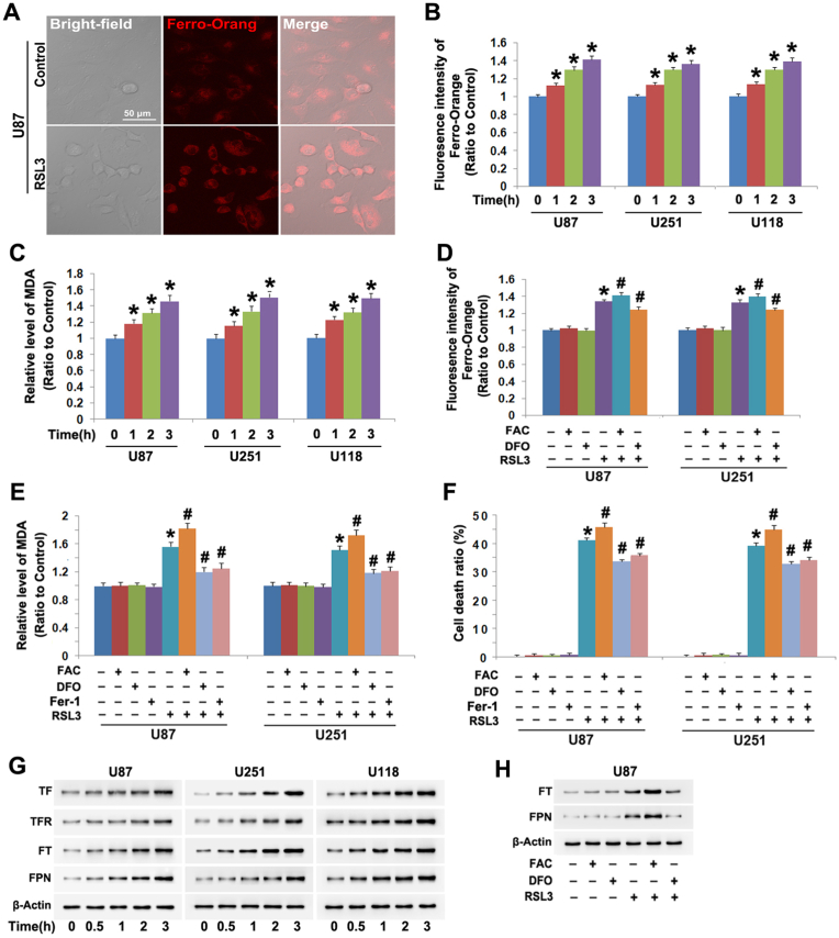
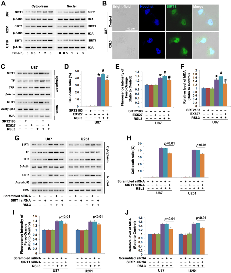
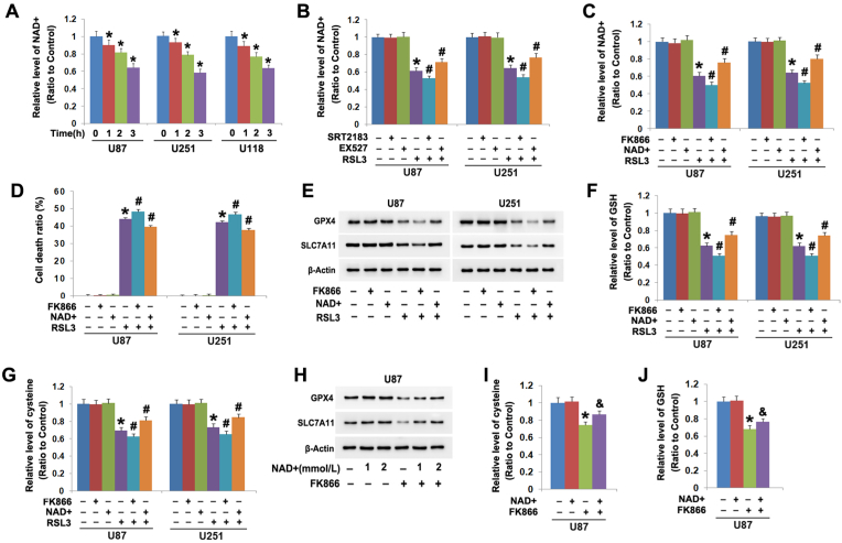
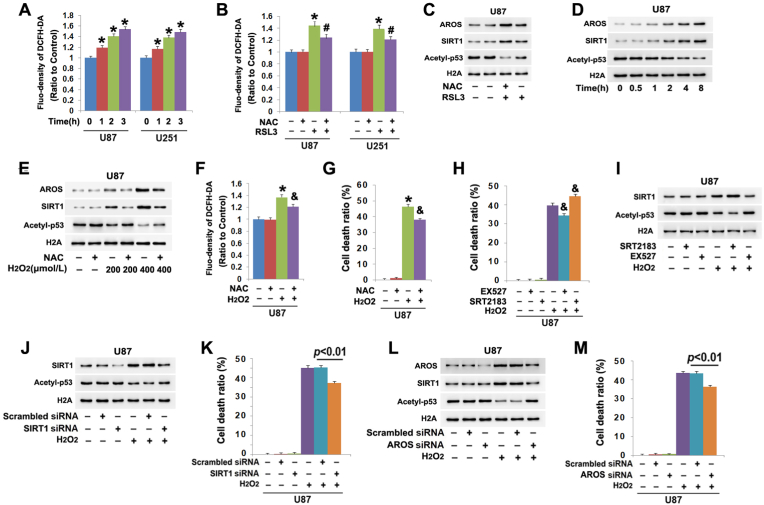
Ferroptosis is a type of programmed cell death resulting from iron overload-dependent lipid peroxidation, and could be promoted by activating transcription factor 3 (ATF3). SIRT1 is an enzyme accounting for removing acetylated lysine residues from target proteins by consuming NAD+, but its role remains elusive in ferroptosis and activating ATF3. In this study, we found SIRT1 was activated during the process of RSL3-induced glioma cell ferroptosis. Moreover, the glioma cell death was aggravated by SIRT1 activator SRT2183, but suppressed by SIRT inhibitor EX527 or when SIRT1 was silenced with siRNA. These indicated SIRT1 sensitized glioma cells to ferroptosis. Furthermore, we found SIRT1 promoted RSL3-induced expressional upregulation and nuclear translocation of ATF3. Silence of ATF3 with siRNA attenuated RSL3-induced increases of ferrous iron and lipid peroxidation, downregulation of SLC7A11 and GPX4 and depletion of cysteine and GSH. Thus, SIRT1 promoted glioma cell ferroptosis by inducting ATF3 activation. Mechanistically, ATF3 activation was reinforced when RSL3-induced decline of NAD+ was aggravated by FK866 that could inhibit NAD + synthesis via salvage pathway, but suppressed when intracellular NAD+ was maintained at higher level by supplement of exogenous NAD+. Notably, the NAD + decline caused by RSL3 was enhanced when SIRT1 was further activated by SRT2183, but attenuated when SIRT1 activation was inhibited by EX527. These indicated SIRT1 promoted ATF3 activation via consumption of NAD+. Finally, we found RSL3 activated SIRT1 by inducing reactive oxygen species-dependent upregulation of AROS. Together, our study revealed SIRT1 activated by AROS sensitizes glioma cells to ferroptosis via activation of ATF3-dependent inhibition of SLC7A11 and GPX4.

Keywords: AROS; ATF3; Ferroptosis; Glioma; NAD+; Oxidative stress; SIRT1.


Conflict of interest statement

Declaration of competing interest We declare that we have no conflict of interests.

Figures

Fig. 1
Fig. 1
RSL3 triggered ferroptosis in glioma cells. (A) Confocal microscopy combined with ferrous iron probe FerroOrang staining showed the fluorescence exhibited by FerroOrang was much stronger in the U87 cells treated with RSL3 (10 μmol/L) for 3 h than that in control cells. (B) Statistical analysis proved that the fluorescence intensity was significantly elevated by RSL3 (10 μmol/L) in U87, U251 and U118 glioma cells at incubation 1 h, which became more apparent when incubation time was extended to 2 h and 3 h. (C) RSL3 (10 μmol/L) triggered generation of lipid peroxidation product MDA in U87, U251 and U118 glioma cells in a time-dependent manner. (D) Pretreatment with FAC (500 μmol/L) for 1 h enhanced, but with FAC (500 μmol/L) inhibited RSL3-induced elevation in the fluorescence exhibited by FerroOrange. (E) The elevation of MDA induced by RSL3 (10 μmol/L) at 3 h was significantly enhanced by FAC, but apparently alleviated by DFO or Fer-1 (30 μmol/L). (F) LDH release assay revealed the glioma cell death provoked by RSL3 (10 μmol/L) was exacerbated by FAC, but inhibited by DFO or Fer-1. (G) Western blotting demonstrated RSL3 (10 μmol/L) upregulated the protein levels of transferrin (TF), transferrin receptor (TFR), ferritin (FT) and ferroportin (FPN). (H) Western blotting showed that FAC reinforced, but DFO attenuated RSL3-indcued upregulation of ferritin (FT) and ferroportin (FPN). *: p < 0.01 versus control group; #: p < 0.01 versus RSL3 group. The values are expressed as mean ± SD (n = 5 per group).
Fig. 2
Fig. 2
SIRT1 sensitized glioma cells to RSL3-induced ferroptosis. (A)Western blotting showed the protein level of SIRT1 was time-dependently upregulated in both cytoplasmic and nuclear fractions isolated from U87, U251 and U118 glioma cells treated with RSL3 (10 μmol/L), when compared with that in control cells. (B) Confocal microscopy combined with immunocytochemical staining showed that SIRT1 accumulated more apparently in the nuclei of the U87 cells treated with RSL3 for 3 h than that in control cells. (C) Western blotting revealed that the upregulation of SIRT1, transferrin (TF) and transferrin receptor (TFR) and the downregulation of acetyl-p53 caused by RSL3 were all reinforced in the cells pretreated 1 h with SIRT1 activator SRT2183 (20 μmol/L), but suppressed by SIRT1 inhibitor EX527 (200 μmol/L). (D) LDH release assay proved that SRT2183 exacerbated, but EX527 inhibited the glioma cell death caused by RSL3 (10 μmol/L). (E) Statistical analysis of the fluorescence exhibited by FerroOrange revealed that the elevated ferrous iron at 3 h due to RSL3 (10 μmol/L) treatment was aggravated by SRT2183, but prevented by EX527. (F) MDA assay showed the lipid peroxidation triggered by RSL3 (10 μmol/L) was exacerbated by SRT2183, but attenuated by EX527. (G) Western blotting proved that SIRT1 knockdown with siRNA not only obviously inhibited RSL3-induced upregulation of SIRT1, transferrin (TF) and transferrin receptor (TFR), but also prevented the downregulation of acetyl-p53. (H) LDH release assay showed that knockdown of SIRT1 with siRNA inhibited RSL3-induced glioma cell death. (I and J) RSL3-trigged ferrous iron increase and MDA elevation were both suppressed when SIRT1 was silenced by siRNA. *: p < 0.01 versus control group; #: p < 0.01 versus RSL3 group. The values are expressed as mean ± SD (n = 5 per group).
Fig. 3
Fig. 3
SIRT1 promoted RSL3-induced downregulation of SLC7A11 and GPX4. (A) Western blotting showed that RSL3 (10 μmol/L) induced time-dependent downregulation of SLC7A11 and GPX4 in U87, U251 and U118 glioma cells. (B and C) RSL3-triggered declines of cysteine and GSH became more apparent in the cells pretreated 1 h with SRT2183 (20 μmol/L), but suppressed in the cells pretreated with EX527 (200 μmol/L). (D) Western blotting revealed SRT2183 exacerbated, but EX527 attenuated RSL3-induced downregulation of SLC7A11 and GPX4. (E)Western blotting demonstrated silence of SIRT1 with siRNA prevented the downregulation of SLC7A11 and GPX4 caused by RSL3 (10 μmol/L). (F and G) Knockdown of SIRT1 with siRNA partially reversed RSL3-indcued declines of cysteine and GSH. (H) Western blotting showed treatment with SRT2183 (40 μmol/L) could obviously upregulate SIRT1 in both cytoplasmic and nuclear fractions, but downregulate acetyl-p53, SLC7A11 and GPX4 in a time-dependent manner. (I) LDH release assay showed the glioma cell death caused by SRT2183 (40 μmol/L) at 24 h was attenuated when the cells were pretreated 1 h with EX527 (200 μmol/L) or supplemented with exogenous NAD+ (2 mmol/L). (J and K) The declines of cysteine and GSH induced by SRT2183 (40 μmol/L) at 24 h were both suppressed by pretreating the cells 1 h with EX527 or supplementing exogenous NAD+. (L) Western blotting proved pretreatment with EX527 or supplement of exogenous NAD + for 1 h not only alleviated SIRT1 upregulation in both cytoplasmic and nuclear fractions, but also inhibited the downregulation of acetyl-p53, SLC7A11 and GPX4 provoked by SRT2183 (40 μmol/L) at 24 h. (M) NAD + assay showed that NAD + decline triggered by SRT2183 (40 μmol/L) at 24 h was inhibited in the cells pretreated 1 h with EX527 or supplemented with exogenous NAD+. *: p < 0.01 versus control group; #: p < 0.01 versus RSL3 group; &: p < 0.01 versus SRT2183 group The values are expressed as mean ± SD (n = 5 per group).
Fig. 4
Fig. 4
SIRT1 triggered downregulation of SLC7A11 and GPX4 by depletion of NAD+. (A) RSL3 (10 μmol/L) triggered time-dependent decline of NAD+ in U87, U251 and U118 cells. (B)The NAD + decline caused by RSL3 (10 μmol/L) at 3 h was aggravated by pretreating the cells 1 h with SRT2183 (20 μmol/L), but attenuated by EX527 (200 μmol/L). (C) Pretreatment with FK866 (500 μmol/L) for 1 h exacerbated, but supplement of exogenous NAD+ (2 mmol/L) prevented NAD + decline in the cells treated with RSL3 (10 μmol/L) for 3 h. (D) LDH release assay showed RSL3-induced glioma cell death was aggravated by FK866, but inhibited by supplement of exogenous NAD+. (E) Western blotting revealed RSL3-induced downregulation of SLC7A11 and GPX4 was enhanced by FK866, but attenuated by exogenous NAD + supplement. (F and G) Pretreatment with FK866 reinforced, but NAD + supplement attenuated RSL3-triggered declines of cysteine and GSH. (H) Western blotting showed that the downregulation of SLC7A11 and GPX4 at 24 h in the cells treated with FK866 (500 μmol/L) alone was inhibited by supplement of 1 mmol/L NAD+, and the inhibition became more apparent when NAD + dosage was increased to 2 mmol/L (I and J) The declines of cysteine and GSH in the cells treated with FK866 (500 μmol/L) was attenuated significantly by supplement of exogenous NAD + at 2 mmol/L *: p < 0.01 versus control group; #: p < 0.01 versus RSL3 group; &: p < 0.01 versus FK866 group. The values are expressed as mean ± SD (n = 5 per group).
Fig. 5
Fig. 5
NAD + depletion accounted for SIRT1-dependent activation of ATF3 (A) Western blotting revealed the protein level of ATF3 was time-dependently upregulated in both cytoplasmic and nuclear fractions isolated from the U87, U251 and U118 glioma cells treated with RSL3 (10 μmol/L), when compared with that in control cells. (B) Confocal microscopy combined with immunocytochemical staining showed that ATF3 accumulated more apparently at 3 h in the nuclei of the U87 cells treated with RSL3 (10 μmol/L) than that in control cells. (C) Knockdown of ATF3 with siRNA prevented RSL3-induced downregulation of SLC7A11 and GPX4. (D) LDH release assay showed ATF3 knockdown with siRNA suppressed RSL3-induced glioma cell death. (E and F) The declines of cysteine and GSH caused by RSL (10 μmol/L) at 3 h were both partially reversed when ATF3 was knocked down with siRNA. (G) Pretreatment with FK866 (500 μmol/L) or supplement of exogenous NAD + at 2 mmol/L for 1 h prevented RSL3-induced upregulation of ATF3 in both cytoplasmic and nuclear fractions. (H) Supplement of exogenous NAD + at 1 mmol/L could inhibit ATF3 upregulation in the cells treated with FK866 (500 μmol/L) for 24 h, and this inhibition became more apparent by increasing NAD + dosage to 2 mmol/L. (I) The upregulation of ATF3 caused by RSL3 was aggravated by SRT2183, but suppressed by EX527. (J) Pretreatment with EX527 (200 μmol/L) or supplement of exogenous NAD + at 2 mmol/L for 1 h prevented ATF3 upregulation in the cells treated with SRT2183 (40 μmol/L) for 24 h. The values are expressed as mean ± SD (n = 5 per group).
Fig. 6
Fig. 6
AROS contributed RSL3-induced activation of SIRT1. (A) Western blotting showed AROS and DBC-1 were both upregulated in the U87, U251 and U118 cells treated with RSL3 (10 μmol/L) in a time-dependent manner. (B) Confocal microscopy combined with immunocytochemical staining showed AROS was increased more apparently at 3 h in the U87 cells treated with RSL3 (10 μmol/L) than that in control cells. (C) Western blotting showed that the upregulation of AROS and downregulation of DBC-1 caused by RSL3 were both aggravated in the cells pretreated 1 h with SRT2183 (20 μmol/L), but inhibited by EX527 (200 μmol/L). (C) Co-immunoprecipitation combined with western blotting revealed that the AROS level co-immunoprecipitated with SIRT1 was elevated with the extension of RSL3 treatment time. (D) Knockdown of AROS with siRNA apparently prevented RSL3-induced upregulation of SIRT1 and ATF3 and downregulation of acetyl-p53, SLC7A11 and GPX4. (F) AROS knockdown with siRNA prevented RSL-induced decline of NAD+. (G) RSL3-triggered increase of ferrous iron was inhibited when AROS was silenced with siRNA. (H and I) Knockdown of AROS suppressed RSL3-induced decline of cysteine and GSH. (J) LDH release assay proved the glioma cell death caused by RSL3 was attenuated by knocking AROS down with siRNA. The values are expressed as mean ± SD (n = 5 per group).
Fig. 7
Fig. 7
ROS contributed to AROS-dependent activation of SIRT1. (A) Treatment with RSL3 (10 μmol/L) provoked time-dependent increases of intracellular ROS in the U87 and U251 cells. (B) Pretreatment with antioxidant NAC (5 mmol/L) for 1 h significantly prevented the increase of ROS at 3 h in the cells treated with RSL3 (10 μmol/L). (C) Western blotting showed that NAC obviously suppressed RSL3-induced upregulation of AROs and SIRT1 and downregulation of acetyl-p53. (D) Western blotting revealed AROS and SIRT were both upregulated, but acetyl-p53 was downregulated time-dependently in the U87 cells treated with 400 μmol/L H2O2. (E)Western blotting showed H2O2 triggered dosage-dependent upregulation of AROS and SIRT1 and downregulation of acetyl-p53, which were effectively inhibited by NAC. (F and G) Pretreatment 1 h with NAC (5 mmol/L) obviously prevented H2O2-induced increases of ROS and the glioma cell death. (H) LDH release assay showed that EX527 inhibited, but SRT2183 aggravated the glioma cell death provoked by H2O2. (I) SRT2183 enhanced, but EX527 inhibited H2O2-induced upregulation of SIRT1 and the downregulation of acetyl-p53. (J and K) Knockdown of SIRT1 with siRNA prevented H2O2-induced acetyl-p53 downregulation and glioma cell death. (L and M) Silence of AROS with siRNA suppressed H2O2-induced SIRT1 upregulation, acetyl-p53 downregulation and glioma cell death. *: p < 0.01 versus control group; #: p < 0.01 versus RSL3 group; &: p < 0.01 versus H2O2 group. The values are expressed as mean ± SD (n = 5 per group).
Fig. 8
Fig. 8
Schematic diagram for the role of SIRT1 in glioma cell ferroptosis. As a specific inhibitor of GPX4, RSL3 not only induces glioma cell ferroptosis, but also improves intracellular ROS levels. The increased ROS activates SIRT1 by upregulation of SIRT1 activator AROS and downregulation of SIRT1 inhibitor DBC-1. Then, the activated SIRT1 consumes its substrate NAD + excessively, which results in ATF3 accumulation in nuclei. Within nuclei, ATF3 acts as a transcription repressor to inhibit SLC7A11 transcription and expression, which decreases intracellular levels of cysteine and GSH. GSH depletion not only aggravates glioma cell ferroptosis via promoting lipid peroxidation, but also further improves ROS levels. Thus, SIRT1 activated by AROS sensitize RSL3-induced glioma cell ferroptosis by induction of NAD + depletion-dependent activation of ATF3.

References

    1. Dixon S.J., Lemberg K.M., Lamprecht M.R., Skouta R., Zaitsev E.M., Gleason C.E., et al. Ferroptosis: an iron-dependent form of nonapoptotic cell death. Cell. 2012;149:1060–1072. - PMC - PubMed
    1. Wang Z., Ding Y., Wang X., Lu S., Wang C., He C., et al. Pseudolaric acid B triggers ferroptosis in glioma cells via activation of Nox4 and inhibition of xCT. Cancer Lett. 2018;428:21–33. - PubMed
    1. Lu S., Wang X.Z., He C., Wang L., Liang S.P., Wang C.C., et al. ATF3 contributes to brucine-triggered glioma cell ferroptosis via promotion of hydrogen peroxide and iron. Acta Pharmacol. Sin. 2021;42:1690–1702. - PMC - PubMed
    1. Liu X., Chen C., Han D., Zhou W., Cui Y., Tang X., et al. SLC7A11/GPX4 inactivation-mediated ferroptosis contributes to the pathogenesis of triptolide-induced cardiotoxicity. Oxid. Med. Cell. Longev. 2022;2022 - PMC - PubMed
    1. Liu X.Y., Wei D.G., Li R.S. Capsaicin induces ferroptosis of NSCLC by regulating SLC7A11/GPX4 signaling in vitro. Sci. Rep. 2022;12 - PMC - PubMed

LinkOut - more resources