. 2024 Feb:69:103023.
doi: 10.1016/j.redox.2023.103023. Epub 2023 Dec 30.

PINK1 restrains periodontitis-induced bone loss by preventing osteoclast mitophagy impairment

Affiliations

PINK1 restrains periodontitis-induced bone loss by preventing osteoclast mitophagy impairment

Ji Sun Jang et al. Redox Biol. 2024 Feb.

Abstract

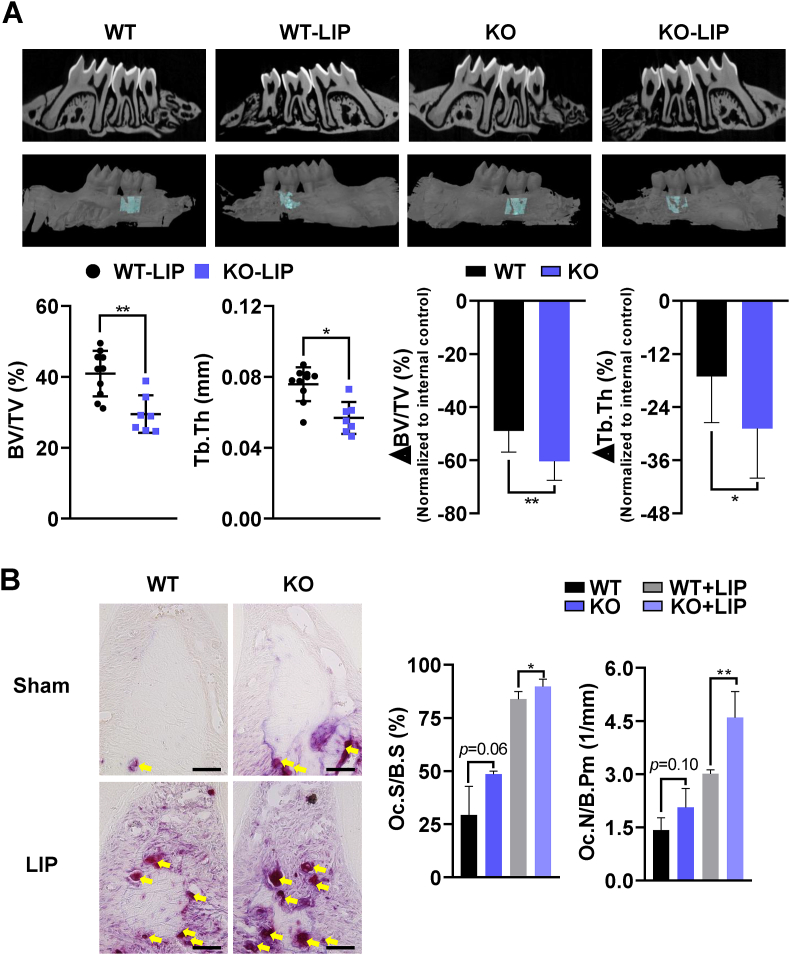
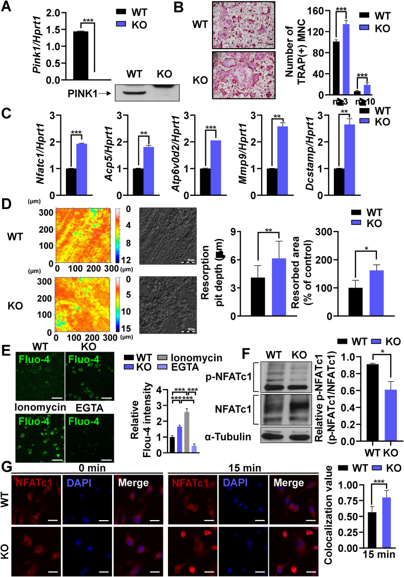
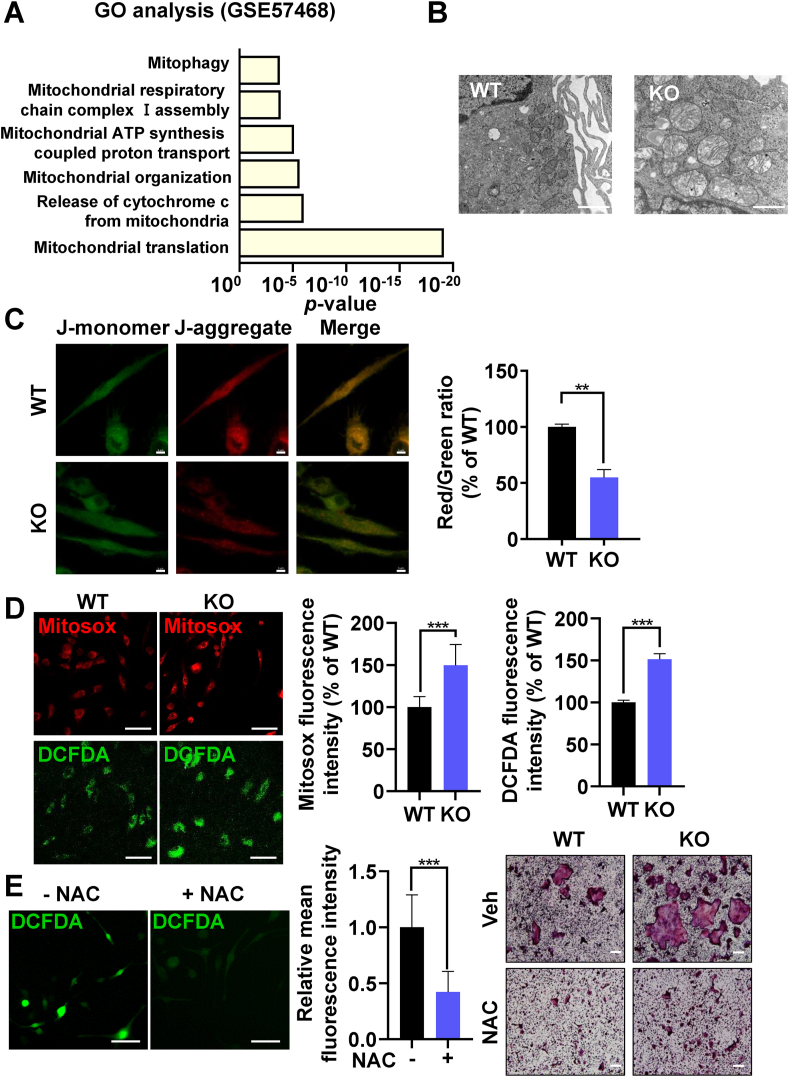
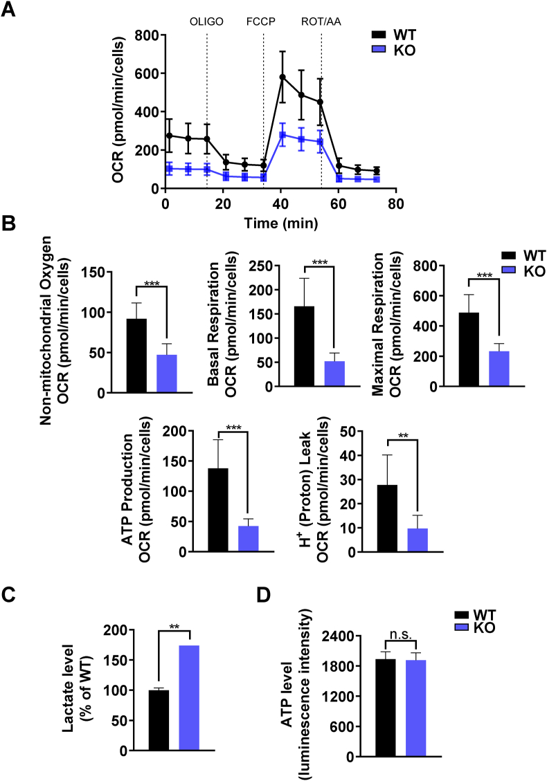
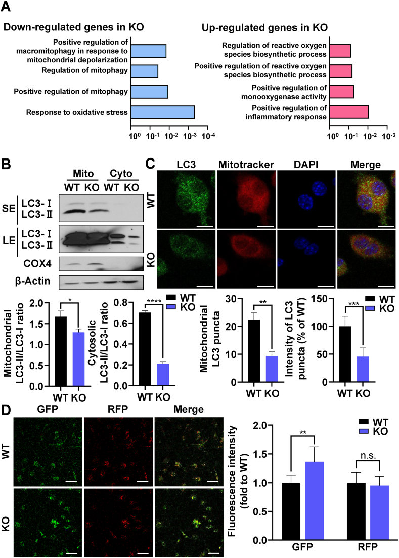
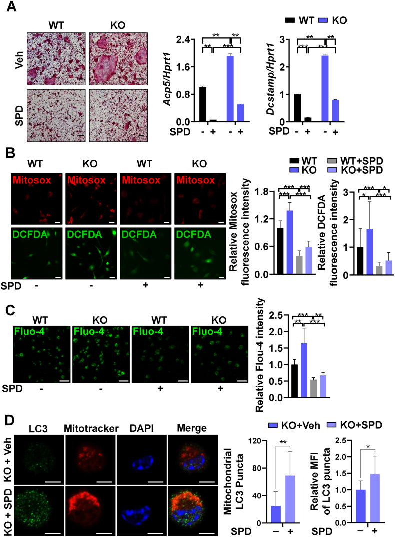
The oral colonization of periodontal pathogens onto gingival tissues establishes hypoxic microenvironment, often disrupting periodontal homeostasis in conjunction with oxidative stress. The association between reactive oxygen species (ROS) and osteolytic periodontitis have been suggested by recent studies. PTEN-induced kinase 1 (PINK1), a mitochondrial serine/threonine kinase, is an essential protein for mitochondrial quality control as it protects cells from oxidative stress by promoting degradation of damaged mitochondria through mitophagy. However, the pathophysiological roles of PINK1 in osteoclast-mediated bone loss have not been explored. Here we aimed to determine whether PINK1 plays a role in the regulation of osteoclastogenesis and alveolar bone resorption associated with periodontitis. C57BL/6 wild type (WT) and Pink1 knockout (KO) mice were subjected to ligature-induced periodontitis (LIP), and alveolar bones were evaluated by μCT-analysis and tartrate-resistant acid phosphatase (TRAP) staining. The μCT-analysis showed that bone volume fraction and travecular thickness were lower in Pink1 KO compared to WT mice. The number of TRAP-positive osteoclasts was markedly increased in the periodontal tissues of Pink1 KO mice with LIP. The genetic silencing or deletion of Pink1 promoted excessive osteoclast differentiation and bone resorption in vitro, as respectively indicated by TRAP staining and resorption pits on dentin slices. PINK1 deficiency led to mitochondrial instabilities as indicated by confocal microscopy of mitochondrial ROS, mitochondrial oxygen consumption rate (OCR) analysis, and transmission electron microscopy (TEM). Consequently, a significant increase in Ca2+-nuclear factor of activated T cells 1 (NFATc1) signaling was also found. On the other hand, restoration of mitophagy and autophagy by spermidine (SPD) treatment and the resolution of oxidative stress by N-acetyl-l-cysteine (NAC) treatment protected PINK1 deficiency-induced excessive generation of osteoclasts. Taken together, our findings demonstrate that PINK1 is essential for maintaining mitochondrial homeostasis during osteoclast differentiation. Therefore, targeting PINK1 may provide a novel therapeutic strategy for severe periodontitis with fulminant osteolysis.

Keywords: Mitophagy; Osteoclast; PINK1; Periodontitis; ROS.


Conflict of interest statement

Declaration of competing interest The authors declared no potential conflicts of interest with respect to the research, authorship, and/or publication of this article.

Figures

Fig. 1
Fig. 1
Genetic deletion of Pink1 exacerbates ligature-induced periodontitis. (A) Two- and three-dimensional μCT images of sham and silk-ligated Pink1 WT (n = 10) and Pink1 KO (n = 7) littermates. The bone parameters of alveolar bone region of maxillary second molars, indicated by turquoise color, were assessed by μCT-analysis (dot graphs). The relative reductions of bone parameters of ligated sides compared to the contralateral un-ligated sides are shown as bar graphs (B) TRAP staining images of periodontal tissues of Pink1 WT and Pink1 KO littermates (at 400× magnification). TRAP+ cells were indicated by yellow arrows. Scale bar, 50 μm. TRAP+ cells on alveolar bone surfaces were assessed. The osteoclast surfaces and numbers were measured at 200× magnification with the Osteomeasure software. Oc.N/B.Pm, Osteoclast number/bone perimeter, Oc.S/B.S, Osteoclast surface/bone surface. *P < 0.05 and **P < 0.01. (For interpretation of the references to color in this figure legend, the reader is referred to the Web version of this article.)
Fig. 2
Fig. 2
PINK1 inhibits osteoclastogenesis through the Ca2+-NFATc1 axis. (A) BMMs isolated from Pink1 WT or Pink1 KO mice were subjected to RT-PCR and western blotting. ***P < 0.001. (B) Pink1 WT or Pink1 KO BMMs were cultured in the presence of RANKL and M-CSF for 3 days and stained for TRAP. The number of TRAP+ multinucleated cells with more than 3 or 10 nuclei are presented. MNC, multinucleated cells. Scale bar, 200 μm. (C) BMMs isolated from Pink1 WT and Pink1 KO mice were cultured with the osteoclastogenic medium for 2 days. The mRNA levels of indicated genes were analyzed by RT-PCR. (D) Representative images of dentine slices. The BMMs from Pink1 WT and Pink1 KO were cultured on dentin slices, and the images of dentin slices were obtained by using confocal microscope. *P < 0.05, **P < 0.01. Scale bar, 75 μm. (E) Pink1 WT and Pink1 KO BMMs were cultured with osteoclastogenic medium for 2 days and subjected to Fluo-4/AM staining. Scale bar, 50 μm. (F) Total lysates of Pink1 WT and Pink1 KO BMMs cultured under osteoclastogenic conditions for 2 days were subjected to western blotting. The density of bands relative to α-Tubulin was quantified by ImageJ. *P < 0.05 (G) Pink1 WT and Pink1 KO pOCs were serum-starved and stimulated with RANKL (500 ng/ml) for 15 min. Representative confocal images are presented. Scale bar, 20 μm. (For interpretation of the references to color in this figure legend, the reader is referred to the Web version of this article.)
Fig. 3
Fig. 3
PINK1 regulates mitochondrial functions during osteoclastogenesis. (A) Enriched pathway analysis using the database of GSE57468. (B) Representative TEM images of Pink1 WT and Pink1 KO pOCs. Scale bar, 1 μm. (C, D) Relative levels of mitochondrial membrane potential, mitochondrial ROS, and intracellular ROS in Pink1 WT and Pink1 KO pOCs. **P < 0.01, ***P < 0.001. (E) BMMs treated with vehicle (Veh) or NAC (2.5 mM) in the presence of RANKL and M-CSF for 3 days were subjected to confocal analysis for intracellular ROS. ***P < 0.001 (left panel). Scale bar, 50 μm. BMMs were cultured with Veh or NAC (2.5 mM) together with RANKL and M-CSF for 3 days and then stained for TRAP (right panel). Scale bar, 200 μm. (For interpretation of the references to color in this figure legend, the reader is referred to the Web version of this article.)
Fig. 4
Fig. 4
PINK1 deficiency decreased OCR. (A) The OCR from Pink1 WT and Pink1 KO pOCs was assessed using the Seahorse XF96 analyzer. (B) Data were analyzed by using the Wave 2.6.1 software (n = 8). Bar charts represent mean ± SD. **P < 0.01, ***P < 0.001. (C). Intracellular lactate levels of Pink1 WT and Pink1 KO pOCs were measured by using a colorimetric/fluorometric assay kit. **P < 0.01 versus WT. (D) Intracellular ATP levels of Pink1 WT and Pink1 KO pOCs were measured by using a luminescent ATP assay kit. n.s., non-significant. (For interpretation of the references to color in this figure legend, the reader is referred to the Web version of this article.)
Fig. 5
Fig. 5
PINK1 deficiency promotes osteoclastogenesis by regulating mitophagy. (A) RNA sequencing of Pink1 WT and Pink1 KO pOCs was performed and GO functional clusters of up- and down-regulated genes were presented. The length of the bar represents the -log 10-transformed P-value. (B) Representative immunoblots of mitochondrial and cytosolic LC3 of Pink1 WT and Pink1 KO pOCs. The density of bands relative to β-Actin or COX4, were quantified by using ImageJ. *P < 0.05, ****P < 0.0001. SE, short exposure. LE, long exposure. (C) Representative confocal images of LC3 and Mitotracker staining. The number of mitochondrial LC3 puncta and the intensity of mitochondrial LC3 puncta were quantified by using ImageJ. **P < 0.01, ***P < 0.001. Scale bar, 10 μm. (D) Pink1 WT and Pink1 KO BMMs were transfected with the mRFP-GFP-LC3 plasmid. After 2 days of culture in the osteoclastogenic medium, cells were subjected to confocal microscopy. Scale bar, 50 μm. n.s., non-significant. (For interpretation of the references to color in this figure legend, the reader is referred to the Web version of this article.)
Fig. 6
Fig. 6
Induction of mitophagy and autophagy attenuates PINK1 deficiency-induced enhancement of osteoclastogenesis and mitochondria damage. (A) Pink1 WT and Pink1 KO BMMs were cultured with or without SPD (5 μM) in the osteoclastogenic medium, and subjected to TRAP staining and RT-PCR. (B) Representative confocal images for mitochondrial and intracellular ROS of Pink1 WT and Pink1 KO pOCs treated with or without SPD (5 μM). Scale bar, 20 μm. (C) Pink1 WT and Pink1 KO pOCs treated with or without SPD (5 μM) were subjected to Fluo-4/AM staining for intracellular Ca2+. Scale bar, 50 μm. (D) Pink1 KO BMMs were cultured with or without SPD (5 μM) in the osteoclastogenic medium for 2 days and the cells were subjected to confocal microscopy. The number of mitochondrial LC3 puncta and the intensity of mitochondrial LC3 puncta were quantified by using ImageJ. *P < 0.05, **P < 0.01. Scale bar, 5 μm. (For interpretation of the references to color in this figure legend, the reader is referred to the Web version of this article.)

References

    1. Sima C., Viniegra A., Glogauer M. Macrophage immunomodulation in chronic osteolytic diseases-the case of periodontitis. J. Leukoc. Biol. 2019;105(3):473–487. - PMC - PubMed
    1. Liu C., Mo L., Niu Y., Li X., Zhou X., Xu X. The role of reactive oxygen species and autophagy in periodontitis and their potential linkage. Front. Physiol. 2017;8:439. - PMC - PubMed
    1. Akalin F.A., Baltacioglu E., Alver A., Karabulut E. Lipid peroxidation levels and total oxidant status in serum, saliva and gingival crevicular fluid in patients with chronic periodontitis. J. Clin. Periodontol. 2007;34(7):558–565. - PubMed
    1. Sheikhi M., Gustafsson A., Jarstrand C. Cytokine, elastase and oxygen radical release by fusobacterium nucleatum-activated leukocytes: a possible pathogenic factor in periodontitis. J. Clin. Periodontol. 2000;27(10):758–762. - PubMed
    1. Zhu X.Q., Lu W., Chen Y., Cheng X.F., Qiu J.Y., Xu Y., Sun Y. Effects of porphyromonas gingivalis lipopolysaccharide tolerized monocytes on inflammatory responses in neutrophils. PLoS One. 2016;11(8) - PMC - PubMed