. 2024 Jan 4;187(1):79-94.e24.
doi: 10.1016/j.cell.2023.12.002.

Vaccine induction of CD4-mimicking HIV-1 broadly neutralizing antibody precursors in macaques

Affiliations

Vaccine induction of CD4-mimicking HIV-1 broadly neutralizing antibody precursors in macaques

Kevin O Saunders et al. Cell. .

Abstract

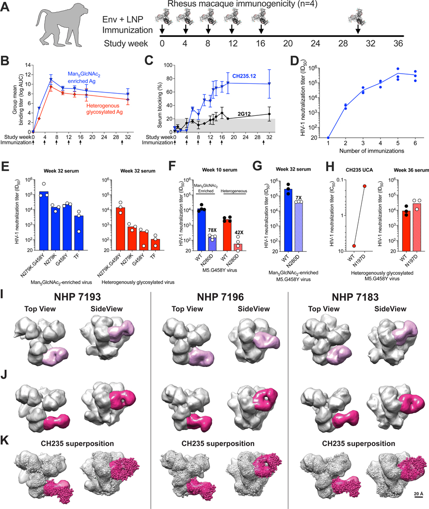
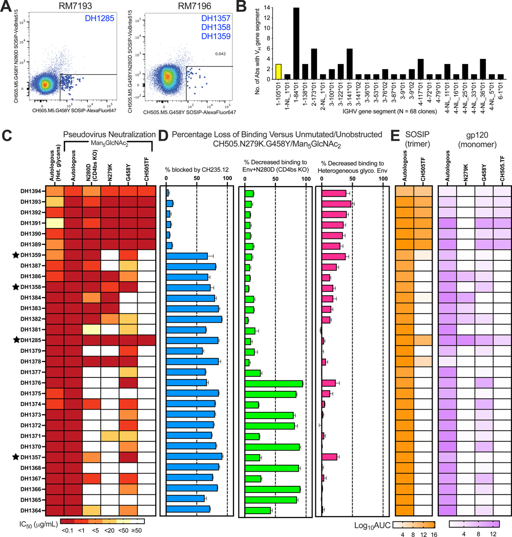
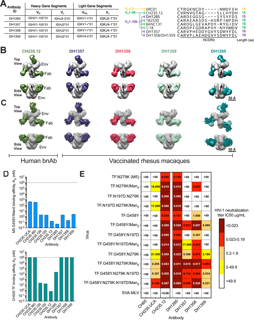
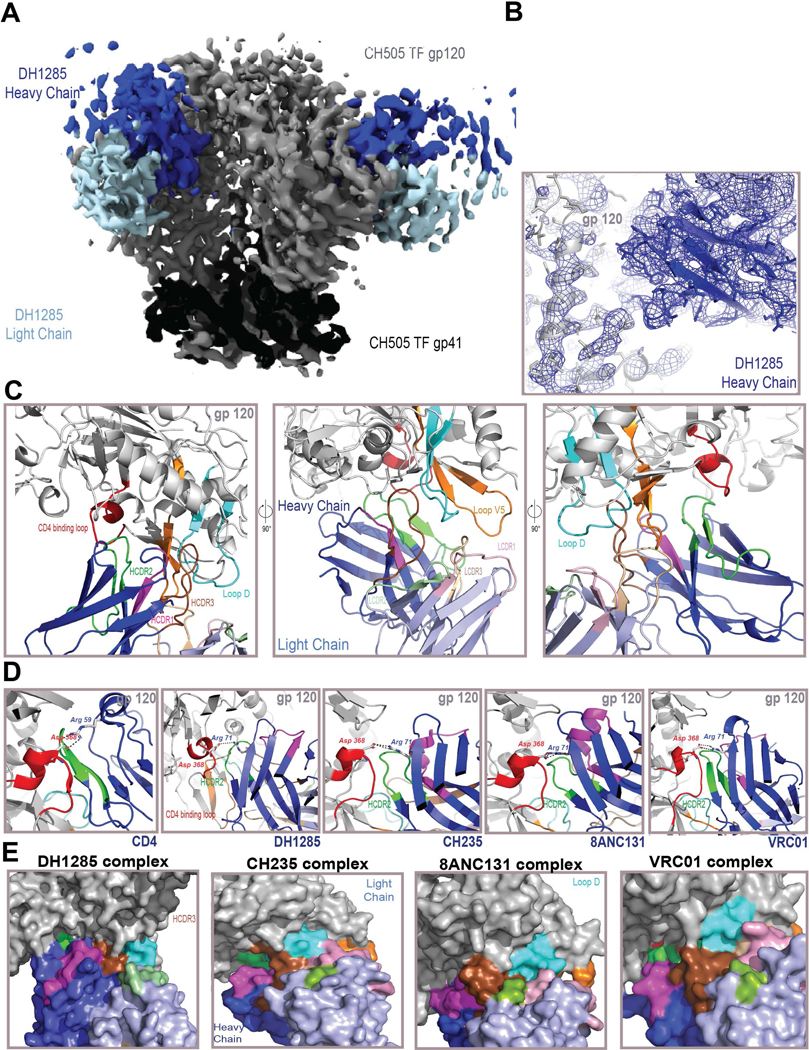
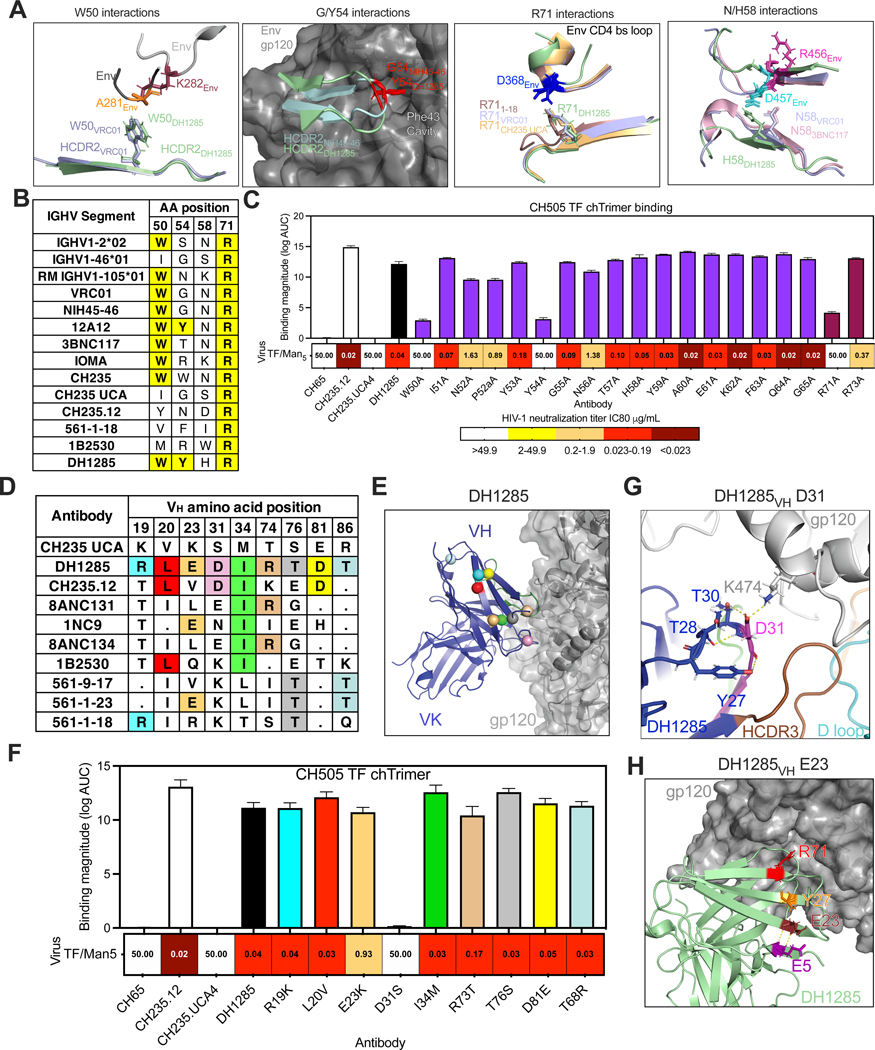
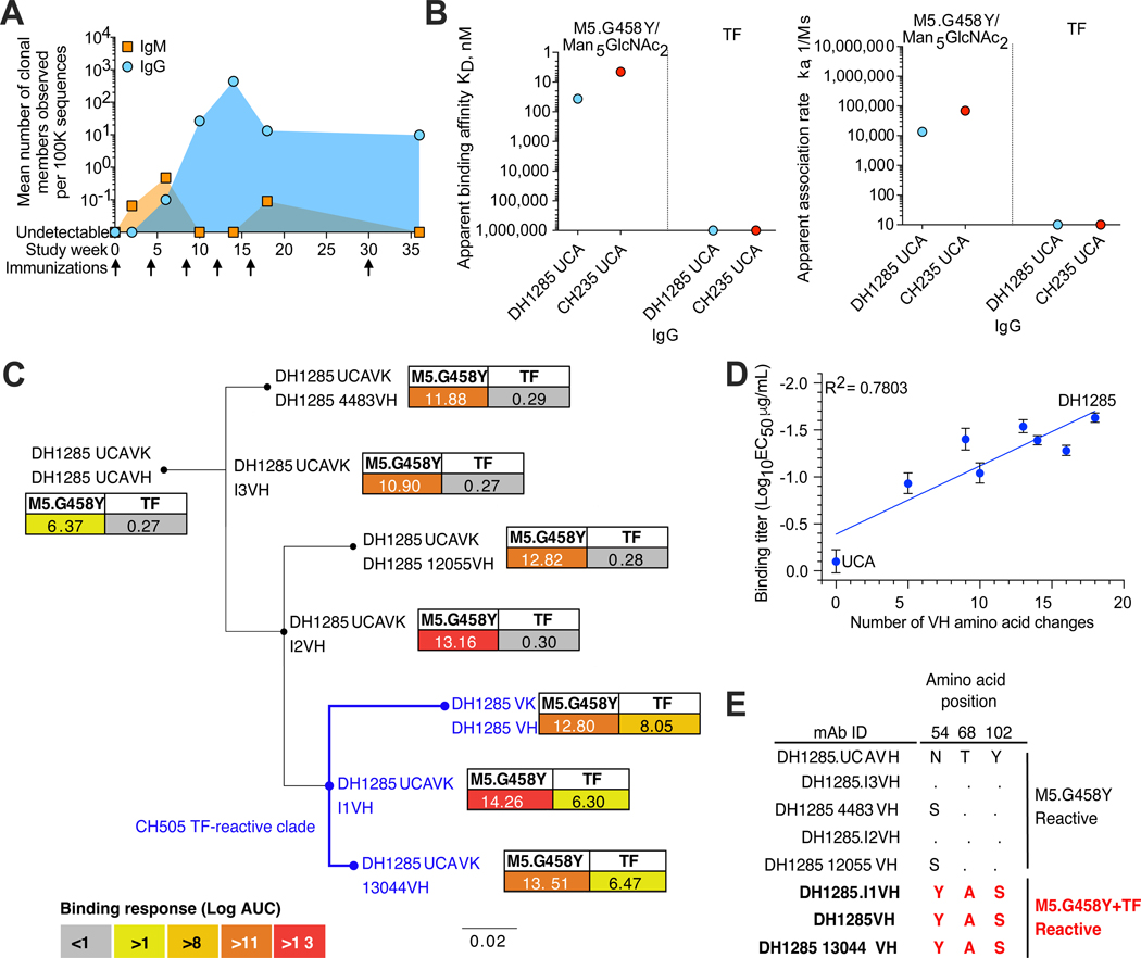
The CD4-binding site (CD4bs) is a conserved epitope on HIV-1 envelope (Env) that can be targeted by protective broadly neutralizing antibodies (bnAbs). HIV-1 vaccines have not elicited CD4bs bnAbs for many reasons, including the occlusion of CD4bs by glycans, expansion of appropriate naive B cells with immunogens, and selection of functional antibody mutations. Here, we demonstrate that immunization of macaques with a CD4bs-targeting immunogen elicits neutralizing bnAb precursors with structural and genetic features of CD4-mimicking bnAbs. Structures of the CD4bs nAb bound to HIV-1 Env demonstrated binding angles and heavy-chain interactions characteristic of all known human CD4-mimicking bnAbs. Macaque nAb were derived from variable and joining gene segments orthologous to the genes of human VH1-46-class bnAb. This vaccine study initiated in primates the B cells from which CD4bs bnAbs can derive, accomplishing the key first step in the development of an effective HIV-1 vaccine.

Keywords: CD4 binding site; CD4-mimetic antibody; EM polyclonal epitope mapping; EMPEM; HIV vaccine; broadly neutralizing antibodies; cryo-EM; cryo-electron microscopy; rhesus macaques.


Conflict of interest statement

Declaration of interests Y.K.T. and C.B. are employees of Acuitas Therapeutics. D.W. and Y.K.T. are named on a patent describing the use of nucleoside-modified mRNA in lipid nanoparticles as a vaccine platform. D.W. is named on patents that describe the use of nucleoside-modified mRNA as a platform to deliver therapeutic proteins. K.O.S., D.C.M., R.H., P.A., and B.F.H. have patents concerning the envelope immunogens used in this study.

Figures

Figure 1.
Figure 1.. CH505.M5.G458Y stabilized gp140 Trimers Induce Serum CD4 Binding Site-Directed Antibodies In Rhesus Macaques.
(A) Vaccination of rhesus macaques with Man5GlcNAc2-enriched CH505.M5.G458Y Env trimers formulated with ionizable lipid nanoparticles. (B) Serum IgG binding magnitude to Man5GlcNAc2-enriched (blue) and heterogeneously glycosylated (red) vaccine-matched Env. Values are reported as the group mean and standard error for the area under the log10 transformed concentration curve (logAUC) over time. Immunization time points indicated by arrows. (C) Serum blocking of CD4bs bnAb CH235.12 (blue) and N332 glycan bnAb 2G12 (gray) binding to CH505.M5.G458Y Env. (D) Serum neutralization ID50 titer against vaccine-matched tier 2 pseudotyped virus increases over the course of vaccination. Trend line shows group geometric mean (n=3 macaques). (E) Week 32 (post-6 immunizations) neutralization activity depends upon germline-targeting mutations N279K (M5) and G458Y, as well as Man5GlcNAc2 enrichment. In E-H, different glycoforms of pseudovirus are color-coded as in B. Bars represent group geometric mean titers. Values for individual macaques are shown as symbols. (F,G) Weeks 10 (F, post 3 immunizations) and 32 (G) serum neutralization of CH505.M5.G458Y pseudovirus is greatly diminished in the presence of CD4bs KO mutation N280D. Values reported above the bars in F and G are fold change in the group geometric mean titer. (H) Removal of the N-linked glycosylation site at Env position 197, which shields the CD4bs, improves serum neutralization of heterogeneously glycosylated CH505.M5.G458Y pseudovirus by both the CH235 UCA and vaccinated macaque serum. (I-K) 3D reconstruction of negative stain electron microscopy images of serum-derived Fabs bound to CH505.M5.G458Y Env trimers. (I)Top and side view of serum CD4bs antibodies with binding orientations such that both Fab chains are visible when looking down from the trimer. (J) Top and side view of serum CD4bs antibodies with binding orientations similar to CH235. The Fab orientation is rotated 90 degrees compared to the Fabs in I. (K) Superposition of the CH235-bound Env structure (spheres representation) into the observed density for serum Fabs bound to Env (surface representation). See also Figures S1, S2, S3, and S4.
Figure 2.
Figure 2.. CH505.M5.G458Y-Specific Monoclonal Antibodies from Vaccinated Macaques Demonstrate CD4bs-Directed Binding and Neutralization.
(A) Fluorescence-activated sorting of Env-reactive single B cells that lack binding in the presence of CD4 KO substitution N280D. (B) IGHV gene segments used by unique B cell clones isolated from NHP7193 and 7196. IGHV1–105 is highlighted yellow since it is orthologous to the human IGHV1–46 gene segment used by CD4bs bnAbs. (C) Pseudotyped virus neutralization IC50 titer against autologous virus with and without Man5GlcNAc2 enrichment. Neutralization was mapped to the CD4bs by removal of CD4bs bnAb targeting substitutions (N279K or G458Y) or CD4bs knockout substitution (N280D). (D) Percent loss of macaque antibody binding due to competition with CH235.12 binding to Env (blue), the presence of CD4bs knockout substitution N280D (green), or heterogenous Env glycosylation (pink) as determined by ELISA. Blocking magnitude or decrease in binding is determined relative to binding to CH505.M5.G458Y/GnT1-. Error bars represent the standard deviation of three replicates. (E) Binding reactivity, reported as logAUC, for SOSIP trimers (orange) (N=3 independent experiments) or gp120 versions of HIV-1 Env by ELISA (N=2 independent experiments). NAbs that are derived from IGHV1–105 and compete with CH235.12 for binding to envelope are marked with stars beside their names. See related Figures S6 - S8.
Figure 3.
Figure 3.. Characterization of Four CH235-like Precursors Isolated from Two Vaccinated Macaques.
(A) (Left) Immunogenetics of macaque CD4bs antibodies and (right) HCDR3 alignment of human CD4bs bnAbs and macaque CD4bs antibodies. (B) Negative stain electron microscopy shows approach angles for different Fabs binding to the CD4bs of CH505.M5.G458Y. Human CD4-mimicking bnAb CH235.12 is shown for comparison. The gp120 axis is indicated by a black arrow. Note the gp120 is rotated in the DH1285 bound Env. (C) Superposition of CH235.12 and rhesus Fab in complex with Env. CH235.12 bound to Env is the structure shown in spheres representation. (D) Comparison of Fab binding affinity for M5.G458Y (top) or TF (bottom) stabilized gp140 Env trimers. Dashed lines indicate limit of detection. (E) Macaque nAb IC50 neutralization titers against CH505 pseudovirus variants with different CH235 enabling substitutions. See also Figures S9 and S10.
Figure 4.
Figure 4.. Cryo-EM Structure of DH1285 Bound to CH505 TF Env Demonstrates Antibody Mimicry of CD4-like Human BnAbs.
(A) Cryo-EM reconstruction of DH1285 Fab (heavy chain in dark blue light chain in light blue) in complex with CH505 TF Env SOSIP (gp120 in light gray, gp41 in black). (B) Cryo-EM reconstruction (shown as a blue mesh) from local refinement of the gp120/Fab interface, with underlying fitted model shown in cartoon representation. (C) Three views of DH1285 bound to CH505 TF gp120; gp120 colored gray with the CD4 binding loop colored red, Loop D colored cyan, and V5 Loop colored orange. DH1285 heavy chain colored dark blue with HCDR1, HCDR2 and HCDR3 colored magenta, green and brown, respectively. DH1285 heavy chain colored dark blue with HCDR1, HCDR2 and HCDR3 colored magenta, green and brown, respectively. DH1285 light chain colored light blue with LCDR1, LCDR2 and LCDR3 colored pink, light green, and light brown, respectively. (D) Interactions of the CD4 binding loop (red) shown with (from left to right) CD4, DH1285, CH235, 8ANC131, and VRC01. Residue Asp368 in gp120, Arg71 in the antibody heavy chains and Arg59 in CD4 are shown in stick representation. The salt bridge between Asp368 and Arg71 in the antibodies or between Asp368 and Arg59 in CD4 are shown as dashed lines. (E) Surface representation showing the interactions of gp120 Loop D (cyan) with the bound antibody. Antibody heavy chain is shown in dark blue and light chain in light blue; HCDR1, HCDR2 and HCDR3 colored magenta, green and brown, respectively. See also related Figures S11-S13 and Tables S1 and S2.
Figure 5.
Figure 5.. Molecular Features Are Conserved Between DH1285 and Human CD4-Mimicking BnAbs.
(A,B) DH1285 has the key amino acids for interaction with the CD4bs (antibody amino acids 50, 54, 58, and 71) that were previously identified in human CD4-mimicking bnAbs. (A) Antibody amino acid interactions by human bnAbs and DH1285 with Env are superimposed (PDBs:6UDA, 5V8M, 6UDJ, 5WDU, 6V8X). Of note are the interactions between the D loop and W50, the Phe43 cavity and Y54, the CD4bs loop and R71, and the V5 loop and H58. (B) Comparison of residue identity at key sites listed in (A) and their conservation among VH gene segment-restricted CD4bs bnAbs. Relevant human and rhesus germlines are shown in the first three rows. Shared amino acids with DH1285 are shown in bold and highlighted yellow. (C) Binding and neutralization activity of the DH1285 HCDR2, R71, and R73 alanine mutants. CH505.TF Env trimer binding mean log AUC of three independent experiments and standard error of mean are shown. Man5GlcNAc2-enriched CH505 TF pseudovirus neutralization IC80 titers for the DH1285 alanine mutant antibodies are shown under the binding magnitude graph. (D) VH amino acids encoded for by somatic mutations that are conserved between DH1285 and VH1–46 bnAbs are shown in colored boxes. Boxes with periods indicate amino acids that are identical to the CH235 UCA. (E) Structural location of conserved DH1285 and VH1–46 bnAb mutations relative to the HIV-1 gp120 interface. The conserved amino acids are shown as spheres within the DH1285 variable region and are color-coded as in (D). (F) CH505 TF Env trimer binding by antibodies with conserved amino acids reverted to the VH1–46 germline amino acid. Binding magnitude is shown as the mean logAUC from triplicate experiments with standard error of the mean. The heatmap under the bar graph shows the neutralization IC80 titer for the same mutant antibodies against Man5GlcNAc2-enriched CH505 TF pseudovirus. Neutralization titer is color-coded as in C. (G) DH1285 cryo-EM structure showing D31DH1285 contacts with K474Env. (H) DH1285 cryo-EM structure showing positioning of E23DH1285 relative to other framework region residues with potential structural coordination of the R71 for contact with the CD4bs loop. See also Figure S9.
Figure 6.
Figure 6.. DH1285 Ontogeny Shows the Putative DH1285 Precursor Antibody Binds the Vaccine Immunogen and the Emergence of CH505 TF Env Recognition.
(A) DH1285 clonal sequences detected by MiSeq sequencing of peripheral blood B cells over time. The number of DH1285 clonal sequences measured in two independent sequencing runs were averaged and shown as the mean number of sequences per 100,000 sequencing reads for each timepoint. (B) (left) Apparent binding affinity and (right) association-rate of DH1285 UCA and CH235 UCA IgG binding to Man5GlcNAc2-enriched CH505.M5.G458Y or TF Env trimer. (C) Phylogenetic tree of DH1285 clonally-related VH sequences from week 36. Terminal nodes show observed sequences with internal nodes indicating inferred intermediates. Binding magnitude for Man5GlcNAc2-enriched CH505.M5.G458Y and CH505.TF gp140 was determined for each DH1285 clonal member when paired with the DH1285 UCA light chain. Nodes colored blue indicate antibodies that react with CH505 TF Env trimer. Values are the mean of 2 independent experiments. (D) Spearman’s correlation of M5.G458Y (blue) Env trimer log10 EC50 binding titer and the number of DH1285 VH amino acid substitutions. (E) Amino acid residues at three sites that mutate when CH505 TF Env trimer binding is first observed in the DH1285 lineage. See also Figure S10.

Similar articles

Cited by

References

    1. Corey L, Gilbert PB, Juraska M, Montefiori DC, Morris L, Karuna ST, Edupuganti S, Mgodi NM, deCamp AC, Rudnicki E, et al. (2021). Two Randomized Trials of Neutralizing Antibodies to Prevent HIV-1 Acquisition. N Engl J Med 384, 1003–1014. 10.1056/NEJMoa2031738. - DOI - PMC - PubMed
    1. Moldt B, Rakasz EG, Schultz N, Chan-Hui PY, Swiderek K, Weisgrau KL, Piaskowski SM, Bergman Z, Watkins DI, Poignard P, and Burton DR. (2012). Highly potent HIV-specific antibody neutralization in vitro translates into effective protection against mucosal SHIV challenge in vivo. Proc Natl Acad Sci U S A 109, 18921–18925. 10.1073/pnas.1214785109. - DOI - PMC - PubMed
    1. Mascola JR, Stiegler G, VanCott TC, Katinger H, Carpenter CB, Hanson CE, Beary H, Hayes D, Frankel SS, Birx DL, and Lewis MG. (2000). Protection of macaques against vaginal transmission of a pathogenic HIV-1/SIV chimeric virus by passive infusion of neutralizing antibodies. Nat Med 6, 207–210. 10.1038/72318. - DOI - PubMed
    1. Ko SY, Pegu A, Rudicell RS, Yang ZY, Joyce MG, Chen X, Wang K, Bao S, Kraemer TD, Rath T, et al. (2014). Enhanced neonatal Fc receptor function improves protection against primate SHIV infection. Nature 514, 642–645. 10.1038/nature13612. - DOI - PMC - PubMed
    1. Haynes BF, Wiehe K, Borrrow P, Saunders KO, Korber B, Wagh K, McMichael AJ, Kelsoe G, Hahn BH, Alt F, and Shaw GM. (2022). Strategies for HIV-1 vaccines that induce broadly neutralizing antibodies. Nat Rev Immunol, 1–17. 10.1038/s41577-022-00753-w. - DOI - PMC - PubMed

Publication types

LinkOut - more resources