Review
. 2024 Jan 4;31(1):7-24.
doi: 10.1016/j.stem.2023.12.006.

Hallmarks of stemness in mammalian tissues

Affiliations
Review

Hallmarks of stemness in mammalian tissues

Joep Beumer et al. Cell Stem Cell. .

Abstract

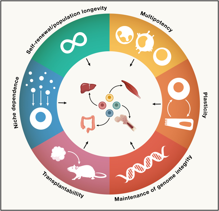
All adult tissues experience wear and tear. Most tissues can compensate for cell loss through the activity of resident stem cells. Although the cellular maintenance strategies vary greatly between different adult (read: postnatal) tissues, the function of stem cells is best defined by their capacity to replace lost tissue through division. We discuss a set of six complementary hallmarks that are key enabling features of this basic function. These include longevity and self-renewal, multipotency, transplantability, plasticity, dependence on niche signals, and maintenance of genome integrity. We discuss these hallmarks in the context of some of the best-understood adult stem cell niches.

Keywords: adult stem cells; hallmarks; lineage tracing; longevity; niche; organoids; plasticity; regeneration.


Conflict of interest statement

Declaration of interests H.C. and J.B. are inventors on patents held by the Royal Netherlands Academy of Arts and Sciences that cover organoid technology. H.C. is currently head of pharma Research and Early Development (pRED) at Roche, Basel, Switzerland. H.C.’s full disclosure is given at https://www.uu.nl/staff/JCClevers/.

Figures

Figure 1
Figure 1
Hallmarks of stemness in mammalian tissues We describe a set of six complementary hallmarks that can be observed among tissue resident stem cells. These collectively enable the stem cell’s function to replace lost cells in the corresponding tissues.
Figure 2
Figure 2
Maintaining individual stem cells or stem cell populations (A) Example of a “neutral drift” adult stem cell model. In the intestinal epithelium, stem cells residing at the crypt bottom divide symmetrically and compete for the limited niche space. Stem cell maintenance is guaranteed at the level of the population, but individual ASC clones are stochastically outcompeted while others gain dominance. (B) In the fibers of skeletal muscles, satellite cells can divide asymmetrically when the mitotic axis is perpendicular to the muscle fiber. The daughter cell retaining access to the basal lamina will maintain stem cell potential, whereas the other daughter cells commit to differentiation. In this example, the ASC displays asymmetry at the single-cell level, and individual clones are maintained.
Figure 3
Figure 3
Determining stem cell potential in dividing and quiescent cells (A) Proliferating cells can be traced exploiting expression of KI67. KI67 is restricted to all cell-cycle phases except G0. A KI67-expression controlled Cre-recombinase can be used to trace dividing cells to assess their long-term clonogenicity and multipotency. When reporter recombination is initiated in a dividing stem cell (labeled in red, first scenario), long-term tracing will be observed. When the dividing cell is a committed progenitor, labeling is transient and will eventually be lost (second scenario). (B) Strategy to perform lineage tracing from slowly dividing or post-mitotic label-retaining cells (LRCs) in the murine small intestine. Cre-recombinase is expressed as two parts: the ubiquitously produced C-terminal CreB (in yellow) and the N-terminal CreA that is fused to histone 2B (H2B) (in blue). CreA expression can be induced by the addition of β-naphthoflavone (βNF), followed by cell-cycle-dependent dilution of the H2B-CreA protein. Both Cre-halves are additionally fused to an FKHB domain, of which fusion can be induced by a small molecule in order to reconstitute a functional enzyme and induce lineage tracing (traced cells will be red). CreB is constitutively expressed (yellow cells, left). Induction of CreA expression by βNF followed by a 3–13 days dilution will lead to a scenario in which only slowly dividing LRCs and post-mitotic cells retain both parts of the enzyme (blue and yellow cells, middle). The induction of dimerization does not yield long-term clone formation, suggesting that slowly dividing cells are not homeostatic stem cells (red cells, top). When epithelial damage is induced before dimerization of the Cre-domains, long-term tracing can be observed (bottom). These experiments evidence that LRCs are not homeostatic stem cells, but regain stemness upon damage.
Figure 4
Figure 4
Plasticity in tissues with fast and slow turnover (A) Waddington landscape of the intestinal crypt in homeostasis (left) and upon damage to the stem cell compartment (right). In homeostasis, cells gradually commit and differentiate while leaving the stem cell zone and losing access to stem cell niche factors. Yet, during damage and loss of stem cells, the majority of crypt cells can readily reverse their commitment due to permissive chromatin when exposed to stem cell signals. Stem cells at the “edge” of the stem cell niche (numbers indicate the cell position counting from the bottom of the crypt) are more likely to lose access to key stem cell signals and differentiate. (B) Examples of tissues that employ self-replicating mature cells to sustain tissue renewal. In the damaged pancreatic acini, acinar cells can increase self-renewal and induce an accelerated rate of acini fission. Damage to the liver causes the emergence of oval cells from cholangiocytes that can both generate both new cholangiocytes as well as hepatocytes. Chief cells at the bottom of the stomach glands can self-replicate during homeostasis to sustain their pool, but damage endows these cells with expanded lineage potential.

Similar articles

Cited by

References

    1. Post Y., Clevers H. Defining adult stem cell function at its simplest: the ability to replace lost cells through mitosis. Cell Stem Cell. 2019;25:174–183. - PubMed
    1. Clevers H., Watt F.M. Defining adult stem cells by function, not by phenotype. Annu. Rev. Biochem. 2018;87:1015–1027. - PubMed
    1. Knoblich J.A. Mechanisms of asymmetric stem cell division. Cell. 2008;132:P583–P597. - PubMed
    1. Loeffler D., Schroeder T. Symmetric and asymmetric activation of hematopoietic stem cells. Curr. Opin. Hematol. 2021;28:262–268. - PubMed
    1. Loeffler D., Wehling A., Schneiter F., Zhang Y., Müller-Bötticher N., Hoppe P.S., Hilsenbeck O., Kokkaliaris K.D., Endele M., Schroeder T. Asymmetric lysosome inheritance predicts activation of haematopoietic stem cells. Nature. 2019;573:426–429. - PubMed

LinkOut - more resources