. 2024 Jan 5;15(1):11.
doi: 10.1038/s41419-023-06375-x.

Protein tyrosine phosphatase PTPRO represses lung adenocarcinoma progression by inducing mitochondria-dependent apoptosis and restraining tumor metastasis

Affiliations

Protein tyrosine phosphatase PTPRO represses lung adenocarcinoma progression by inducing mitochondria-dependent apoptosis and restraining tumor metastasis

Yuan Dai et al. Cell Death Dis. .

Abstract

Emerging evidence indicates that protein activities regulated by receptor protein tyrosine phosphatases (RPTPs) are crucial for a variety of cellular processes, such as proliferation, apoptosis, and immunological response. Protein tyrosine phosphatase receptor type O (PTPRO), an RPTP, has been revealed as a putative suppressor in the development of particular tumors. However, the function and the underlying mechanisms of PTPRO in regulating of lung adenocarcinoma (LUAD) are not well understood. In this view, the present work investigated the role of PTPRO in LUAD. Analysis of 90 pairs of clinical LUAD specimens revealed significantly lower PTPRO levels in LUAD compared with adjacent non-tumor tissue, as well as a negative correlation of PTPRO expression with tumor size and TNM stage. Survival analyses demonstrated that PTPRO level can help stratify the prognosis of LUAD patients. Furthermore, PTPRO overexpression was found to suppress the progression of LUAD both in vitro and in vivo by inducing cell death via mitochondria-dependent apoptosis, downregulating protein expression of molecules (Bcl-2, Bax, caspase 3, cleaved-caspase 3/9, cleaved-PARP and Bid) essential in cell survival. Additionally, PTPRO decreased LUAD migration and invasion by regulating proteins involved in the epithelial-to-mesenchymal transition (E-cadherin, N-cadherin, and Snail). Moreover, PTPRO was shown to restrain JAK2/STAT3 signaling pathways. Expression of PTPRO was negatively correlated with p-JAK2, p-STAT3, Bcl-2, and Snail levels in LUAD tumor samples. Furthermore, the anti-tumor effect of PTPRO in LUAD was significant but compromised in STAT3-deficient cells. These data support the remarkable suppressive role of PTPRO in LUAD, which may represent a viable therapeutic target for LUAD patients.


Conflict of interest statement

The authors declare no competing interests.

Figures

Fig. 1
Fig. 1. Low PTPRO expression is associated with advanced TNM stage and poor prognosis of LUAD patients.
A Pan-cancer analysis of PTPRO gene expression in different tumor types from TCGA datasets. B Different PTPRO gene expression in LUAD and adjacent normal lung specimens in GEO datasets (GSE19188 and GSE 19804). C PTPRO gene expression difference between LUAD and normal lung tissues in TCGA cohort. D PTPRO gene expression in LUAD with TNM stage I–II vs. those with TNM stage III–IV in TCGA cohort. E PTPRO gene expression in LUAD with T stage T1–2 vs. T stage T3–4 in TCGA cohort. F, G Kaplan–Meier overall survival analyses of LUAD patients according to the gene level of PTPRO in TCGA cohort and a combined cohort (TCGA, EGA, GEO), respectively. H Representative IHC staining of PTPRO protein expression in paired LUAD tissues and adjacent normal specimens in microarray. Statistical comparison of PTPRO immunoreactivity scores were exhibited in the right panels. I Representative IHC staining of high expression and low expression of PTPRO in LUAD specimens. J Kaplan–Meier overall survival analysis of LUAD patients according to PTPRO protein expression level. ACC adrenocortical carcinoma, BLCA adrenocortical carcinoma, BRCA breast invasive carcinoma, CESC cervical squamous cell carcinoma and endocervical adenocarcinoma, CHOL cholangiocarcinoma, COAD colon adenocarcinoma, DLBC lymphoid neoplasm diffuse large B-cell lymphoma, ESCA esophageal carcinoma, GBM glioblastoma multiforme, HNSC head and neck squamous cell carcinoma, KICH kidney chromophobe, KIRC kidney renal clear cell carcinoma, KIRP kidney renal papillary cell carcinoma, LAML acute myeloid leukemia, LGG brain lower grade glioma, LIHC liver hepatocellular carcinoma, LUAD lung adenocarcinoma, LUSC lung squamous cell carcinoma, MESO mesothelioma, OV ovarian serous cystadenocarcinoma, PAAD pancreatic adenocarcinoma, PCPG pheochromocytoma and paraganglioma, PRAD prostate adenocarcinoma, READ rectum adenocarcinoma, SARC sarcoma, SKCM skin cutaneous melanoma, STAD stomach adenocarcinoma, TGCT testicular germ cell tumors, THCA thyroid carcinoma, THYM thymoma, UCEC uterine corpus endometrial carcinoma, UCS uterine carcinosarcoma, UVM uveal melanoma.
Fig. 2
Fig. 2. PTPRO overexpression attenuates LUAD growth both in vitro and in vivo.
A The transfection efficiency was validated by western blotting in HCC827, PC9 and H1975 cells, after transfected with vector and PTPRO plasmids. B MTT assay was performed to examine the cell proliferation abilities of transfected LUAD cell lines. C Colony-formation assays of LUAD cells transfected with vector or PTPRO plasmids. The colonies were numbered and statistically compared in the right panel. D Stable transfected PC9 cells were subcutaneously injected into nude mice. After 3 weeks, mice were sacrificed, and xenografts were excised. E Excised tumor weight was measured as mean ± SD for all xenografts in two independently repeated experiments (n = 9, the other four paired xenografts are presented in Supplementary Fig. 1). F Tumor volume was calculated every 5 days to monitor tumor growth (n = 9). Data are represented as mean ± SD. *p < 0.05, **p < 0.01, ***p < 0.001.
Fig. 3
Fig. 3. PTPRO induces cells apoptosis in LUAD.
A The GSEA enrichment analysis of PTPRO in TCGA-LUAD cohort. B GSEA enrichment analysis of PTPRO regarding apoptosis pathway in LUAD. C Cell apoptosis was evaluated by flow cytometry strategy in HCC827, PC9 and H1975 cells between PTPRO-overexpression and vector groups. Statistical analyses were presented in the right panel. *p < 0.05, **p < 0.01.
Fig. 4
Fig. 4. PTPRO induces mitochondria-dependent apoptosis of LUAD cells in vitro.
A Western blotting analysis of mitochondria-dependent apoptosis pathway-related proteins in HCC827, PC9 and H1975 cells, including Bcl-2, Bax, cleaved-caspase 3, caspase 3, cleaved PARP, cleaved-caspase 9 and Bid. B Representative images of JC-1 staining for mitochondrial membrane potential (Δψm). Quantitative analysis of Red/Green fluorescence ratio were presented as mean ± SD (n = 3). C Representative images of MPTP opening staining (weaker green fluorescence indicated more MPTP opening), and statistically quantitative analysis of green fluorescence was demonstrated as mean ± SD (n = 3). *p < 0.05, **p < 0.01, ***p < 0.001.
Fig. 5
Fig. 5. PTPRO overexpression induces mitochondria-dependent apoptosis of LUAD in vivo.
A Representative IHC staining targeting PTPRO, Ki67, Bax, Bcl-2, cleaved-caspase 3 and Bid in the xenografts formed by subcutaneously injection of stably-transfected LUAD cells, with statistical analyses in the right panels. B Western blotting showed the expression of mitochondria apoptosis related proteins in resected xenografts. C Statistically analyses of western blotting results were indicated as mean ± SD (n = 9). *p < 0.05, **p < 0.01, ***p < 0.001.
Fig. 6
Fig. 6. PTPRO suppresses LUAD metastasis both in vitro and in vivo.
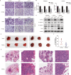
A Transwell assays were used to evaluate the migration and invasion abilities of HCC827, PC9 and H1975 cells transfected with vector or PTPRO plasmids. B Statistically analyses of transwell assays were indicated as mean ± SD (n = 3). C Western blotting demonstrated the expression of metastasis related proteins including E-cadherin, N-cadherin and Snail in LUAD cells with transfection of vector or PTPRO plasmids. D Stably-transfected PC9 cells were injected into tail vein, and mice were cultured for 4 weeks before sacrificed. Then mice lungs were resected to observe tumors on the lung surfaces. Two independent replicate experiments were conducted, each involving three pairs of mice. E Representative H&E staining of the abovementioned lung tissue slices. F Morphometry of lung sections (n = 6). *p < 0.05, **p < 0.01.
Fig. 7
Fig. 7. JAK2/STAT3 signaling pathway is involved in PTPRO anti-tumor activity in LUAD.
A KEGG pathway analysis for significantly correlated genes of PTPRO in TCGA-LUAD dataset. B Phosphorylation levels of JAK2 and STAT3 were tested by western blotting in HCC827, PC9 and H1975 cells transfected with vector or PTPRO plasmids. C Western blotting analyses of p-JAK2 and p-STAT3 in mice LUAD xenografts. D Representative IHC staining of PTPRO, p-JAK2, p-STAT3, Bcl-2 and Snail in LUAD tissues from ten patients. E Spearman’s rank correlation analysis was performed to unveil the relationships between PTPRO and p-JAK2, p-STAT3, Bcl-2, Snail, respectively.
Fig. 8
Fig. 8. PTPRO exerts slight anti-tumor effects in STAT3-deficient LUAD cells.
A The knockout efficiency was verified through Western blotting in PC9 cells after transfection with LentiCRISPRv2 targeting STAT3. B MTT assay was conducted to assess the effect of PTPRO on cell proliferation in STAT3-deficient PC9 cells. The data are represented as mean ± SD. C Flow cytometry strategy was used to assess cell apoptosis in STAT3-deficient cells transfected with vector or PTPRO plasmids. D Transwell assays were employed to evaluate the migration and invasion abilities of STAT3-deficient cells following vector or PTPRO plasmids transfection. Statistical analyses are depicted in the right panels. E The proposed functional mechanisms of PTPRO’s anti-tumor effects in LUAD (dotted line indicates the possible signaling pathway whereas need further validation). *p < 0.05 and **p < 0.01 indicate statistically significant by unpaired Student’s t test, respectively.

Similar articles

Cited by

References

    1. Arimura Y, Yagi J. Comprehensive expression profiles of genes for protein tyrosine phosphatases in immune cells. Sci Signal. 2010;3:rs1. doi: 10.1126/scisignal.2000966. - DOI - PubMed
    1. He R, Zeng LF, He Y, Zhang S, Zhang ZY. Small molecule tools for functional interrogation of protein tyrosine phosphatases. FEBS J. 2013;280:731–50. doi: 10.1111/j.1742-4658.2012.08718.x. - DOI - PMC - PubMed
    1. Chen L, Juszczynski P, Takeyama K, Aguiar RC, Shipp MA. Protein tyrosine phosphatase receptor-type O truncated (PTPROt) regulates SYK phosphorylation, proximal B-cell-receptor signaling, and cellular proliferation. Blood. 2006;108:3428–33. doi: 10.1182/blood-2006-03-013821. - DOI - PubMed
    1. Kang MM, Shan SL, Wen XY, Shan HS, Wang ZJ. Tumor-suppression mechanisms of protein tyrosine phosphatase O and clinical applications. Asian Pac J Cancer Prev. 2015;16:6215–23. doi: 10.7314/APJCP.2015.16.15.6215. - DOI - PubMed
    1. Thomas PE, Wharram BL, Goyal M, Wiggins JE, Holzman LB, Wiggins RC. GLEPP1, a renal glomerular epithelial cell (podocyte) membrane protein-tyrosine phosphatase. Identification, molecular cloning, and characterization in rabbit. J Biol Chem. 1994;269:19953–62. doi: 10.1016/S0021-9258(17)32113-0. - DOI - PubMed

Publication types

MeSH terms

Substances