. 2024 Feb;34(2):140-150.
doi: 10.1038/s41422-023-00917-w. Epub 2024 Jan 5.

LDLR is an entry receptor for Crimean-Congo hemorrhagic fever virus

Affiliations

LDLR is an entry receptor for Crimean-Congo hemorrhagic fever virus

Zhi-Sheng Xu et al. Cell Res. 2024 Feb.

Abstract

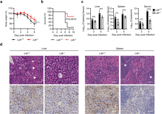
Crimean-Congo hemorrhagic fever virus (CCHFV) is the most widespread tick-born zoonotic bunyavirus that causes severe hemorrhagic fever and death in humans. CCHFV enters the cell via clathrin-mediated endocytosis which is dependent on its surface glycoproteins. However, the cellular receptors that are required for CCHFV entry are unknown. Here we show that the low density lipoprotein receptor (LDLR) is an entry receptor for CCHFV. Genetic knockout of LDLR impairs viral infection in various CCHFV-susceptible human, monkey and mouse cells, which is restored upon reconstitution with ectopically-expressed LDLR. Mutagenesis studies indicate that the ligand binding domain (LBD) of LDLR is necessary for CCHFV infection. LDLR binds directly to CCHFV glycoprotein Gc with high affinity, which supports virus attachment and internalization into host cells. Consistently, a soluble sLDLR-Fc fusion protein or anti-LDLR blocking antibodies impair CCHFV infection into various susceptible cells. Furthermore, genetic knockout of LDLR or administration of an LDLR blocking antibody significantly reduces viral loads, pathological effects and death following CCHFV infection in mice. Our findings suggest that LDLR is an entry receptor for CCHFV and pharmacological targeting of LDLR may provide a strategy to prevent and treat Crimean-Congo hemorrhagic fever.


Conflict of interest statement

The authors declare no competing interests.

Figures

Fig. 1
Fig. 1. LDLR is an important host factor for CCHFV infection.
a Screening of LDLR and LDLR-related proteins (LRPs) that are crucial for CCHFV infection. The HEK293T cells were edited with a control or sgRNAs targeting the genes encoding LDLR and LRPs (two sgRNAs for each gene). After puromycin selection, the cell pools were infected with CCHFV (MOI = 0.05) for 24 h before RT-qPCR was performed. Data were normalized to the relative mRNA level of CCHFV S in the control sgRNA-edited cells. b Surface expression of LDLR in different cell lines. The indicated cell lines were assessed by flow cytometry using the anti-LDLR mAb (R301). c CCHFV infectivity of different cell lines. The indicated cell lines were infected with CCHFV (MOI = 0.05) for 48 h. The CCHFV Gn-positive cells were examined by flow cytometry with a customized anti-Gn monoclonal antibody (7A11). d Overexpression of LDLR enhances CCHFV infection in DLD1 cells. Control and LDLR-overexpressing DLD1 cells were infected with CCHFV (MOI = 0.05) for 24 h (left panel) or 48 h (right panel). The levels of CCHFV S mRNA and NP protein were determined by RT-qPCR (left panel) and immunoblots (right panel), respectively. e Effects of LDLR-deficiency on CCHFV infection in SW13 cells. SW13 cells were edited with a control (gNC) or three individual sgRNAs targeting different regions of LDLR coding sequence (gLDLR). The control and LDLR sgRNA-edited SW13 cell pools were infected with CCHFV (MOI = 0.05). CCHFV NP expression (left panel, 48 hpi), mRNA level of CCHFV S segment (2nd panel, 24 hpi), percentage of Gn-positive cells (3rd panel, 48 hpi) and cell cytopathic effects (right panel, 72 hpi) was measured by immunoblots, RT-qPCR, flow cytometry and crystal violet staining, respectively. For bar graphs, data are normalized to that of the control gRNA-edited cells. f Effects of LDLR-deficiency on production of progeny viruses. SW13 cells were edited with a control (gNC) or three individual sgRNAs targeting different regions of LDLR coding sequence (gLDLR). The sgRNA-edited SW13 cell pools were then infected with CCHFV (MOI = 0.05) for 72 h. Titers of progeny viruses in the supernatants were measured by TCID50 assay. Data are normalized to that of the control gRNA-edited cells. LOD, limit of detection. g Effects of LDLR-deficiency on CCHFV infection in various cells. Huh7, Vero E6 and Hepa1-6 cells were edited with a control gRNA or the indicated numbers of gRNAs targeting LDLR gene. Cells were infected with CCHFV (MOI = 0.05) for 24 h before RT-qPCR was performed. Data are normalized to the CCHFV S mRNA level in the control gRNA-edited cells. h CCHFV infectivity in LDLR-knockout SW13 and Huh7 cells. Single clones of LDLR-knockout SW13 and Huh7 were isolated and confirmed by immunoblots (left). The control (gNC) or LDLR-deficient clone (gLDLR-C1) were infected with CCHFV (MOI = 0.05) for 24 h before RT-qPCR analysis. Data are normalized to that of each control gRNA-edited cells. i CCHFV infectivity in Ldlr−/− primary cells. Primary hepatocytes and lung fibroblasts (MLFs) prepared from WT and Ldlr−/− mice were incubated with CCHFV (MOI = 0.05). The mRNA level of CCHFV S segment (top, 48 hpi) and the viral genomic copies in the supernatant (bottom, 72 hpi) were measured by RT-qPCR. j Effects of LDLR-deficiency on CCHFV, RVFV, EBIV and VSV infection. The control (gNC) or LDLR-deficient clone (gLDLR-C1) were inoculated with the indicated viruses for 24 h before RT-qPCR was performed. Data are represented as mean ± SD. **P < 0.01; ***P < 0.001; ****P < 0.0001.
Fig. 2
Fig. 2. LDLR is required for CCHFV infection.
a A schematic presentation of full-length LDLR and its truncated mutants that lack the ligand binding domain (∆LBD) and epidermal growth factor like domain (∆EGF). b–f The LBD of LDLR is important for CCHFV infection. The control (gNC) or LDLR-deficient (gLDLR-C1) SW13 cells were reconstituted with a control vector, full-length LDLR or the indicated LDLR truncations respectively. The cells were inoculated with CCHFV (MOI = 0.05), and CCHFV NP expression (b 48 hpi), mRNA level of CCHFV S segment (c 24 hpi), percentage of CCHFV Gn-positive cells (d 48 hpi), cell survival (e 72 hpi) and production of CCHFV progeny viruses (f 72 hpi) were measured by RT-qPCR, immunoblots, flow cytometry, crystal violet staining and TCID50 assay, respectively. LOD, limit of detection. Data are represented as mean ± SD. ****P < 0.0001; ns, not significant.
Fig. 3
Fig. 3. LDLR is essential for CCHFV binding to cells.
a Effects of LDLR on CCHFV attachment and internalization. The control (gNC), LDLR-deficient (gLDLR-C1) or LDLRAP1-deficient (gLDLRAP1) SW13 cells were incubated with CCHFV at 4 °C for 1 h (for binding assay), or followed with incubation at 37 °C for 1 h (for internalization assay). The cells were collected and CCHFV S mRNA level was measured by RT-qPCR. Data are normalized to the CCHFV S mRNA level in the control gRNA-edited cells. b Effects of LDLR blocking antibodies on CCHFV infection. SW13, Huh7, Vero E6, primary human PBMCs and HUVECs, and mouse Hepa1-6 cells were pre-incubated with a rabbit anti-hLDLR mAb (R301), a goat anti-hLDLR pAb (#AF2148), a rabbit anti-mLDLR mAb (R004), or their respective control IgGs as indicated for 1 h before CCHFV infection (MOI = 0.05). Twenty-four hours after infection, the cells were collected for RT-qPCR analysis for CCHFV S mRNA level. Data are normalized to that of cells treated with the respective control IgG at 0 μg/mL. c Effects of LDLR blocking antibodies on the entry of RVFV, EBIV and VSV. SW13 cells were pre-incubated with the indicated concentrations of a control rIgG or a rabbit anti-hLDLR mAb (R301) for 1 h before infection RVFV, EBIV or VSV. Twenty-four hours after infection, mRNA levels of RVFV M segment, EBIV S segment, or VSV L gene were measured by RT-qPCR analysis. Data are normalized to that of cells treated with the respective control IgG at 0 μg/mL. d LDL inhibits CCHFV infection. SW13 cells (left) and HUVEC cells (right) were pre-treated with the indicated concentrations of LDL for 1 h and then left uninfected or infected with CCHFV (MOI = 0.05). Twenty-four hours post infection, CCHFV S mRNA level was analyzed by RT-qPCR. Data are normalized to that of CCHFV infected cells without LDL treatment. e The soluble human LDLR-Fc fustion protein (sohLDLR-Fc) inhibits CCHFV infection. CCHFV (MOI = 0.05) was pre-incubated with the indicated concentrations of Fc or sohLDLR-Fc for 1 h before infection of SW13 and Huh7 cells. Twenty-four hours post infection, CCHFV S mRNA level was analyzed by RT-qPCR. Data are normalized to that of cells infected with un-pretreated viruses. f Effects of sohLDLR-Fc on RVFV, EBIV and VSV infection in SW13 cells. RVFV (MOI = 0.1), EBIV (MOI = 0.5) or VSV (MOI = 0.1) were pre-incubated with the indicated concentrations of sohLDLR-Fc or Fc for 1 h before infection of SW13 cells. Twenty-four hours post infection, mRNA levels of RVFV M segment, EBIV S segment, or VSV L gene were measured by RT-qPCR analysis. Data are normalized to that of cells infected with the respective un-pretreated viruses. Data are represented as mean ± SD. *P < 0.05; **P < 0.01; ***P < 0.001; ****P < 0.0001.
Fig. 4
Fig. 4. LDLR binds directly to Gc of CCHFV.
a Pull-down of CCHFV virions by sohLDLR. CCHFV, biotinylated sohLDLR and magnetic streptavidin beads were co-incubated as indicated. Pull-down was performed with a magnet and the pellet was subjected to RT-qPCR analysis. Data are represented as mean ± SD. b Monoclonal antibodies against CCHFV Gc (ADI 36121), Gn (JE12) or their respective control IgG was immobilized on plates. ELISA-based binding assays were performed with CCHFV, biotinylated sohLDLR and Avidin-HRP. Data are represented as mean ± SD. c Recombinant Gc of CCHFV YL16070 and IbAr 10200 strains and biotinylated sohLDLR were co-incubated as indicated. Pull-down assay was performed with magnetic streptavidin beads and the pellets were subjected to immunoblots with the indicated antibodies. d Biotinylated sohLDLR was immobilized onto the streptavidin biosensors. Binding parameters of recombinant Gc or Gn of the YL16070 and IbAr 10200 strain and VSV-G to LDLR were measured by Bio-Layer Interferometry (BLI) in the indicated PBS buffers. Fitted curves are shown with dotted lines. A 1:1 binding model was used to calculate the KD.
Fig. 5
Fig. 5. LDLR is required for CCHFV pathogenesis in mice.
a, b WT (n = 11) and LDLR-knockout (n = 11) mice were pretreated with anti-IFNAR1 monoclonal antibody MAR1-5A3 (200 µg/mouse) 24 h before infection and then infected with CCHFV (10 TCID50) via the intraperitoneal route. Forty-eight hours post infection, 200 µg of MAR1-5A3 was administrated. The body weight (a) and survival (b) of the mice were monitored daily. c, d WT (n = 7) and LDLR-knockout (n = 7) mice were pretreated with anti-IFNAR1 monoclonal antibody MAR1-5A3 (200 µg/mouse) 24 h before infection and then infected with CCHFV (10 TCID50) via the intraperitoneal route. Forty-eight hours post infection, 200 µg of MAR1-5A3 was administrated. Mice were necropsied and the livers and spleens were collected at day 3 and 5 post infection. Viral loads were quantified by RT-qPCR and shown as the number of viral RNA copies per microgram of organs or per mL of sera (c). H&E staining and immunostaining with anti-Gn mAb (7A11) were performed and the pathological changes were indicated (d). The extensive necrosis (white arrowheads) and necrotic cellular debris (white arrows) in the liver were indicated; white pulps in the spleen (white asterisks) were marked. The bars represent 100 µm. Data are represented as mean ± SD. **P < 0.01; ***P < 0.001; ****P < 0.0001.
Fig. 6
Fig. 6. Anti-LDLR treatment protects mice from CCHFV pathogenesis in mice.
a A flowchart of the experiment. C57BL/6 mice were pretreated with anti-IFNAR1 monoclonal antibody MAR1-5A3 (300 µg/mouse) plus control rIgG (n = 10, 100 µg/mouse) or an LDLR blocking antibody (R004) (n = 7, 100 µg/mouse) intraperitoneally 24 h before infection. Another dose of the rIgG or R004 mAb (100 µg) was administrated 1 h prior to infection. Mice were challenged with CCHFV (100 TCID50 per mouse, subcutaneously). Twenty-four hours post infection, MAR1-5A3 (200 µg/mouse) was administrated. The rIgG or R004 mAb was administrated at 100 µg quaque die for 5 days post infection. b, c, Protective effects of LDLR blocking antibody on fatality caused by CCHFV infection. As described in (a), C57BL/6 mice were treated with rIgG (n = 10) or the LDLR blocking antibody (R004) (n = 7) and subjected to CCHFV challenge. Body weight (b) and survival (c) of the mice were monitored daily. d, e Protective effects of LDLR blocking antibody on CCHFV caused pathogenesis. As described in (a), C57BL/6 mice were treated with rIgG (n = 6) or the LDLR blocking antibody (R004) (n = 6) and subjected to CCHFV challenge. Mice were necropsied and the livers and spleens were collected at day 5 post infection. Viral loads were quantified by qRT-PCR and shown as the number of viral RNA copies per microgram of organs or per mL of sera (d). H&E staining and immunostaining with anti-Gn mAb (7A11) were performed and the pathological changes were indicated (e). The extensive necrosis (white arrowheads) and necrotic cellular debris (white arrows) in liver were indicated; white pulps in the spleen (white asterisks) were marked. The bars represent 100 µm. Data are represented as mean ± SD. *P < 0.05; **P < 0.01.

Similar articles

Cited by

References

    1. Ergonul O. Crimean-Congo haemorrhagic fever. Lancet Infect. Dis. 2006;6:203–214. doi: 10.1016/S1473-3099(06)70435-2. - DOI - PMC - PubMed
    1. Bente DA, et al. Crimean-Congo hemorrhagic fever: history, epidemiology, pathogenesis, clinical syndrome and genetic diversity. Antiviral Res. 2013;100:159–189. doi: 10.1016/j.antiviral.2013.07.006. - DOI - PubMed
    1. Akinci E, Bodur H, Leblebicioglu H. Pathogenesis of Crimean-Congo hemorrhagic fever. Vector Borne Zoonotic Dis. 2013;13:429–437. doi: 10.1089/vbz.2012.1061. - DOI - PubMed
    1. Zivcec, M., Scholte, F. E. M., Spiropoulou, C. F., Spengler, J. R. & Bergeron, E. Molecular insights into Crimean-Congo hemorrhagic fever virus. Viruses8, 106 (2016). - PMC - PubMed
    1. Hawman, D. W. & Feldmann, H. Recent advances in understanding Crimean-Congo hemorrhagic fever virus. F1000Res7(F1000 Faculty Rev), 1715 (2018). - PMC - PubMed

Publication types

MeSH terms