. 2024 Jan 5;12(1):1.
doi: 10.1186/s40635-023-00587-3.

Mastering the brain in critical conditions: an update

Affiliations

Mastering the brain in critical conditions: an update

Chiara Robba et al. Intensive Care Med Exp. .

Abstract

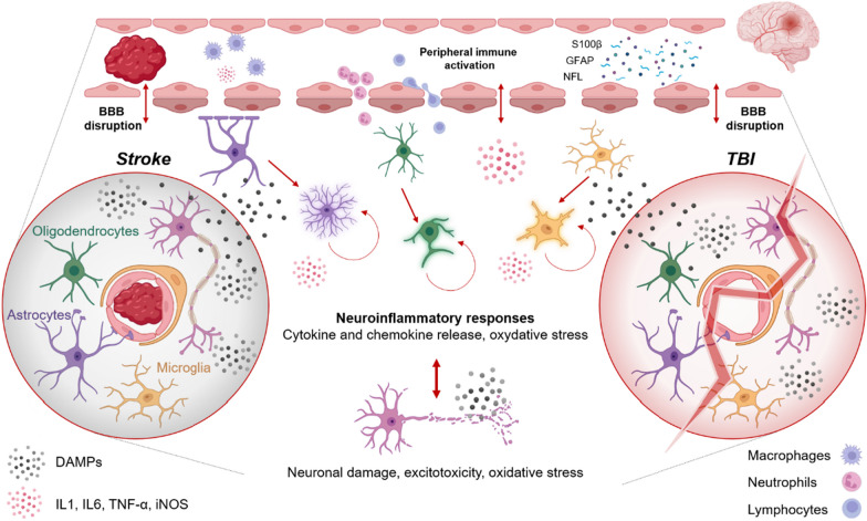
Acute brain injuries, such as traumatic brain injury and ischemic and hemorragic stroke, are a leading cause of death and disability worldwide. While characterized by clearly distict primary events-vascular damage in strokes and biomechanical damage in traumatic brain injuries-they share common secondary injury mechanisms influencing long-term outcomes. Growing evidence suggests that a more personalized approach to optimize energy substrate delivery to the injured brain and prognosticate towards families could be beneficial. In this context, continuous invasive and/or non-invasive neuromonitoring, together with clinical evaluation and neuroimaging to support strategies that optimize cerebral blood flow and metabolic delivery, as well as approaches to neuroprognostication are gaining interest. Recently, the European Society of Intensive Care Medicine organized a 2-day course focused on a practical case-based clinical approach of acute brain-injured patients in different scenarios and on future perspectives to advance the management of this population. The aim of this manuscript is to update clinicians dealing with acute brain injured patients in the intensive care unit, describing current knowledge and clinical practice based on the insights presented during this course.

Keywords: Acute brain injury; Neuroimaging; Neuromonitoring; Outcome; Prognostication.


Conflict of interest statement

None of the authors have any competing interests in the manuscript.

Figures

Fig. 1
Fig. 1
Neuroinflammatory response after ABI. Inflammation triggered by release of damage-associated molecular patterns (DAMPs) and reactive oxygen species (ROS) will cause activation of resident immune cells and release of proinflammatory citokines. Invasion of the CNS by circulating immune cells initially consists of innate immune cells, but is joined by adaptive leukocytes within days (primed T cells). While this response largely wanes, a proportion of patients display a persistent immune dysregulation, directly contributing to tissue damage. BBB blood brain barrier, DAMPS damaged associated molecular patterns, IL interleukin, iNOS inducible nitric oxide synthase, s100 calcium binding protein B (s100B); glial fibrillary acid protein (GFAP) neurofilament light (NFL); TGF transforming growth factor, TNF tumor necrosis factor. Figure created with BioRender.com
Fig. 2
Fig. 2
Conceptual framework of various neuromonitoring devices based on the model of “a house”. Structural assessments of brain parenchyma include pupillometry and neuro-radiology modalities, such as computed tomography (CT) and magnetic resonance (MR) imaging. Plumbing or cerebral hemodynamic assessments include brain tissue perfusion monitors, transcranial Doppler, and near-infrared spectroscopy. This may also include vascular Imaging such as CT angiography and CT perfusion (not shown) and intracranial pressure monitors. An assessment of the electrical compartment may include electroencephalography,somatosensory evoked potentials and nerve conduction velocity/electromyography. Assessment of neurochemistry make constitute the last compartment, including cerebral micro dialysis and serum and cerebro spinal fluid biomarkers. Copyright permission from Rajagopalan S, Sarwal A. Neuromonitoring in critically ill patients. Crit Care Med. 2023 Apr 1;51(4):525–542
Fig. 3
Fig. 3
Clinical magnetic resonance imaging sequences: examples of magnetic resonance imaging sequences commonly used for traumatic brain injury

Similar articles

Cited by

References

    1. Stiles J, Jernigan TL. The basics of brain development. Neuropsychol Rev. 2010;20:327–348. doi: 10.1007/s11065-010-9148-4. - DOI - PMC - PubMed
    1. Castellani G, Croese T, Peralta Ramos JM, Schwartz M. Transforming the understanding of brain immunity. Science. 2023;380:eabo7649. doi: 10.1126/science.abo7649. - DOI - PubMed
    1. Simon DW, McGeachy MJ, Bayır H, et al. The far-reaching scope of neuroinflammation after traumatic brain injury. Nat Rev Neurol. 2017;13:171–191. doi: 10.1038/nrneurol.2017.13. - DOI - PMC - PubMed
    1. Stocchetti N, Zanier ER. Chronic impact of traumatic brain injury on outcome and quality of life: a narrative review. Crit Care. 2016;20:148. doi: 10.1186/s13054-016-1318-1. - DOI - PMC - PubMed
    1. George PM, Steinberg GK. Novel stroke therapeutics: unraveling stroke pathophysiology and its impact on clinical treatments. Neuron. 2015;87:297–309. doi: 10.1016/j.neuron.2015.05.041. - DOI - PMC - PubMed

LinkOut - more resources