

SB431542 partially inhibits high glucose-induced EMT by restoring mitochondrial homeostasis in RPE cells
- PMID: 38183022
- PMCID: PMC10768373
- DOI: 10.1186/s12964-023-01372-1
SB431542 partially inhibits high glucose-induced EMT by restoring mitochondrial homeostasis in RPE cells
Abstract
Background: The epithelial-mesenchymal transition (EMT) of retinal pigment epithelial (RPE) cells participated in the development of retinal fibrosis. SB431542 is a small molecule inhibitor with inhibitory effects on the ALK4, ALK5 and ALK7. Our study aimed to explore the effect of SB431542 on the EMT of RPE cells and to provide new ideas for the treatment of retinal fibrosis.
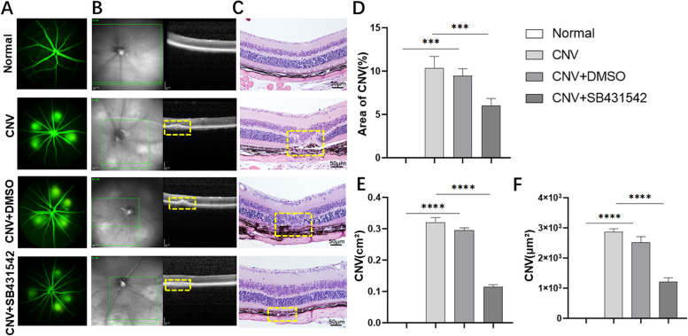
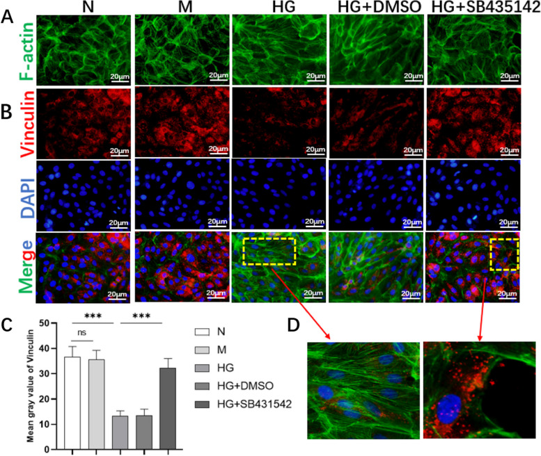
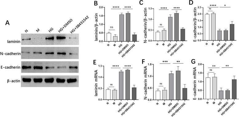
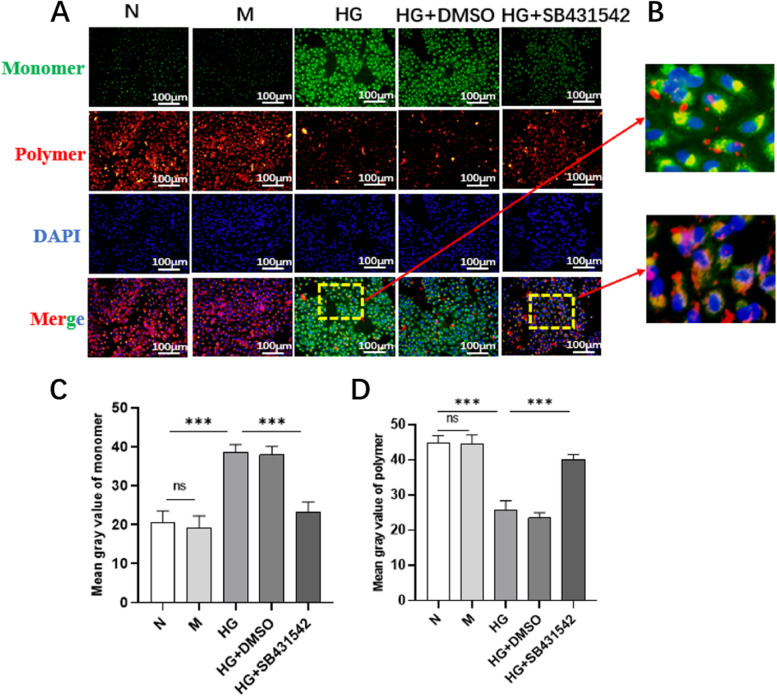
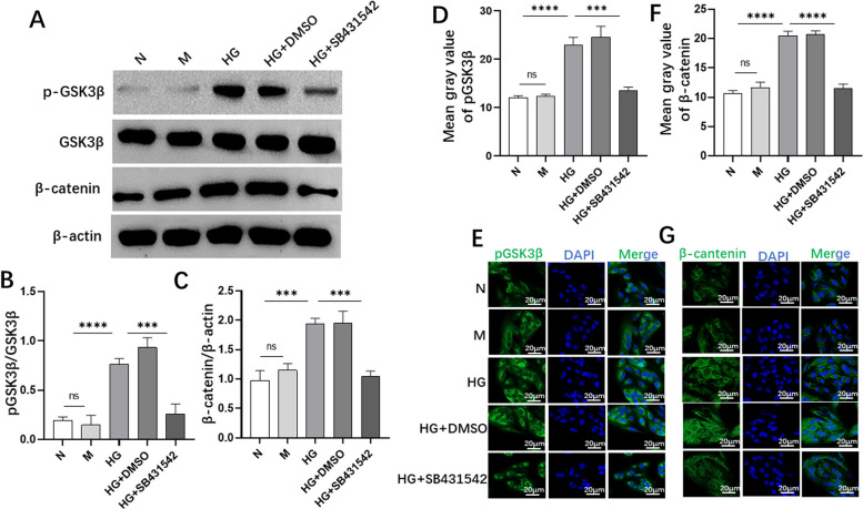
Methods: We performed fundus fluorescein angiography, optical coherence tomography and hematoxylin-eosin staining in vivo to observe the effect of SB431542 on choroidal neovascularization (CNV)-induced retinopathy. The proliferation, migration, cytoskeleton, adhesion, reactive oxygen species (ROS), mitochondrial morphology and membrane potential of RPE cells were observed in vitro through fluorescein diacetate staining, Cell Counting Kit-8 experiment, wound healing assay, phalloidin staining, immunofluorescence, MitoSOX, DCFH-DA, MitoTracker and JC-10 staining. Western blot, reverse transcription quantitative and immunofluorescence were used to detect the expression of EMT-related markers, pERK1/2, pGSK3β and β-catenin.
Results: SB431542 significantly alleviated retinopathy in the CNV model. The proliferation, migration and adhesion in RPE cells decreased to a certain extent in SB431542 treatment. SB431542 partially normalized the structure of RPE cells. The expression levels of E-cadherin increased, while the expression levels of laminin and N-cadherin decreased with SB431542 treatment. SB431542 reduced the production of total ROS, mitochondrial SOX and recovered the mitochondrial membrane potential to a certain degree. In addition, our study showed that SB431542 downregulated the phosphorylation of ERK1/2, GSK3β and the expression of β-catenin.
Conclusion: SB431542 improved EMT in RPE cells by maintaining mitochondrial homeostasis via the ERK1/2 and GSK3β/β-catenin pathways. Video Abstract SB431542 inhibits EMT in RPE cells under high glucose conditions.
Keywords: Epithelial mesenchymal transformation; Mitochondria; Proliferative diabetes retinopathy; RPE; SB431542.
© 2023. The Author(s).
Conflict of interest statement
The authors declare no competing interests.
Figures


Similar articles
-
[Effect of SB431542 on autophagy and epithelial mesenchymal transition in retinal pigment epithelial cells induced by high glucose].Zhonghua Yan Ke Za Zhi. 2025 Mar 11;61(3):202-210. doi: 10.3760/cma.j.cn112142-20240311-00109. Zhonghua Yan Ke Za Zhi. 2025. PMID: 40050106 Chinese.
-
TNF-α mediates choroidal neovascularization by upregulating VEGF expression in RPE through ROS-dependent β-catenin activation.Mol Vis. 2016 Feb 3;22:116-28. eCollection 2016. Mol Vis. 2016. PMID: 26900328 Free PMC article.
-
Complement activation contributes to subretinal fibrosis through the induction of epithelial-to-mesenchymal transition (EMT) in retinal pigment epithelial cells.J Neuroinflammation. 2022 Jul 14;19(1):182. doi: 10.1186/s12974-022-02546-3. J Neuroinflammation. 2022. PMID: 35831910 Free PMC article.
-
Dichloroacetate prevents TGFβ-induced epithelial-mesenchymal transition of retinal pigment epithelial cells.Exp Eye Res. 2020 Aug;197:108072. doi: 10.1016/j.exer.2020.108072. Epub 2020 May 27. Exp Eye Res. 2020. PMID: 32473169
-
The intervention of epithelial-mesenchymal transition in homeostasis of human retinal pigment epithelial cells: a review.J Histotechnol. 2022 Dec;45(4):148-160. doi: 10.1080/01478885.2022.2137665. Epub 2022 Nov 15. J Histotechnol. 2022. PMID: 36377481 Review.
Cited by
-
Rekindling Vision: Innovative Strategies for Treating Retinal Degeneration.Int J Mol Sci. 2025 Apr 25;26(9):4078. doi: 10.3390/ijms26094078. Int J Mol Sci. 2025. PMID: 40362317 Free PMC article. Review.
-
TGF-β Isoforms and Local Environments Greatly Modulate Biological Nature of Human Retinal Pigment Epithelium Cells.Bioengineering (Basel). 2024 Jun 7;11(6):581. doi: 10.3390/bioengineering11060581. Bioengineering (Basel). 2024. PMID: 38927817 Free PMC article.
-
Scale-Up of Human Amniotic Epithelial Cells Through Regulation of Epithelial-Mesenchymal Plasticity Under Defined Conditions.Adv Sci (Weinh). 2025 Mar;12(11):e2408581. doi: 10.1002/advs.202408581. Epub 2025 Jan 13. Adv Sci (Weinh). 2025. PMID: 39804851 Free PMC article.
-
The epigenetic regulation of crosstalk between cardiac fibroblasts and other cardiac cell types during stress.Front Cardiovasc Med. 2025 Apr 8;12:1539826. doi: 10.3389/fcvm.2025.1539826. eCollection 2025. Front Cardiovasc Med. 2025. PMID: 40264508 Free PMC article. Review.
-
Deficient AMPK-SENP1-Sirt3 signaling impairs mitochondrial complex I function in Parkinson's disease model.Transl Neurodegener. 2025 Jul 1;14(1):34. doi: 10.1186/s40035-025-00489-2. Transl Neurodegener. 2025. PMID: 40597361 Free PMC article.
References
Publication types
MeSH terms
Substances
LinkOut - more resources
Full Text Sources
Medical
Research Materials
Miscellaneous
