Review
. 2024 Jan 5;21(1):3.
doi: 10.1186/s12987-023-00478-5.

Blood-brain borders: a proposal to address limitations of historical blood-brain barrier terminology

Affiliations
Review

Blood-brain borders: a proposal to address limitations of historical blood-brain barrier terminology

Jerome Badaut et al. Fluids Barriers CNS. .

Abstract

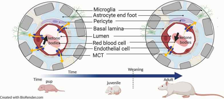
Many neuroscientists use the term Blood-Brain Barrier (BBB) to emphasize restrictiveness, often equating or reducing the notion of BBB properties to tight junction molecules physically sealing cerebral endothelial cells, rather than pointing out the complexity of this biological interface with respect to its selectivity and variety of exchange between the general blood circulation and the central nervous tissue. Several authors in the field find it unfortunate that the exquisitely dynamic interfaces between blood and brain continue to be viewed primarily as obstructive barriers to transport. Although the term blood-brain interface is an excellent descriptor that does not convey the idea of a barrier, it is important and preferable for the spreading of an idea beyond specialist communities to try to appeal to well-chosen metaphors. Recent evidence reviewed here indicates that blood-brain interfaces are more than selective semi-permeable membranes in that they display many dynamic processes and complex mechanisms for communication. They are thus more like 'geopolitical borders'. Furthermore, some authors working on blood-brain interface-relevant issues have started to use the word border, for example in border-associated macrophages. Therefore, we suggest adopting the term Blood-Brain Border to better communicate the flexibility of and movement across blood-brain interfaces.

Keywords: Blood–brain barrier; Blood–brain border; Blood–brain interface; Immune system; Neurovascular unit; Transport.


Conflict of interest statement

The authors declare no competing interests. Robert G Thorne is a paid employee of Denali Therapeutics.

Figures

Fig. 1
Fig. 1
Illustration of key borders between the blood and brain and between the blood and cerebrospinal fluid (CSF). Blood–brain border (neurovascular unit): cerebral endothelial cells in the parenchyma of the brain contain tight junctions and express numerous transporters and receptors that regulate the transfer of substances between the blood and brain (upper right), as shown by the following examples: Efflux transporters (e.g. P-glycoprotein, ABCB1; breast cancer resistance protein, ABCG2) prevent brain entry of many circulating endogenous substances as well as xenobiotics (drugs). The glucose transporter 1 (GLUT1, SLC2A1) and transferrin receptor (TfR, CD71) mediate brain homeostasis of glucose and iron, respectively, through the uptake of circulating glucose and iron-bound transferrin. The major facilitator superfamily domain containing 2a (MFSD2A) is a fatty acid transporter that is specifically expressed in CNS endothelial cells; MFSD2A also serves an important role in inhibiting clathrin-independent caveolae-mediated transcytosis [37, 38]). Outer blood-CSF border (meninges): outer arachnoid epithelial border cells contain tight junctions and express numerous transporters and receptors (not shown) that potentially assist in the regulated transfer of substances between the blood and extraventricular CSF (bottom right). Inner blood-CSF border (choroid plexus): choroid plexus epithelial cells contain tight junctions and express numerous channels, transporters, receptors and enzymes (not shown) that regulate the composition of CSF (bottom left). As examples, a finely tuned interplay between apically and basolaterally located inorganic anion transporters and channels is responsible for CSF production [39]. Transporters of the ABCC, SLC0, SLC21 families control CSF concentration of potentially deleterious endo and xenobiotics [4], helped by efficient enzymatic detoxification mechanisms [40]. The choroidal transport or secretion of growth factors, hormones and proteins into the brain participates in processes essential for brain development and homeostatic balance [33, 41]. Schematics based in part on several sources [, –44]
Fig. 2
Fig. 2
Illustration of the physiological adaptation of a number of transporters on brain endothelial cells with the transition from the use of the ketone bodies during lactation to that of mostly glucose after weaning. The endothelial cells adapt the number of monocarboxylate transporters (MCT in Blue) in function of the need of the energy substrate, with higher number of MCT during lactation to facilitate the ketone bodies to fuel the brain than in the adulthood with the use of glucose from the general blood circulation. It is similar to a country facilitating product importation in function of its needs (Fig. 3)
Fig. 3
Fig. 3
Schematic Illustration of the trade between Switzerland and E.U. with changes and adaptations over time depending on the needs of each partner. The supply exchange at the custom border is controlled and selective with potential taxes fixed (before) by the partners

References

    1. Stern L, Gautier R. Recherches sur le liquide céphalo-rachidien: I. Les rapports entre le liquide céphalorachidien et la circulation sanguine. Archiv Int Physiol. 1921;17(2):138–92.
    1. Reese TS, Karnovsky MJ. Fine structural localization of a blood-brain barrier to exogenous peroxidase. J Cell Biol. 1967;34(1):207–217. doi: 10.1083/jcb.34.1.207. - DOI - PMC - PubMed
    1. Vanlandewijck M, He L, Mae MA, Andrae J, Ando K, Del Gaudio F, et al. A molecular atlas of cell types and zonation in the brain vasculature. Nature. 2018;554(7693):475–480. doi: 10.1038/nature25739. - DOI - PubMed
    1. Strazielle N, Ghersi-Egea JF. Physiology of blood-brain interfaces in relation to brain disposition of small compounds and macromolecules. Mol Pharm. 2013;10(5):1473–1491. doi: 10.1021/mp300518e. - DOI - PubMed
    1. Banks WA. From blood-brain barrier to blood-brain interface: new opportunities for CNS drug delivery. Nat Rev Drug Discovery. 2016;15(4):275–292. doi: 10.1038/nrd.2015.21. - DOI - PubMed

LinkOut - more resources