. 2024 Jan 5;22(1):26.
doi: 10.1186/s12967-023-04846-5.

(Pro)renin receptor mediates tubular epithelial cell pyroptosis in diabetic kidney disease via DPP4-JNK pathway

Affiliations

(Pro)renin receptor mediates tubular epithelial cell pyroptosis in diabetic kidney disease via DPP4-JNK pathway

Shiying Xie et al. J Transl Med. .

Abstract

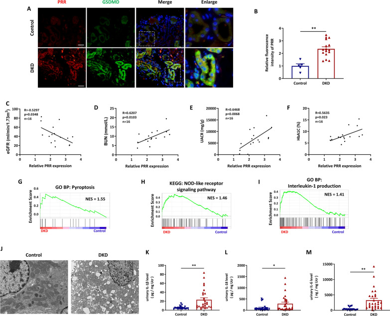
Background: (Pro)renin receptor (PRR) is highly expressed in renal tubules, which is involved in physiological and pathological processes. However, the role of PRR, expressed in renal tubular epithelial cells, in diabetic kidney disease (DKD) remain largely unknown.

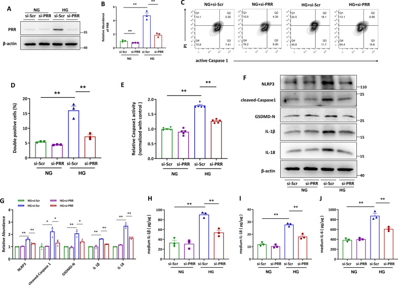
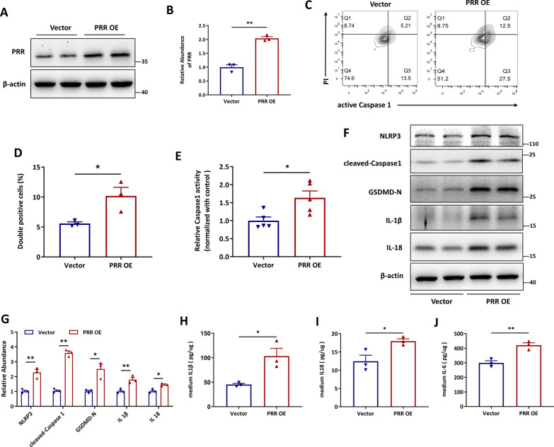
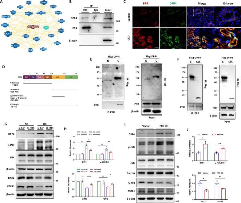
Methods: In this study, kidney biopsies, urine samples, and public RNA-seq data from DKD patients were used to assess PRR expression and cell pyroptosis in tubular epithelial cells. The regulation of tubular epithelial cell pyroptosis by PRR was investigated by in situ renal injection of adeno-associated virus9 (AAV9)-shRNA into db/db mice, and knockdown or overexpression of PRR in HK-2 cells. To reveal the underlined mechanism, the interaction of PRR with potential binding proteins was explored by using BioGrid database. Furthermore, the direct binding of PRR to dipeptidyl peptidase 4 (DPP4), a pleiotropic serine peptidase which increases blood glucose by degrading incretins under diabetic conditions, was confirmed by co-immunoprecipitation assay and immunostaining.

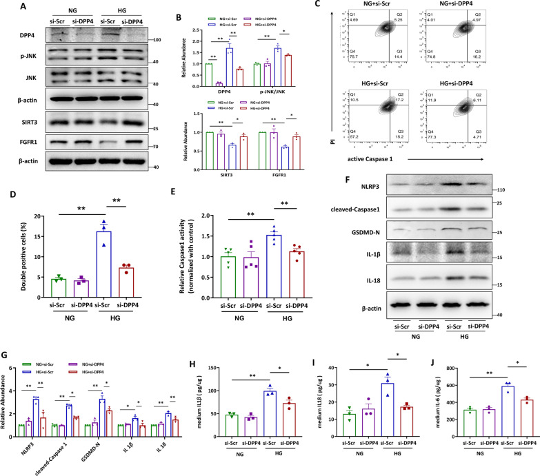
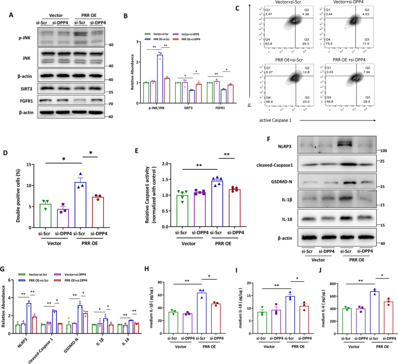
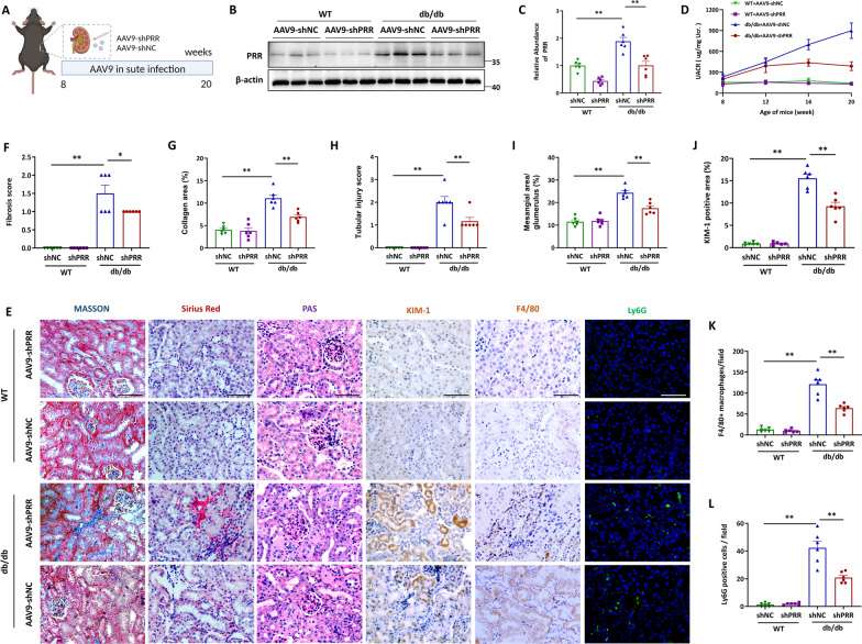
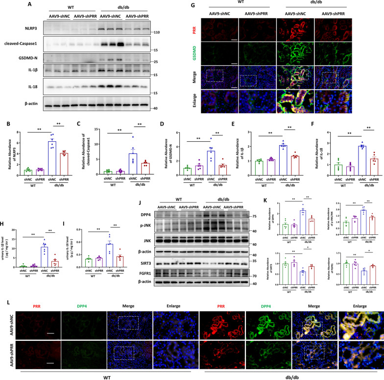
Results: Higher expression of PRR was found in renal tubules and positively correlated with kidney injuries of DKD patients, in parallel with tubular epithelial cells pyroptosis. Knockdown of PRR in kidneys significantly blunted db/db mice to kidney injury by alleviating renal tubular epithelial cells pyroptosis and the resultant interstitial inflammation. Moreover, silencing of PRR blocked high glucose-induced HK-2 pyroptosis, whereas overexpression of PRR enhanced pyroptotic cell death of HK-2 cells. Mechanistically, PRR selectively bound to cysteine-enrich region of C-terminal of DPP4 and augmented the protein abundance of DPP4, leading to the downstream activation of JNK signaling and suppression of SIRT3 signaling and FGFR1 signaling, and then subsequently mediated pyroptotic cell death.

Conclusions: This study identified the significant role of PRR in the pathogenesis of DKD; specifically, PRR promoted tubular epithelial cell pyroptosis via DPP4 mediated signaling, highlighting that PRR could be a promising therapeutic target in DKD.

Keywords: (Pro)renin receptor; Diabetic kidney disease; Dipeptidyl peptidase 4; Renal tubular cell pyroptosis.


Conflict of interest statement

The authors declare that there are no conflicts of interest.

Figures

Fig. 1
Fig. 1
PRR expression and cell pyroptosis were increased in renal tubules of DKD patients. A and B Representative fluorescent images of coimmunostaining PRR (red) with GSDMD (green) in kidney biopsies from DKD patients (n = 16), and the control subjects were from paracancerous tissue (n = 5) (A), and quantitative data showed the upregulation of PRR in renal tubule. Bar = 20 μm. (B). CF Analysis of the association between relative tubular PRR expression and eGFR(C), BUN (D), urinary albumin-to-creatinine ratio (UACR) (E) and Hemoglobin A1c (HbA1c) (F) of patients with DKD. (GI) GSEA analysis of pyroptosis (G), NOD-like receptor signaling pathway (H) and Interleukin-1 production (I) in renal tubules of DKD patients (n = 10) and healthy controls (n = 12) by using public GSE dataset (GSE30529). J Representative microscopy images of tubular cell morphology in renal sections from DKD patients and health controls. Bar = 1 μm. (K and L) Elisa detection of urinary IL-1β (K) and IL-18 (L) from DKD patients (n = 28) and health controls (n = 23). M Elisa detection of urinary IL-6 from DKD patients (n = 28) and health controls (n = 18). Data are presented as mean ± SEM of biologically independent samples. ∗P < 0.05, ∗∗P < 0.01. P values were determined by Student’s t-test for comparison between two groups
Fig. 2
Fig. 2
Knockdown of PRR effectively blocked high glucose (HG)-augmented HK-2 cell pyroptosis. A and B Western blot analyses (A) and quantitative data (B) showed that knocking down by siRNA diminished HG-induced PRR expression. (n = 3). C and D Flow cytometry analysis showed that HG induced active Caspase1+PI+ HK-2 cells were decreased by PRR siRNA (C) and quantitative data (D). (n = 3). E PRR was silenced in HG stimulated HK-2 cells, and the Caspase1 activity in cell lysis was determined by kits (n = 5). F and G Western blot analyses (F) and quantitative data (G) showed that PRR siRNA blocked HG-induced NLRP3, cleaved-Caspase1, GSDMD-N, IL-1β and IL-18 expression in HK-2 cells (n = 3). H and I PRR was ablated in HG treated HK-2 cells, and the IL-1β (H), IL-18 (I) or IL-6 (J) concentration in the culture medium was determined by ELISA, and then normalized by protein concentration in cell lysates (n = 3). Data are presented as mean ± SEM of biologically independent samples. ∗ P < 0.05, ∗∗ P < 0.01. One-way ANOVA was used to analyze the data among multiple groups, followed by Tukey’s post hoc test
Fig. 3
Fig. 3
Overexpression of PRR promotes HK-2 cell pyroptosis. (A and B) Western blot analyses. A and quantitative data (B) showed the increased protein abundance of PRR in PRR overexpression plasmid transfected HK-2 cells. (n = 3). (C and D) Flow cytometry analysis showed that active Caspase1+PI+ HK-2 cells were increased by PRR overexpression (C) and quantitative data (D) (n = 3). E PRR was overexpressed in HK-2 cells, and the Caspase1 activity in cell lysis was determined by kits (n = 5). (F and G) Western blot analyses (F) and quantitative data (G) showed that PRR overexpression increased NLRP3, cleaved-Caspase1, GSDMD-N, IL-1β and IL-18 expression in HK-2 cells. (n = 3). H and I PRR was overexpressed in HK-2 cells, and the IL-1β (H), IL-18 (I) or IL-6 (J) concentration in the culture medium was determined by ELISA, and then normalized by protein concentration in cell lysis. (n = 3). Data are presented as mean ± SEM of biologically independent samples. ∗ P < 0.05, ∗∗ P < 0.01. P values were determined by Student’s t-test
Fig. 4
Fig. 4
PRR interacted with DPP4 to activate JNK signaling and inhibit SIRT3 signaling and FGFR1 signaling in vitro. A BioGRID database was used to analyze the potential interaction between PRR and DPP4. B Co-immunoprecipitation (Co-IP) to detect the interaction between PRR and DPP4. The protein lysates isolated from HK-2 cells were immunoprecipitated with anti-PRR antibody and were immunoblotted with indicated antibodies. C Representative fluorescent images of coimmunostaining PRR (red) with DPP4 (green) in renal sections from DKD patients and the healthy control was from paracancerous tissue. Bar = 20 μm. D Schematic representation of the DPP4 domain. E Co-IP was used to detect the interaction between PRR and Flag-DPP4. HK-2 cells were transfected with Flag-tagged truncation of DPP4 C-terminal 325–766 amino acids (Flag-DPP4 C), or 1–324 amino acids (Flag-DPP4 N). Protein lysates were immunoprecipitated with anti-Flag antibody and immunoblotted with the indicated antibodies. F Co-IP was used to detect the interaction between PRR and Flag-DPP4. HK-2 cells were transfected with Flag-tagged truncation of DPP4 C-terminal 325–766 amino acids (Flag-DPP4 C), or 325–551 amino acids (Flag-DPP4 CYS). Protein lysates were immunoprecipitated with anti-Flag antibody and immunoblotted with the indicated antibodies. G and H Western blot analyses (G) and quantitative data (H) showed that PRR siRNA blocked HG-induced DPP4 abundance and phosphorylation of JNK, and restored HG-suppressed SIRT3 and FGFR1 expression in HK-2 cells (n = 3). I and J Representative Western blot (I) and quantitative data (J) showed increased DPP4, upregulated phosphorylation of JNK, suppressed SIRT3 and reduced FGFR1 in PRR overexpressed HK-2 cells (n = 3). Data are presented as mean ± SEM of biologically independent samples. ∗ P < 0.05, ∗∗ P < 0.01. One-way ANOVA was used to analyze the data among multiple groups, followed by Tukey’s post hoc test
Fig. 5
Fig. 5
DPP4 facilitated HG-induced HK-2 cell pyroptosis by activating JNK and suppressing SIRT3 and FGFR1. A and B Western blot analyses. A and quantitative data (B) showed that knocking down by siRNA diminished HG-induced DPP4 expression and phosphorylated JNK, and restored HG-reduced SIRT3 and FGFR1 abundance. (n = 3). C and D Flow cytometry analysis showed that knocking down DPP4 reduced HG increased active Caspase1+PI+ HK-2 cells (C) and quantitative data (D) (n = 3). E DPP4 was ablated in HG stimulated HK-2 cells, and the Caspase1 activity in cell lysis was determined by kits. F and G Western blot analyses (F) and quantitative data (G) showed that DPP4 siRNA attenuated HG-induced NLRP3, cleaved-Caspase1, GSDMD-N, IL-1β and IL-18 expression in HK-2 cells (n = 3). ( Hand I) DPP4 was knocked down in HG treated HK-2 cells, and the IL-1β (H), IL-18 (I) or IL-6 (J) concentration in the culture medium was determined by ELISA, and then normalized by protein concentration in cell lysates (n = 3). Data are presented as mean ± SEM of biologically independent samples. ∗ P < 0.05, ∗∗ P < 0.01. One-way ANOVA was used to analyze the data among multiple groups, followed by Tukey’s post hoc test
Fig. 6
Fig. 6
PRR exerted pyroptotic effects through DPP4-mediated signaling. A and B Representative western blot analyses (A) and quantitative data (B) showed that knocking down DPP4 by siRNA in HK-2 cells diminished PRR overexpression induced phosphorylation of JNK, and restored PRR overexpression reduced SIRT3 and FGFR1 expression (n = 3). C and D Flow cytometry analysis showed that knocking down DPP4 reduced PRR overexpression induced frequency of active Caspase1+PI+ HK-2 cells (C) and quantitative data (D) (n = 3). E PRR was silenced in HG stimulated HK-2 cells, and the Caspase1 activity in cell lysis was determined by kits. f and g Western blot analyses (F) and quantitative data (G) showed that DPP4 siRNA blocked HG-induced NLRP3, cleaved-Caspase1, GSDMD-N, IL-1β and IL-18 expression in HK-2 cells (n = 3). H and I DPP4 was knocked down in PRR overexpressed HK-2 cells, and the IL-1β (H), IL-18 (I) or IL-6 (J) concentration in the culture medium was determined by ELISA, and then normalized by protein concentration in cell lysates (n = 3). Data are presented as mean ± SEM of biologically independent samples. ∗ P < 0.05, ∗∗ P < 0.01. One-way ANOVA was used to analyze the data among multiple groups, followed by Tukey’s post hoc test
Fig. 7
Fig. 7
Silencing of PRR ameliorated kidney injury and inflammation in db/db mice. A Schematic diagram of the AAV injection surgery. B and C The protein level of PRR was assessed by Western blot. Representative Western blot (B) and quantitative data (C) are showed (n = 6). D Measured of urinary albumin concentration by using albumin detection kit, the quantitative data was normalized with urinary creatinine. EL Representative images of Masson, Sirius Red, PAS, KIM-1, F4/80 and Ly6G staining of renal section of WT mice and db/db mice with or without PRR silence (E) and quantitative analysis (FL) (n = 6). Bar = 20 μm. Data are presented as mean ± SEM of biologically independent samples. ∗ P < 0.05, ∗∗ P < 0.01. One-way ANOVA was used to analyze the data among multiple groups, followed by Tukey’s post hoc test
Fig. 8
Fig. 8
PRR knockdown blunted pyroptotic cell death and fibrotic response in db/db mice via DPP4-mediated signaling. AF The protein levels of NLRP3, cleaved-Caspase1, GSDMD-N, IL-1β, IL-18 were assessed by Western blot. Representative Western blot (A) and quantitative data (BF) are showed (n = 6). C Representative immunofluorescence microscopy images of coimmunostaining PRR (red) with GSDMD (green) in renal sections from WT and db/db mice with or without PRR ablation. Bar = 20 μm. H and I Elisa detection of urinary IL-1β (H) and IL-18 (I) concentration, the quantitative data were normalized with urinary creatine (n = 6). JK western blot analyses (J) and quantitative data (K) showed the abundance of DPP4, phosphorylation level of JNK, SIRT3 and FGFR1 (n = 6). L Representative fluorescent images of renal sections from mice with or without PRR silence and stained with PRR (red), DPP4 (green). Bar = 20 μm. Data are presented as mean ± SEM of biologically independent samples. ∗ P < 0.05, ∗∗ P < 0.01. One-way ANOVA was used to analyze the data among multiple groups, followed by Tukey’s post hoc test

Similar articles

Cited by

References

    1. Doshi SM, Friedman AN. Diagnosis and management of type 2 diabetic kidney disease. Clin J Am Soc Nephrol. 2017;128:1366–1373. doi: 10.2215/CJN.11111016. - DOI - PMC - PubMed
    1. Broz P, Pelegrin P, Shao F. The gasdermins, a protein family executing cell death and inflammation. Nat Rev Immunol. 2020;203:143–157. doi: 10.1038/s41577-019-0228-2. - DOI - PubMed
    1. Li J, Liu H, Takagi S, Nitta K, Kitada M, Srivastava SP, et al. Renal protective effects of empagliflozin via inhibition of EMT and aberrant glycolysis in proximal tubules. JCI Insight. 2020;56:e129034. doi: 10.1172/jci.insight.129034. - DOI - PMC - PubMed
    1. Li J, Shi S, Srivastava SP, Kitada M, Nagai T, Nitta K, et al. FGFR1 is critical for the anti-endothelial mesenchymal transition effect of N-acetyl-seryl-aspartyl-lysyl-proline via induction of the MAP4K4 pathway. Cell Death Dis. 2017;88:e2965. doi: 10.1038/cddis.2017.353. - DOI - PMC - PubMed
    1. Shi S, Srivastava SP, Kanasaki M, He J, Kitada M, Nagai T, et al. Interactions of DPP-4 and integrin beta1 influences endothelial-to-mesenchymal transition. Kidney Int. 2015;883:479–489. doi: 10.1038/ki.2015.103. - DOI - PubMed

Publication types