. 2024 Jan 5;23(1):10.
doi: 10.1186/s12936-023-04833-7.

Immunofluorescence study of cytoskeleton in endothelial cells induced with malaria sera

Affiliations

Immunofluorescence study of cytoskeleton in endothelial cells induced with malaria sera

Mathusorn Wongsawat et al. Malar J. .

Abstract

Background: Endothelial cells (ECs) play a major role in malaria pathogenesis, as a point of direct contact of parasitized red blood cells to the blood vessel wall. The study of cytoskeleton structures of ECs, whose main functions are to maintain shape and provide strength to the EC membrane is important in determining the severe sequelae of Plasmodium falciparum malaria. The work investigated the cytoskeletal changes (microfilaments-actin, microtubules-tubulin and intermediate filaments-vimentin) in ECs induced by malaria sera (Plasmodium vivax, uncomplicated P. falciparum and complicated P. falciparum), in relation to the levels of pro-inflammatory cytokines.

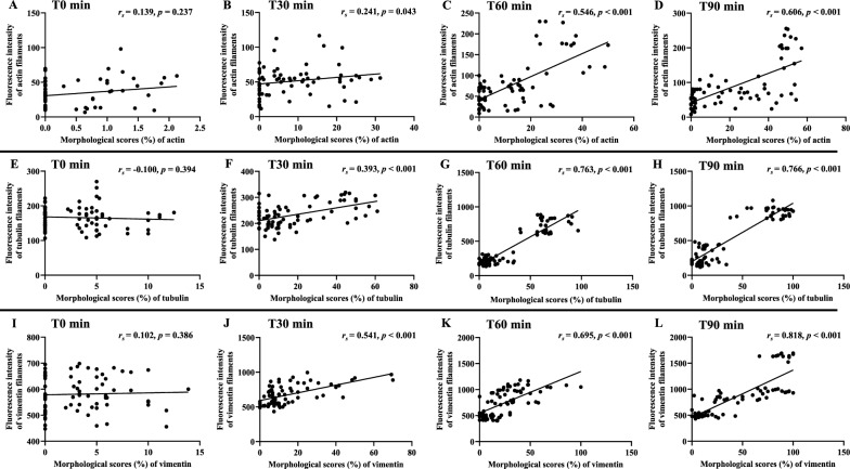
Methods: Morphology and fluorescence intensity of EC cytoskeleton stimulated with malaria sera were evaluated using immunofluorescence technique. Levels of tumour necrosis factor (TNF) and interferon (IFN)-gamma (γ) were determined using enzyme-linked immunosorbent assay (ELISA). Control experimental groups included ECs incubated with media alone and non-malaria patient sera. Experimental groups consisted of ECs incubated with malaria sera from P. vivax, uncomplicated P. falciparum and complicated P. falciparum. Morphological scores of cytoskeletal alterations and fluorescence intensity were compared across each experiment group, and correlated with TNF and IFN-γ.

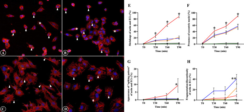
Results: The four morphological changes of cytoskeleton included (1) shrinkage of cytoskeleton and ECs with cortical condensation, (2) appearance of eccentric nuclei, (3) presence of "spiking pattern" of cytoskeleton and EC membrane, and (4) fragmentation and discontinuity of cytoskeleton and ECs. Significant damages were noted in actin filaments compared to tubulin and vimentin filaments in ECs stimulated with sera from complicated P. falciparum malaria. Morphological damages to cytoskeleton was positively correlated with fluorescence intensity and the levels of TNF and IFN-γ.

Conclusions: ECs stimulated with sera from complicated P. falciparum malaria showed cytoskeletal alterations and increased in fluorescence intensity, which was associated with high levels of TNF and IFN-γ. Cytoskeletal changes of ECs incubated with complicated P. falciparum malaria sera can lead to EC junctional alteration and permeability changes, which is mediated through apoptotic pathway. The findings can serve as a basis to explore measures to strengthen EC cytoskeleton and alleviate severe malaria complications such as pulmonary oedema and cerebral malaria. In addition, immunofluorescence intensity of cytoskeleton could be investigated as potential prognostic indicator for malaria severity.

Keywords: Actin; Cytoskeleton; Endothelial cells; IFN-γ; Immunofluorescence; Malaria; Plasmodium falciparum; TNF; Tubulin; Vimentin.


Conflict of interest statement

The authors declare that they have no competing interests.

Figures

Fig. 1
Fig. 1
The alterations of actin filaments and ECs induced by malaria sera. A and E: Shrinkage of actin filaments and ECs with cortical condensation (asterisk- normal ECs, arrowheads- shrinkage); B and F: Appearance of eccentric nuclei (asterisks- normal ECs, arrowheads- eccentric nuclei); C and G: Presence of “spiking pattern” of actin filaments and EC membrane (arrowheads); D and H: Fragmentation and discontinuity of actin filaments and ECs (arrowheads). AD: ECs induced by sera from complicated P. falciparum (magnification X200). ECs were incubated with media only (green line), non-malaria patient sera (grey line), malaria sera from P. vivax (orange line), uncomplicated P. falciparum (blue line) and complicated P. falciparum (red line). *Significant difference of complicated P. falciparum compared with all experimental groups (p < 0.05). **Significant difference of uncomplicated P. falciparum compared with all experimental groups (p < 0.05). Data are presented as mean ± SEM
Fig. 2
Fig. 2
Comparative morphological alterations of actin filaments in different experimental groups at different time points. Severe morphological damages are mostly illustrated in ECs induced with sera from complicated P. falciparum malaria (last column). (magnification X200)
Fig. 3
Fig. 3
The morphology of tubulin filaments and EC induced by malaria sera. A and C: Shrinkage of tubulin filaments and ECs with cortical condensation (arrowheads); B and D: Appearance of eccentric nuclei (arrowheads). Normal ECs are shown in asterisks. A, B: ECs induced by sera from complicated P. falciparum. (magnification X200). ECs were incubated with media only (green line), non-malaria patient sera (grey line), malaria sera from P. vivax (orange line), uncomplicated P. falciparum (blue line) and complicated P. falciparum (red line). **Significant difference of P. falciparum compared with P. vivax and control groups (p < 0.05). Data are presented as mean ± SEM
Fig. 4
Fig. 4
Comparative morphological alterations of tubulin filaments in different experimental groups at different time points. Shrinkage of tubulin filaments and ECs, and appearance of eccentric nuclei are mostly observed in ECs induced with sera from P. falciparum malaria. (magnification X200)
Fig. 5
Fig. 5
The morphology of vimentin filaments and ECs induced by malaria sera. A and C: Shrinkage of vimentin filaments and ECs with cortical condensation (arrowheads); B and D: Appearance of eccentric nuclei (arrowheads). Normal ECs are shown in asterisks. A, B: ECs induced by sera from complicated P. falciparum. (magnification X200). ECs were incubated with media only (green line), non-malaria patient sera (grey line), malaria sera from P. vivax (orange line), uncomplicated P. falciparum (blue line) and complicated P. falciparum (red line). *Significant difference of complicated P. falciparum compared with all experimental groups (p < 0.05). **Significant difference of P. falciparum compared with P. vivax and control groups (p < 0.05). Data are presented as mean ± SEM
Fig. 6
Fig. 6
Comparative morphological alterations of vimentin filaments in different experimental groups at different time points. Shrinkage of vimentin filaments and ECs, and appearance of eccentric nuclei are mostly noted in ECs induced with sera from P. falciparum malaria at T90 min. (magnification X200)
Fig. 7
Fig. 7
The overall morphological changes for cytoskeleton at different time points of different experimental groups. A: actin filaments; B: tubulin filaments and C: vimentin filaments. ECs were incubated with media only (green line), non-malaria patient sera (grey line), malaria sera from P. vivax (orange line), uncomplicated P. falciparum (blue line) and complicated P. falciparum (red line). *Significant difference of complicated P. falciparum compared with all experimental groups (p < 0.05). **Significant difference of P. falciparum compared with P. vivax and control groups (p < 0.05). Data are presented as mean ± SEM
Fig. 8
Fig. 8
The fluorescence intensity of cytoskeleton in ECs induced by malaria sera. A: F-actin; B: G-actin; C: F:G actin ratio; D: tubulin filaments and E: vimentin filaments. ECs were incubated with media only (green line), non-malaria patient sera (grey line), malaria sera from P. vivax (orange line), uncomplicated P. falciparum (blue line) and complicated P. falciparum (red line). *Significant difference of complicated P. falciparum compared with all experimental groups (p < 0.05). Data are presented as mean ± SEM
Fig. 9
Fig. 9
Correlation between morphological changes induced by malaria sera and fluorescence intensity of cytoskeleton at different time points. AD: actin filaments; EH: tubulin filaments and IL: vimentin filaments at different time points
Fig. 10
Fig. 10
Correlation between morphological changes induced by malaria sera and TNF. AD: actin filaments; EH: tubulin filaments and IL: vimentin filaments at different time points
Fig. 11
Fig. 11
Correlation between morphological changes induced by malaria sera and IFN-γ. AD: actin filaments; EH: tubulin filaments and IL: vimentin filaments at different time points

References

    1. WHO . World malaria report 2022. Geneva: World Health Organization; 2022.
    1. World Health Organization Severe malaria. Trop Med Int Health. 2014;19(Suppl 1):7–131. - PubMed
    1. Craig AG, Khairul MFM, Patil PR. Cytoadherence and severe malaria. Malays J Med Sci. 2012;19:5–18. - PMC - PubMed
    1. Miller LH, Baruch DI, Marsh K, Doumbo OK. The pathogenic basis of malaria. Nature. 2002;415:673–679. doi: 10.1038/415673a. - DOI - PubMed
    1. Armah H, Wiredu EK, Dodoo AK, Adjei AA, Tettey Y, Gyasi R. Cytokines and adhesion molecules expression in the brain in human cerebral malaria. Int J Environ Res Public Health. 2005;2:123–131. doi: 10.3390/ijerph2005010123. - DOI - PMC - PubMed

LinkOut - more resources