. 2024 Jan 23;43(1):113636.
doi: 10.1016/j.celrep.2023.113636. Epub 2024 Jan 5.

TDP-43-stratified single-cell proteomics of postmortem human spinal motor neurons reveals protein dynamics in amyotrophic lateral sclerosis

Affiliations

TDP-43-stratified single-cell proteomics of postmortem human spinal motor neurons reveals protein dynamics in amyotrophic lateral sclerosis

Amanda J Guise et al. Cell Rep. .

Abstract

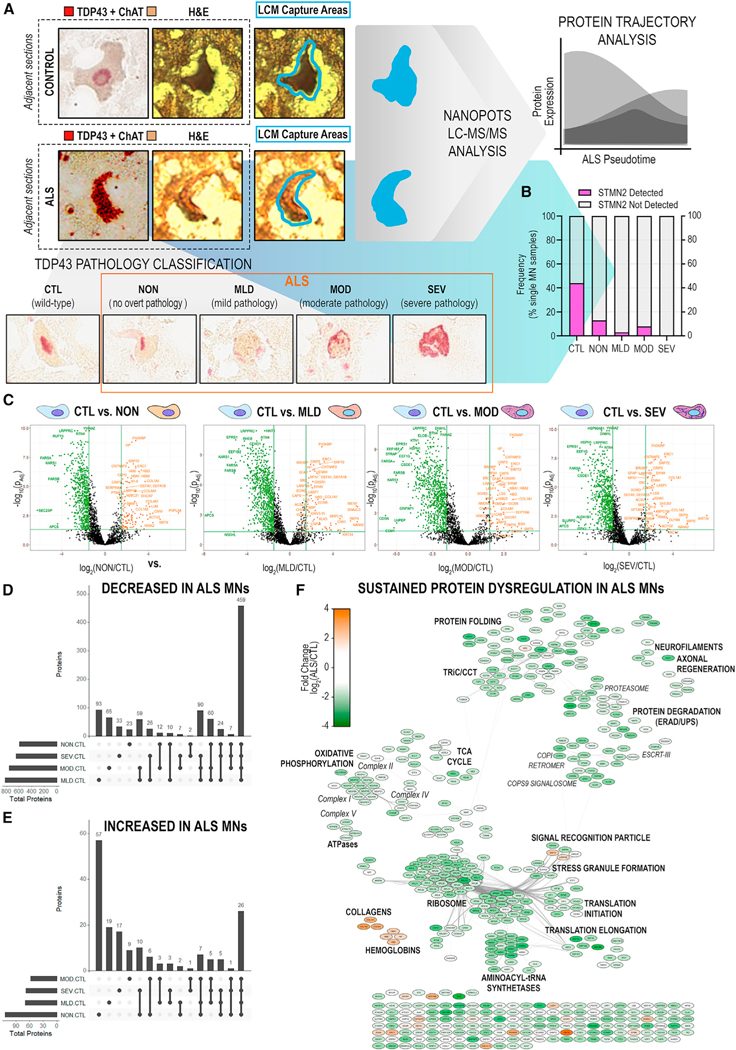
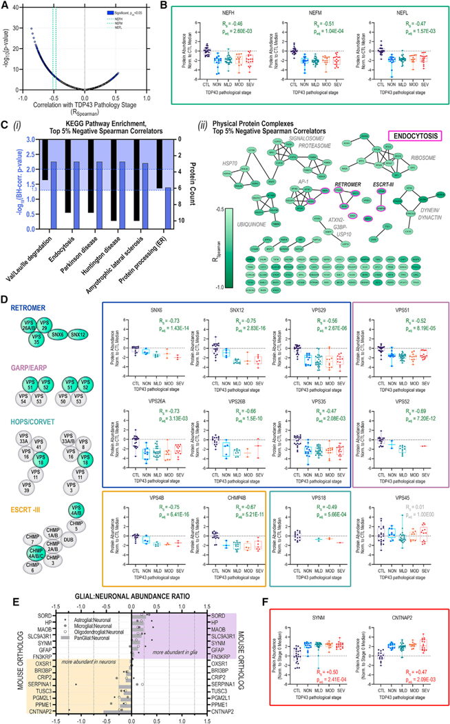
A limitation of conventional bulk-tissue proteome studies in amyotrophic lateral sclerosis (ALS) is the confounding of motor neuron (MN) signals by admixed non-MN proteins. Here, we leverage laser capture microdissection and nanoPOTS single-cell mass spectrometry-based proteomics to query changes in protein expression in single MNs from postmortem ALS and control tissues. In a follow-up analysis, we examine the impact of stratification of MNs based on cytoplasmic transactive response DNA-binding protein 43 (TDP-43)+ inclusion pathology on the profiles of 2,238 proteins. We report extensive overlap in differentially abundant proteins identified in ALS MNs with or without overt TDP-43 pathology, suggesting early and sustained dysregulation of cellular respiration, mRNA splicing, translation, and vesicular transport in ALS. Together, these data provide insights into proteome-level changes associated with TDP-43 proteinopathy and begin to demonstrate the utility of pathology-stratified trace sample proteomics for understanding single-cell protein dynamics in human neurologic diseases.

Keywords: ALS; CP: Molecular biology; CP: Neuroscience; STMN2; TDP-43 proteinopathy; laser capture; motor neuron; nanoPOTS; retromer; single-cell proteomics.


Conflict of interest statement

Declaration of interests A.J.G., J.-H.C., S.X., J.G., and E.D.P. are employees and shareholders of Biogen.

Figures

Figure 1.
Figure 1.. Ultrasensitive single-cell proteomic mapping of ALS motor neurons
(A) Proteins (high-confidence master proteins, HCMPs; 5% global FDR) identified across single MNs (n = 6/donor) laser excised from ALS or CTL donor tissues (n = 3/diagnosis). (B) Principal-component analysis (PCA) dimensionality reduction of the top 500 most-variable proteins measured across ALS and CTL MNs. (C) Differential protein abundance in ALS vs. CTL MNs with correction for donor origin. Significant DAPs (|log2(ALS/CTL)| ≥ 1.5 ∩ p-adj < 0.05) indicated in purple. (D) (Left) Age and PMI distributions for ALS and CTL donors; median ± range. (Right) Differential protein abundance with correction for additional covariates (age, sex, PMI). Significant DAPs indicated in purple. (E) PPI network of significant DAPs. Node color indicates fold change in normalized protein abundance; edges denote high-confidence PPIs.
Figure 2.
Figure 2.. Absence of STMN2 protein detection in human ALS MNs parallels decreased abundance of Stathmin-2 (STMN2) RNA and absence of functional TDP-43 interaction partners
(A) Rank-ordered median protein intensities in single CTL or ALS MNs. (B) Uniquely identified and overlapping proteins detected in ALS or CTL single MNs. (C) PPI network of proteins uniquely detected in CTL (left) or ALS (right) single MNs; TDP-43 node added manually. Node border: magenta, BioGRID-reported direct or indirect TDP-43 PPIs; purple, downstream TDP-43 splice target STMN2; edges indicate STRING-annotated physical PPIs. (D) Prevalence of TDP-43 interactors among proteins uniquely detected in CTL MNs, proteins uniquely detected in ALS MNs, or detected in the background human proteome (two-sided Fisher’s exact test). (E) Dual expression of STMN2 RNA (red) and TDP-43 protein (teal) in ALS and CTL human spinal MNs. (F) Quantitation of STMN2 RNA abundance in ALS (n = 5) and CTL (n = 10) donors; violin plots show median (dashed line), interquartile range (IQR; dotted lines), range, and Mann-Whitney U test p values. (G) Cryptic and canonical STMN2 transcript visualization in adjacent ALS MN cross-sections.
Figure 3.
Figure 3.. Significant disruption of proteostasis, mitochondrial dysfunction, and induction of pro-apoptotic signaling are apparent prior to overt TDP-43 aggregation
(A) Schematic of single-MN selection for laser-capture microdissection by dual detection of TDP-43 and ChAT in immediately adjacent tissue sections. Captured MNs were stratified based on TDP-43+ NCI status. (B) Frequency of STMN2 protein detection in single MNs across TDP-43 strata. (C) Differential protein abundances identified across TDP-43+ NCI strata (NON/MLD/MOD/SEV vs. CTL). Significant DAPs (|log2(ALS/CTL)| ≥ 1.5 ∩ p-adj < 0.05) shown in color. (D and E) Shared or unique proteins across TDP-43 strata with significantly (D) decreased or (E) increased abundance in ALS MNs. (F) PPI network of significant DAPs (|log2(ALS/CTL)| ≥ 1.5 ∩ p-adj < 0.05) common to all TDP-43 stages. Node color indicates fold change in protein abundance; edges denote high-confidence PPIs.
Figure 4.
Figure 4.. Neurofilament protein and retromer complex component abundances track inversely with increasing TDP-43 aggregation in postmortem human MNs
(A) Volcano plot of Spearman rank correlations (Rs) for abundance of individual proteins vs. corresponding TDP-43 stratum; purple highlight, significantly (p-adj < 0.05, Bonferroni) correlated proteins; green dashed lines indicate NfP correlations. (B) Distribution of NEFH, NEFM, and NEFL protein abundances in individual MNs across TDP-43+ NCI strata. (C) (i) Over-represented (blue highlight; p-adj < 0.05, p-adj < 0.01, Benjamini-Hochberg) KEGG pathways and (ii) physical protein complex assignments for the top 5% negative TDP-43-correlators; median ± IQR; whiskers indicate range. (D) TDP-43-stratified protein abundances trajectories for retromer (blue), GARP/EARP (mauve), HOPS/CORVET (teal), and ESCRT-III (gold); median ± IQR; whiskers indicate range. (E) Glial:neuronal abundance ratios for proteins with significant, positive RS. (F) TDP-43-stratified protein abundance trajectories for SYNM and CNTNAP2; median ± IQR; whiskers indicate range.

Update of

Similar articles

Cited by

  • Optogenetic induction of TDP-43 aggregation impairs neuronal integrity and behavior in Caenorhabditis elegans.
    Park KH, Yu E, Choi S, Kim S, Park C, Lee JE, Kim KW. Park KH, et al. Transl Neurodegener. 2025 Apr 16;14(1):20. doi: 10.1186/s40035-025-00480-x. Transl Neurodegener. 2025. PMID: 40234916 Free PMC article.
  • Digital Microfluidics for Sample Preparation in Low-Input Proteomics.
    Steinbach MK, Leipert J, Matzanke T, Tholey A. Steinbach MK, et al. Small Methods. 2025 Jan;9(1):e2400495. doi: 10.1002/smtd.202400495. Epub 2024 Aug 29. Small Methods. 2025. PMID: 39205538 Free PMC article. Review.
  • Deciphering distinct genetic risk factors for FTLD-TDP pathological subtypes via whole-genome sequencing.
    Pottier C, Küçükali F, Baker M, Batzler A, Jenkins GD, van Blitterswijk M, Vicente CT, De Coster W, Wynants S, Van de Walle P, Ross OA, Murray ME, Faura J, Haggarty SJ, van Rooij JG, Mol MO, Hsiung GR, Graff C, Öijerstedt L, Neumann M, Asmann Y, McDonnell SK, Baheti S, Josephs KA, Whitwell JL, Bieniek KF, Forsberg L, Heuer H, Lago AL, Geier EG, Yokoyama JS, Oddi AP, Flanagan M, Mao Q, Hodges JR, Kwok JB, Domoto-Reilly K, Synofzik M, Wilke C, Onyike C, Dickerson BC, Evers BM, Dugger BN, Munoz DG, Keith J, Zinman L, Rogaeva E, Suh E, Gefen T, Geula C, Weintraub S, Diehl-Schmid J, Farlow MR, Edbauer D, Woodruff BK, Caselli RJ, Donker Kaat LL, Huey ED, Reiman EM, Mead S, King A, Roeber S, Nana AL, Ertekin-Taner N, Knopman DS, Petersen RC, Petrucelli L, Uitti RJ, Wszolek ZK, Ramos EM, Grinberg LT, Tempini MLG, Rosen HJ, Spina S, Piguet O, Grossman M, Trojanowski JQ, Keene CD, Jin LW, Prudlo J, Geschwind DH, Rissman RA, Cruchaga C, Ghetti B, Halliday GM, Beach TG, Serrano GE, Arzberger T, Herms J, Boxer AL, Honig LS, Vonsattel JP, Lopez OL, Kofler J, White CL 3rd, Gearing M, Glass J, Rohrer JD, Irwin DJ, Lee EB, Van Deerlin V, Castellani R, Mesulam MM, Tartaglia MC, Finger EC, Troakes C… See abstract for full author list ➔ Pottier C, et al. Nat Commun. 2025 Apr 25;16(1):3914. doi: 10.1038/s41467-025-59216-0. Nat Commun. 2025. PMID: 40280976 Free PMC article.
  • Unlocking Disease-Modifying Treatments for TDP-43-Mediated Neurodegeneration.
    San Gil R, Walker AK. San Gil R, et al. Bioessays. 2025 Apr;47(4):e202400257. doi: 10.1002/bies.202400257. Epub 2025 Feb 3. Bioessays. 2025. PMID: 39901378 Free PMC article. Review.
  • Exosomes in Regulating miRNAs for Biomarkers of Neurodegenerative Disorders.
    Sivalingam AM, Sureshkumar DD. Sivalingam AM, et al. Mol Neurobiol. 2025 Jun;62(6):7576-7596. doi: 10.1007/s12035-025-04733-8. Epub 2025 Feb 7. Mol Neurobiol. 2025. PMID: 39918711 Review.

References

    1. Miller RG, Mitchell JD, and Moore DH (2012). Riluzole for Amyotrophic Lateral Sclerosis (ALS)/motor Neuron Disease (MND) (Cochrane Database Syst Rev). CD001447. - PubMed
    1. Witzel S, Maier A, Steinbach R, Grosskreutz J, Koch JC, Sarikidi A, Petri S, Günther R, Wolf J, Hermann A, et al. (2022). Safety and Effectiveness of Long-term Intravenous Administration of Edaravone for Treatment of Patients With Amyotrophic Lateral Sclerosis. JAMA Neurol. 79, 121–130. - PMC - PubMed
    1. Paganoni S, Macklin EA, Hendrix S, Berry JD, Elliott MA, Maiser S, Karam C, Caress JB, Owegi MA, Quick A, et al. (2020). Trial of Sodium Phenylbutyrate-Taurursodiol for Amyotrophic Lateral Sclerosis. N. Engl. J. Med. 383, 919–930. - PMC - PubMed
    1. Miller TM, Cudkowicz ME, Genge A, Shaw PJ, Sobue G, Bucelli RC, Chiò A, Van Damme P, Ludolph AC, Glass JD, et al. (2022). Trial of Antisense Oligonucleotide Tofersen for SOD1 ALS. N. Engl. J. Med. 387, 1099–1110. - PubMed
    1. Arai T, Hasegawa M, Akiyama H, Ikeda K, Nonaka T, Mori H, Mann D, Tsuchiya K, Yoshida M, Hashizume Y, and Oda T (2006). TDP-43 is a component of ubiquitin-positive tau-negative inclusions in frontotemporal lobar degeneration and amyotrophic lateral sclerosis. Biochem. Biophys. Res. Commun. 351, 602–611. - PubMed