Review
. 2024 May;168(5):910-954.
doi: 10.1111/jnc.16032. Epub 2024 Jan 6.

Brain energy metabolism: A roadmap for future research

Affiliations
Review

Brain energy metabolism: A roadmap for future research

Caroline D Rae et al. J Neurochem. 2024 May.

Abstract

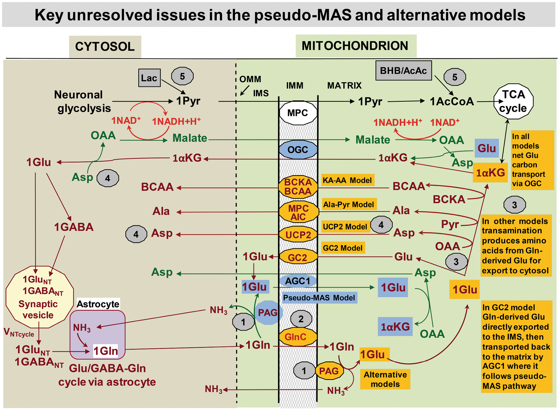
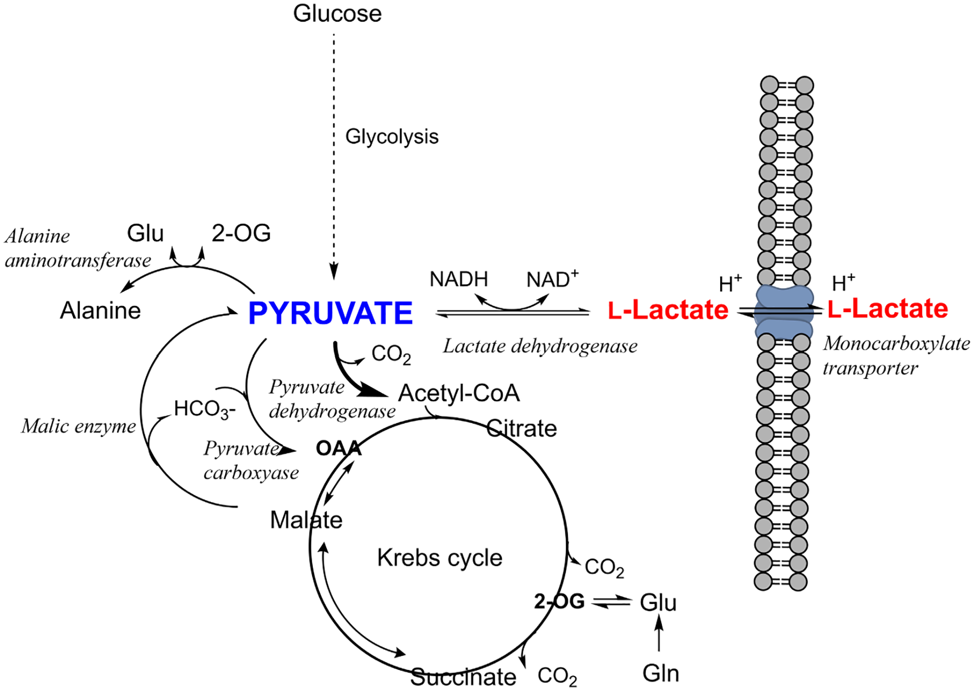
Although we have learned much about how the brain fuels its functions over the last decades, there remains much still to discover in an organ that is so complex. This article lays out major gaps in our knowledge of interrelationships between brain metabolism and brain function, including biochemical, cellular, and subcellular aspects of functional metabolism and its imaging in adult brain, as well as during development, aging, and disease. The focus is on unknowns in metabolism of major brain substrates and associated transporters, the roles of insulin and of lipid droplets, the emerging role of metabolism in microglia, mysteries about the major brain cofactor and signaling molecule NAD+, as well as unsolved problems underlying brain metabolism in pathologies such as traumatic brain injury, epilepsy, and metabolic downregulation during hibernation. It describes our current level of understanding of these facets of brain energy metabolism as well as a roadmap for future research.

Keywords: GLUT4; acetate; aerobic glycolysis; insulin; noradrenaline.


Conflict of interest statement

Caroline Rae is President of the International Society for Neurochemistry, the owner of this journal. Mary C. McKenna and João M.N. Duarte are Editors for the Journal of Neurochemistry. Kelly Drew has shown financial interest in Be Cool Pharmaceutics. Karin Borges declares consulting fees received from Ultragenyx Pharmaceuticals Inc. and Nestlé Purina PetCare in the past. Joseph Baur has received research funding, materials, and consulting fees from Pfizer, Cytokinetics, Elysium Health, and Metro International Biotech; he holds a patent for using NAD+ precursors in liver injury. Robert Zorec is Director and Founder of Celica Biomedical, a concessionaire for research funded by the government of R. Slovenia. The following state that they have no conflicts of interest: Gerald Dienel, Carlos Manlio Díaz-García, Kelly Drew, Timothy A Ryan, Douglas L Rothman, Michael B Robinson, Oliver Kann, Raymond A Swanson, Tibor Kristian, Dasfne Lee-Liu, Gökhan Uruk, Benjamin D Rowlands, Jordi Duran, Nina Vardjan, Britta Lindquist, William Shuttleworth, Ewan McNay, and Starlette Douglass.

Figures

FIGURE 1
FIGURE 1
The pseudo-malate–aspartate shuttle (pseudo-MAS) and proposed alternative models for coupling of neuronal glucose oxidation with the Glu/GABA-Gln cycle. The stepwise process of the glutamate/GABA-glutamine (Glu/GABA-Gln) cycle begins with Glu or GABA release from neurons followed by uptake into astrocytes, conversion to Gln that is released, taken up by neurons, converted to Glu/GABA, and incorporated into synaptic vesicles. The pseudo-MAS model illustrated by the blue-background fill shows Gln converted to Glu by phosphate-activated glutaminase (PAG). PAG was assumed to be located on the cytosolic side of the inner mitochondrial membrane (IMM; intermembrane space = IMS) to which Glu is accessible because the outer mitochondrial membrane (OMM) is permeable to metabolites (dashed line). The proposed coupling of the Glu-Gln cycle with glucose oxidation required Glu entry into the matrix in exchange for aspartate (Asp) via the aspartate–glutamate carrier 1 (AGC1), a component of the MAS. After Glu transamination, the α-ketoglutarate (αKG) is exchanged with malate and exits to the cytosol via the oxoglutarate carrier (OGC), another major component of the MAS. The cytosolic αKG is converted to Glu by transamination followed by packaging in synaptic vesicles, or Glu is decarboxylated to produce GABA followed by insertion into a vesicle. The overall mechanistic stoichiometry is 1:1:1 for Glu or GABA:Gln:neurotransmitter. Reduction of cytosolic oxaloacetate to malate regenerates NAD+, sustains glycolytic flux, and produces pyruvate (Pyr). Pyr enters the matrix via the mitochondrial pyruvate carrier (MPC) for oxidation in the tricarboxylic acid (TCA) cycle via acetyl CoA (AcCoA). To sum up, the pseudo-MAS model proposes transfer of reducing equivalents from the cytosol to mitochondria with generation of the oxidative substrate from glucose, linking glucose oxidation to the Glu/GABA-Gln cycle. Alternative models (orange-background fill) include a Gln carrier (GlnC) to transport Gln to the matrix where it is the substrate for PAG. The Glu can directly exit to the cytosol via the Glu carrier 2 (GC2) or be transaminated to αKG that exits via OGC. Coupling of the GC2 model to AGC1 transport of Glu back into the mitochondria becomes equivalent to the pseudo-MAS model. Various aminotransferases and carriers in alternative models generate and shuttle amino acids that are exported to the cytosol: uncoupling protein 2 (UCP2), MCP and alanine (Ala) carrier (AlC), or branched-chain keto acid (BCKA) and branched-chain amino acid (BCAA) carriers. The grey-background circles with numbers identify key unresolved steps in the Glu/GABA-Gln cycle and its coupling with glucose oxidation (see text). Modified from Figure 1 in (Rothman, Behar, & Dienel, 2022) © 2022 International Society for Neurochemistry, with re-use permission as authors of original figure.
FIGURE 2
FIGURE 2
Relations between (a) cerebral metabolic rate of oxidative glucose consumption (CMRglc(ox)) and (b) flux through pyruvate carboxylase (VPC) and through the glutamate/GABA-glutamine neurotransmitter cycle (VNT). Data are from studies using 13C MRS and similar compartmentalized mathematical modelling in the brain of rats (back symbols), mice (green), tree shrews (magenta), or humans (blue). Two experiments on rats were conducted at near-isoelectricity induced by pentobarbital (Choi et al., 2002) or thiopental (Sonnay et al., 2017). One experiment was conducted in awake rats (Öz et al., 2004). (c) VPC is on average 16.8% of CMRglc(ox). (d) Acute cortical stimulation results in increased CMRglc(ox) but negligible VPC changes (rest = closed symbols; stimulation = open symbols). Circles and squares represent fluxes estimated with a 2-compartment or 3-compartment model, respectively. Metabolic fluxes are shown in μmol/g/min with associated SD (see Table 1).
FIGURE 3
FIGURE 3
Relationship of lactate to the key metabolic crossroad occupied by pyruvate. Pyruvate is involved in several different reactions, some of which are far-equilibrium reactions with high flux control coefficients, while lactate is in fast exchange with pyruvate and the external milieu. Lactate therefore reflects the availability of its source substrate, pyruvate. OAA, oxaloacetate; 2-OG, 2-oxoglutarate (α-ketoglutarate).
FIGURE 4
FIGURE 4
The transport of acetate into the brain exceeds the brain’s capacity to metabolize acetate. Acetate primarily enters the brain through monocarboxylate transporter 1 (MCT1) located in the astrocyte end feet in contact with blood vessels, a small portion of acetate can passively diffuse across the BBB in the form of acetic acid. Intracellular acetate is actively transported into the mitochondrial compartment and converted into acetyl-CoA by acetyl-CoA synthetase 2 (AceCS2; ACSS1, acyl-CoA synthetase short-chain family member 1). Entry of Acetyl-CoA into the astrocytic Krebs cycle can be used to synthesize glutamine which is transported into neurons for glutamate synthesis via the N and A transport systems (green arrows indicate primary use in brain). Regulation of this pathway is thought to be limited as both the protein levels of silent information regulator 3 (SIRT3), which regulates AceCS2 activity, and acyl-CoA thioesterase 9’s (ACOT9) hydrolytic activity is low compared to neurons. Acetate may also be exported through MCT1 located near the surface of neurons that can actively uptake acetate through MCT2/sodium MCT1. Neuronal acetate can be converted to acetyl-CoA in the cytosolic or nuclear compartments by AceCS1 (ACSS2, acyl-CoA synthetase short-chain family member 2), where it can be used by lysine acetyltransferases (KAT)s to acetylate histones for gene regulation, non-histone proteins, and metabolites and play a role in protein synthesis through Nα-acetylation. Acetyl-CoA may also be used for several biosynthetic pathways including the synthesis of acetylcholine and N-acetylaspartate for use in fatty acid synthesis. Acetate may also be actively transported into the neuronal mitochondrial compartment; however, the ability for neurons to use acetate for acetyl-CoA production may be limited in neurons due to the relatively low protein levels of AceCS2 and the relatively high levels of acetyl-CoA hydrolysis activity (ACOT9). However, as neurons are capable of using acetate under metabolically challenged conditions, neuronal capacity to convert acetate into Krebs cycle intermediates may be linked to the energy status of the cell, with neurons possessing relatively high SIRT3 protein levels, which would play a role in deacetylating and activating AceCS2 within neurons. Silent information regulator 1, SIRT1; acyl-CoA thioesterase 12, ACOT12; α-KG, α-Ketoglutarate; GABA, γ aminobutyric acid.

References

    1. Abate MG, Trivedi M, Fryer TD, Smielewski P, Chatfield DA, Williams GB, Aigbirhio F, Carpenter TA, Pickard JD, Menon DK, & Coles JP (2008). Early derangements in oxygen and glucose metabolism following head injury: The ischemic penumbra and pathophysiological heterogeneity. Neurocritical Care, 9, 319–325. - PubMed
    1. Abdelhafiz AH, Rodríguez-Mañas L, Morley JE, & Sinclair AJ (2015). Hypoglycemia in older people-a less well recognized risk factor for frailty. Aging and Disease, 6, 156–167. - PMC - PubMed
    1. Aboufares El Alaoui A, Jackson M, Fabri M, De Vivo L, & Bellesi M (2021). Characterization of subcellular organelles in cortical perisynaptic astrocytes. Frontiers in Cellular Neuroscience, 14, 573944. - PMC - PubMed
    1. Acevedo A, Torres F, Kiwi M, Baeza-Lehnert F, Barros LF, Lee-Liu D, & González-Billault C (2023). Metabolic switch in the aging astrocyte supported via integrative approach comprising network and transcriptome analyses. Aging (Albany NY), 15, 9912. - PMC - PubMed
    1. Adalbert R, & Coleman MP (2013). Review: Axon pathology in age-related neurodegenerative disorders. Neuropathology and Applied Neurobiology, 39, 90–108. - PubMed

Publication types