. 2024 May 16;31(5):1011-1022.e6.
doi: 10.1016/j.chembiol.2023.12.006. Epub 2024 Jan 5.

An acyl-CoA thioesterase is essential for the biosynthesis of a key dauer pheromone in C. elegans

Affiliations

An acyl-CoA thioesterase is essential for the biosynthesis of a key dauer pheromone in C. elegans

Subhradeep Bhar et al. Cell Chem Biol. .

Abstract

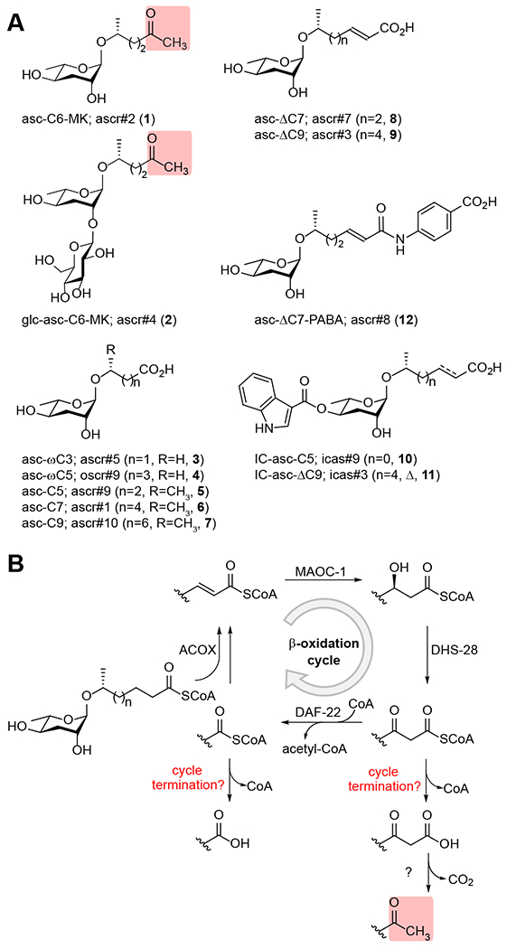
Methyl ketone (MK)-ascarosides represent essential components of several pheromones in Caenorhabditis elegans, including the dauer pheromone, which triggers the stress-resistant dauer larval stage, and the male-attracting sex pheromone. Here, we identify an acyl-CoA thioesterase, ACOT-15, that is required for the biosynthesis of MK-ascarosides. We propose a model in which ACOT-15 hydrolyzes the β-keto acyl-CoA side chain of an ascaroside intermediate during β-oxidation, leading to decarboxylation and formation of the MK. Using comparative metabolomics, we identify additional ACOT-15-dependent metabolites, including an unusual piperidyl-modified ascaroside, reminiscent of the alkaloid pelletierine. The β-keto acid generated by ACOT-15 likely couples to 1-piperideine to produce the piperidyl ascaroside, which is much less dauer-inducing than the dauer pheromone, asc-C6-MK (ascr#2, 1). The bacterial food provided influences production of the piperidyl ascaroside by the worm. Our work shows how the biosynthesis of MK- and piperidyl ascarosides intersect and how bacterial food may impact chemical signaling in the worm.

Keywords: ACOT-15; Caenorhabditis elegans; acyl-CoA thioesterase; anaferine; ascaroside; ascarosides; ascr#2; attraction; avoidance; beta-oxidation; dauer; hydroxylamine; lifespan; metabolomics; methyl ketone; natural products; pelletierine; pheromone; piperideine; piperidine; structure elucidation.


Conflict of interest statement

Declaration of interests The authors declare no competing interests.

Figures

Figure 1.
Figure 1.. Structures of some ascarosides, the role of β-oxidation in ascaroside biosynthesis, and candidate mechanisms for β-oxidation termination.
(A) The chemical structures of some abundant ascarosides in the C. elegans exometabolome. These ascarosides include the MK-containing ascarosides, asc-C6-MK (1) and glc-asc-C6-MK (2). The nomenclature used in this study for the ascarosides is based on their modular structure: head group-asc-(ω)(Δ)C#-terminus group. (B) Ascaroside pheromones are synthesized from long-chain ascarosides which have their side chains shortened through β-oxidation. The mechanisms used to terminate the β-oxidation process and generate side chains terminating in either carboxylic acids or methyl ketones have not been identified. A candidate mechanism for MK biosynthesis is shown.
Figure 2.
Figure 2.. Ascaroside production in the acot-15 mutant and overexpression strains.
(A) The production of ascarosides by the acot-15(ttTi876) mutant relative to wild type. (B) Model for the role of ACOT-15 in the biosynthesis of asc-C6-MK (1) and asc-C7 (6). (C) The production of ascarosides in the acot-15(ttTi876) mutant upon rescue with acot-15p::acot-15. The ratio of the ascaroside in the mutant or rescue strain is shown relative to wild type. Two lines were generated and gave similar results. (D) The production of ascarosides in the acot-15p::acot-15 overexpression strain relative to wild type. (E) The production of ascarosides in the acot-15p::acot-15::SL2::mcherry overexpression strain (generated by injecting the overexpression construct at 20 ng/μL and at 50 ng/μL) relative to wild type. In A, C, D, and E, data represent the mean ± standard deviation of three biological replicates. In A and D, p values were calculated using an unpaired t test, and in C and E, p values were calculated using one-way ANOVA with Dunnett’s post hoc test (*p ≤ 0.05, **p ≤ 0.01, ***p ≤ 0.001, ****p ≤ 0.0001).
Figure 3.
Figure 3.. Analysis of the hydroxylamine-treated endometabolome of wild-type versus acot-15 mutant worms.
(A) Examples of different CoA-thioesters reacted with hydroxylamine (HA), including HA-modified saturated side chains (HA), HA-modified α,β-unsaturated side chains (Δ-HA), HA-modified β-hydroxy side chains (bh-HA), and HA-modified β-keto side chains (bk-HA). (B) The production of ascarosides and HA-modified ascarosides in the endometabolome of acot-15 mutant worms relative to wild-type worms, with relevant structures shown below. HA-modified α,β-unsaturated side chains where the HA group has been reduced are referred to as “Δ-HAR” (e.g., 20, 21). (C) Extracted ion chromatogram of asc-C6-MK-OX (25) in the HA-treated wild-type and acot-15 endometabolomes. (D) Extracted ion chromatogram of asc-C6-MK (1) in the wild-type and acot-15 endometabolomes. (E) Relative amounts of asc-C6-MK (1) in the wild-type and acot-15 endometabolomes prepared under neutral or basic conditions. (F) Extracted ion chromatogram of glc-asc-C6-MK-OX (26) in the HA-treated wild-type and acot-15 endometabolomes. (G) Extracted ion chromatogram of ph-asc-C6-MK-OX (27) in the HA-treated wild-type and acot-15 endometabolomes. For B, C, F, and G, see Data S2.
Figure 4.
Figure 4.. Comparative metabolomics of the exometabolome of wild-type versus acot-15 mutant worms.
Volcano plots of wild-type versus acot-15(ttTi876) exometabolome analyzed in positive (ESI+) (top) and negative mode (ESI−) (bottom). Fold change is indicated on the x-axis, with mass features that are less abundant in the acot-15 exometabolome towards the left and those that are more abundant towards the right. Statistical significance (p value) is indicated on the y-axis. Highlighted features in the volcano plot are numbered, and these numbers correspond to the numbers indicated for the ascaroside structures. The chemical structures are proposed based on the fragmentation pattern by LC-HR-MS/MS. Some features could not be further characterized due to low signal and/or absence of fragmentation by LC-HR-MS/MS. See Data S3. The chemical structure of 29 is elucidated in Fig. 5.
Figure 5.
Figure 5.. Structural characterization of an ascaroside with a piperidine ring.
(A) MS/MS fragmentation pattern of asc-C6-pip (29) analyzed in positive mode (ESI+). (B) Key NMR correlations from COSY and HMBC spectra of asc-C6-pip (29). See Data S4 and Table S1. (C) The structure of asc-C6-pip (29), with the structures of the alkaloids pelletierine (35) and anaferine (36) for comparison. (D) Dauer formation assay in response to asc-C6-MK (1) and asc-C6-pip (29). (E) Lifespan assay in response to asc-C6-pip (29) in both wild-type and acot-15 worms. (F, G) Chemotaxis assay with different ascarosides in hermaphrodites (F) and males (G). In D, F, and G, data represent the mean ± standard deviation of three biological replicates. In F and G, p values were calculated using one-way ANOVA with Dunnett’s post hoc test, and in E, p values were calculated using a log-rank test (*p ≤ 0.05, **p ≤ 0.01, ***p ≤ 0.001, ****p ≤ 0.0001).
Figure 6.
Figure 6.. Biosynthesis of pelletierine and asc-C6-pip.
(A) Biosynthetic pathway to pelletierine (35). (B) Proposed biosynthetic pathway to asc-C6-pip (29). (C) Biosynthetic pathway to 1-piperideine in club mosses (top) and bacteria (bottom). (D) Relative ascaroside production in mixed stage wild-type worms fed bacteria supplemented with different amino acids. (E) Unlabeled and labeled asc-C6-pip (29) production in wild-type worms fed worm powder supplemented with unlabeled and labeled lysine. (F) Extracted ion chromatograms for unlabeled and labeled asc-C6-pip (29) produced by arrested L1 worms treated with unlabeled and labeled lysine in the absence of bacterial food. (G) Production of asc-C6-pip (29) and other ascarosides in wild-type worms fed either OP50, HB101, or various bacterial strains isolated from the C. elegans microbiome. Data represent the mean ± standard deviation of two biological replicates (E, G) or three biological replicates (D). In D and G, p values were calculated using one-way ANOVA with Dunnett’s post hoc test (*p ≤ 0.05, **p ≤ 0.01, ***p ≤ 0.001, ****p ≤ 0.0001). In G, p values are only shown for asc-C6-pip (29) for the different bacterial strains relative to the control bacteria (OP50).

Similar articles

Cited by

References

    1. Butcher RA, Fujita M, Schroeder FC, and Clardy J (2007). Small-molecule pheromones that control dauer development in Caenorhabditis elegans. Nat Chem Biol 3, 420–422. 10.1038/nchembio.2007.3. - DOI - PubMed
    1. Butcher RA, Ragains JR, Kim E, and Clardy J (2008). A potent dauer pheromone component in C. elegans that acts synergistically with other components. Proc Natl Acad Sci U S A 105, 14288–14292. 10.1073/pnas.0806676105. - DOI - PMC - PubMed
    1. Butcher RA, Ragains JR, and Clardy J (2009). An indole-containing dauer pheromone component with unusual dauer inhibitory activity at higher concentrations. Org Lett 11, 3100–3103. 10.1021/ol901011c. - DOI - PMC - PubMed
    1. Srinivasan J, Kaplan F, Ajredini R, Zachariah C, Alborn HT, Teal PE, Malik RU, Edison AS, Sternberg PW, and Schroeder FC (2008). A blend of small molecules regulates both mating and development in Caenorhabditis elegans. Nature 454, 1115–1118. 10.1038/nature07168. - DOI - PMC - PubMed
    1. Pungaliya C, Srinivasan J, Fox BW, Malik RU, Ludewig AH, Sternberg PW, and Schroeder FC (2009). A shortcut to identifying small molecule signals that regulate behavior and development in Caenorhabditis elegans. Proc Natl Acad Sci U S A 106, 7708–7713. 10.1073/pnas.0811918106. - DOI - PMC - PubMed

MeSH terms

LinkOut - more resources