

Ocular biomarkers of cognitive decline based on deep-learning retinal vessel segmentation
- PMID: 38184539
- PMCID: PMC10770952
- DOI: 10.1186/s12877-023-04593-8
Ocular biomarkers of cognitive decline based on deep-learning retinal vessel segmentation
Abstract
Background: The current literature shows a strong relationship between retinal neuronal and vascular alterations in dementia. The purpose of the study was to use NFN+ deep learning models to analyze retinal vessel characteristics for cognitive impairment (CI) recognition.
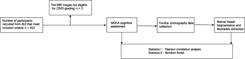
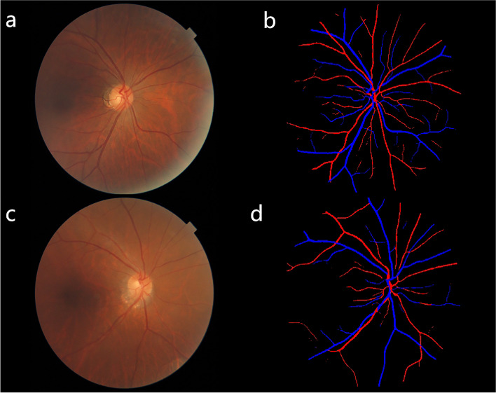
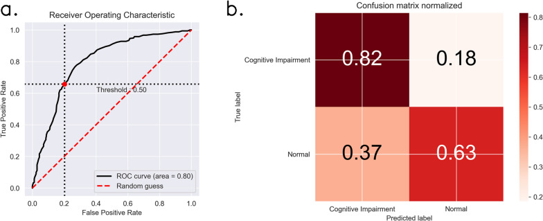
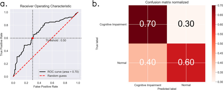
Methods: We included 908 participants from a community-based cohort followed for over 15 years (the prospective KaiLuan Study) who underwent brain magnetic resonance imaging (MRI) and fundus photography between 2021 and 2022. The cohort consisted of both cognitively healthy individuals (N = 417) and those with cognitive impairment (N = 491). We employed the NFN+ deep learning framework for retinal vessel segmentation and measurement. Associations between Retinal microvascular parameters (RMPs: central retinal arteriolar / venular equivalents, arteriole to venular ratio, fractal dimension) and CI were assessed by Pearson correlation. P < 0.05 was considered statistically significant. The correlation between the CI and RMPs were explored, then the correlation coefficients between CI and RMPs were analyzed. Random Forest nonlinear classification model was used to predict whether one having cognitive decline or not. The assessment criterion was the AUC value derived from the working characteristic curve.
Results: The fractal dimension (FD) and global vein width were significantly correlated with the CI (P < 0.05). Age (0.193), BMI (0.154), global vein width (0.106), retinal vessel FD (0.099), and CRAE (0.098) were the variables in this model that were ranked in order of feature importance. The AUC values of the model were 0.799.
Conclusions: Establishment of a predictive model based on the extraction of vascular features from fundus images has a high recognizability and predictive power for cognitive function and can be used as a screening method for CI.
Keywords: Cognitive function; Deep learning segmentation; Fundus photography; Retinal microvascular parameters.
© 2024. The Author(s).
Conflict of interest statement
The authors declare no competing interests.
Figures
References
-
- Lewis M, Peiris CL, Shields N. Long-term home and community-based exercise programs improve function in community-dwelling older people with cognitive impairment: a systematic review. J Phys. 2017;63(1):23–29. - PubMed
-
- Sur S, Lin Z, Li Y, Yasar S, Rosenberg P, Moghekar A, Hou X, Kalyani R, Hazel K, Pottanat G, Xu C, van Zijl P, Pillai J, Liu P, Albert M, Lu H. Association of cerebrovascular reactivity and Alzheimer pathologic markers with cognitive performance. Neurology. 2020;95(8):e962–e972. doi: 10.1212/WNL.0000000000010133. - DOI - PMC - PubMed
-
- István L, Czakó C, Élő Á, Mihály Z, Sótonyi P, Varga A, Ungvári Z, Csiszár A, Yabluchanskiy A, Conley S, Csipő T, Lipecz Á, Kovács I, Nagy ZZ. Imaging retinal microvascular manifestations of carotid artery disease in older adults: from diagnosis of ocular complications to understanding microvascular contributions to cognitive impairment. Geroscience. 2021;43(4):1703–1723. doi: 10.1007/s11357-021-00392-4. - DOI - PMC - PubMed
-
- Mutlu U, Colijn JM, Ikram MA, Bonnemaijer PWM, Licher S, Wolters FJ, Tiemeier H, Koudstaal PJ, Klaver CCW, Ikram MK. Association of Retinal Neurodegeneration on optical coherence tomography with dementia: a population-based study. JAMA Neurol. 2018;75(10):1256–1263. doi: 10.1001/jamaneurol.2018.1563. - DOI - PMC - PubMed
Publication types
MeSH terms
Substances
LinkOut - more resources
Full Text Sources
