. 2024 Jan 6;23(1):4.
doi: 10.1186/s12943-023-01912-w.

CircPPAP2B controls metastasis of clear cell renal cell carcinoma via HNRNPC-dependent alternative splicing and targeting the miR-182-5p/CYP1B1 axis

Affiliations

CircPPAP2B controls metastasis of clear cell renal cell carcinoma via HNRNPC-dependent alternative splicing and targeting the miR-182-5p/CYP1B1 axis

Zaosong Zheng et al. Mol Cancer. .

Abstract

Background: Renal cell carcinoma (RCC) is one of the most common malignant tumor worldwide. Metastasis is a leading case of cancer-related deaths of RCC. Circular RNAs (circRNAs), a class of noncoding RNAs, have emerged as important regulators in cancer metastasis. However, the functional effects and regulatory mechanisms of circRNAs on RCC metastasis remain largely unknown.

Methods: High-throughput RNA sequencing techniques were performed to analyze the expression profiles of circRNAs and mRNAs in highly and poorly invasive clear cell renal cell carcinoma (ccRCC) cell lines. Functional experiments were performed to unveil the regulatory role of circPPAP2B in the proliferation and metastatic capabilities of ccRCC cells. RNA pulldown, Mass spectrometry analysis, RNA methylation immunoprecipitation (MeRIP), RNA immunoprecipitation (RIP), co-immunoprecipitation (CoIP), next-generation RNA-sequencing and double luciferase experiments were employed to clarify the molecular mechanisms by which circPPAP2B promotes ccRCC metastasis.

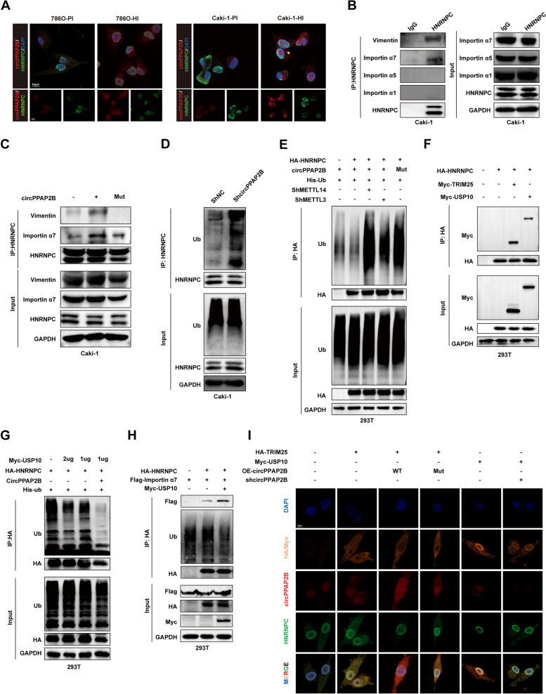
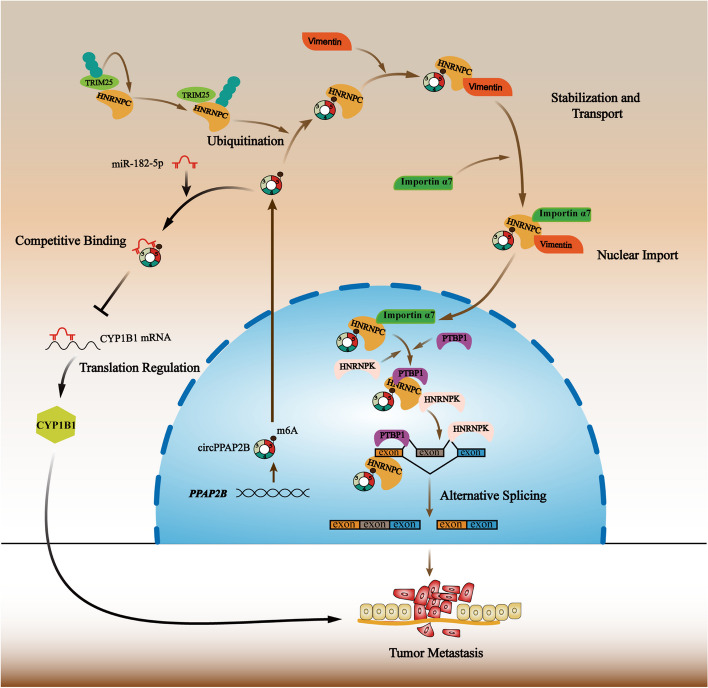
Results: In this study, we describe a newly identified circular RNA called circPPAP2B, which is overexpressed in highly invasive ccRCC cells, as determined through advanced high-throughput RNA sequencing techniques. Furthermore, we observed elevated circPPAP2B in ccRCC tissues, particularly in metastatic ccRCC tissues, and found it to be associated with poor prognosis. Functional experiments unveiled that circPPAP2B actively stimulates the proliferation and metastatic capabilities of ccRCC cells. Mechanistically, circPPAP2B interacts with HNRNPC in a m6A-dependent manner to facilitate HNRNPC nuclear translocation. Subcellular relocalization was dependent upon nondegradable ubiquitination of HNRNPC and stabilization of an HNRNPC/Vimentin/Importin α7 ternary complex. Moreover, we found that circPPAP2B modulates the interaction between HNRNPC and splicing factors, PTBP1 and HNPNPK, and regulates pre-mRNA alternative splicing. Finally, our studies demonstrate that circPPAP2B functions as a miRNA sponge to directly bind to miR-182-5p and increase CYP1B1 expression in ccRCC.

Conclusions: Collectively, our study provides comprehensive evidence that circPPAP2B promotes proliferation and metastasis of ccRCC via HNRNPC-dependent alternative splicing and miR-182-5p/CYP1B1 axis and highlights circPPAP2B as a potential therapeutic target for ccRCC intervention.

Keywords: Alternative splicing; CircPPAP2B; HNRNPC; Nuclear translocation; m6A; miRNA sponge.


Conflict of interest statement

The authors declare no competing interests.

Figures

Fig. 1
Fig. 1
Identification of circPPAP2B in highly invasive ccRCC cell lines. A Establishment of highly invasive and poorly invasive ccRCC cell lines. The highly invasive and poorly invasive ccRCC cell lines were generated through experimental protocols described in the Methods section. B Assessment of the invasive capabilities of invasive and poorly invasive ccRCC cell lines using transwell assay. Representative images of invasive cells and quantification of invasive cell numbers are shown. C High-throughput RNA sequencing to analyze the expression profiles of circRNAs in highly and poorly invasive ccRCC cell lines. D A Venn diagram illustrates the overlapping upregulated circRNAs between highly and poorly invasive ccRCC cell lines. E qPCR validated the upregulation of circPPAP2B in ccRCC tissues. F A schematic illustration of circPPAP2B originates from exons 3 to 5 of the PPAP2B gene. G Sanger sequencing validated the circPPAP2B junction site in circBase. H RNase R assay evaluates the stability of circPPAP2B in the ccRCC cell line. I Actinomycin D assay evaluates the stability of circPPAP2B in the ccRCC cell line. J RNA FISH was performed to determine the subcellular localization of circPPAP2B in ccRCC cell lines. K RNA FISH was performed to detect circPPAP2B expression in ccRCC tissues and normal tissues. L-M qPCR was performed to detect circPPAP2B expression on a cohort of 78 pairs of ccRCC tissues and adjacent non-tumorous tissues. N Kaplan–Meier survival analysis was conducted to evaluate the prognostic value of circPPAP2B in ccRCC. Data are represented as mean ± SEM. ***P < 0.001 vs. WT group
Fig. 2
Fig. 2
CircPPAP2B promotes ccRCC proliferation and metastasis in vivo and in vitro A-C Colony formation assays, CCK8 assays, and Edu assays were performed to evaluate the proliferation ability of ccRCC cells treated with shNC or shcircPPAP2B. D-E Transwell assays and wound-healing assays were conducted to detect the invasive ability of ccRCC cells treated with shNC or shcircPPAP2B. The representative images of nude mice subcutaneously injected with caki-1 stably expressed shNC or shcircPPAP2B. G The volume of tumors was measured every week. The weight of tumors was measured 5 weeks after injection. I The representative images of lung metastases in nude mice by HE staining. Tumor burdens were reflected by the percentages of tumor area in each slide. J Transwell assays were conducted to detect the invasive ability of ccRCC cells treated with OE-NC or OE-circPPAP2B. K-L CCK8 assays and Edu assays were performed to evaluate the proliferation ability of ccRCC cells treated with OE-NC or OE-circPPAP2B. Data are represented as mean ± SEM. *P < 0.05, **P < 0.01, ***P < 0.001 vs. WT group
Fig. 3
Fig. 3
CircPPAP2B directly interacts with HNRNPC in a m6A-dependent manner A. RNA pulldown assay was performed using a biotin-labeled sense or antisense probe of circPPAP2B. The proteins enriched by sense or antisense probe of circPPAP2B were separated by SDS-PAGE and subjected to silver staining. B Mass spectrometry analysis identified HNRNPC as one of the most abundant proteins enriched by the sense probe of circPPAP2B. C RNA pulldown assay was performed to confirm the direct interaction between circPPAP2B and HNRNPC in ccRCC cell lines. D RNA pulldown assay was performed to confirm the direct interaction between circPPAP2B and HNRNPC in 293 T. E RIP assay was performed to validate direct interaction between circPPAP2B and HNRNPC in ccRCC cell lines using anti-HNRNPC antibody. F RNA FISH was performed to determine the co-localization of circPPAP2B and HNRNPC in ccRCC cell lines. G Full-length and truncated flag-labeled HNRNPC was constructed and RIP assays were performed to identify the specific domain of HNRNPC which interacts with circPPAP2B. H RNA pulldown assays were performed to identify the specific domain of HNRNPC which interacts with circPPAP2B. I MeRIP assays were performed to investigate whether circPPAP2B undergoes m6A modification. J Graphic illustration of the secondary structure of circPPAP2B and its m6A modification site predicted by RNA-fold software. Construction of WT and m6A modification site mutant circPPAP2B. K RNA pulldown assay and dot blot assay were performed to validate the m6A modification site in circPPAP2B. L RIP assay was performed to validate the m6A modification site in circPPAP2B and its role in the interaction between circPPAP2B and HNRNPC. M Transwell assay was performed on the role of m6A modification site in circPPAP2B in promoting ccRCC invasion. I The representative images of lung metastases in nude mice by HE staining. Tumor burdens were reflected by the percentages of tumor area in each slide. Data are represented as mean ± SEM. *P < 0.05, **P < 0.01, ***P < 0.001 vs. WT group
Fig. 4
Fig. 4
CircPPAP2B facilitates HNRNPC nuclear translocation via regulating HNRNPC nondegradable ubiquitination and stabilizing HNRNPC/Vimentin/Importin α7 ternary complex A RNA FISH was performed to determine subcellular localization of HNRNPC in highly and poorly invasive ccRCC cell lines. B CoIP assay was performed to determine the direct binding between HNRNPC with Vimentin and Importin α. C Endogenous CoIP assay was performed to confirm the direct interaction between HNRNPC and Vimentin with Importin α7, and the role of the m6A modification site in their interaction. D CoIP assay was performed to explore whether circPPAP2B regulates the ubiquitination of HNRNPC in ccRCC cell lines. E CoIP assay was performed to determine whether the regulatory of circPPAP2B on HNRNPC ubiquitination is dependent on m6A modification. F CoIP assay was performed to confirm the direct interaction between HNRNPC and TRIM25 or USP10. G CoIP assay was performed to determine the role of USP10 on ubiquitination levels of HNRNPC. H CoIP assay was performed to explore whether HNRNPC ubiquitination regulates the interaction between HNRNPC and Importin α7. I FISH was performed to determine the role of TRIM25 or USP10 on the subcellular localization of HNRNPC in ccRCC cells
Fig. 5
Fig. 5
CircPPAP2B modulates the interaction between HNRNPC and splicing factors to regulate pre-mRNA alternative splicing A GSEA enrichment analysis was performed to reveal the relationship between HNRNPC and RNA splicing with mRNA processing in ccRCC. B-C Mass Spectrometry analysis and STRING prediction were employed to identify splicing factors HNRNPC regulated in ccRCC. D CoIP assay was performed to confirm the direct interaction between PTBP1, HNRNPK, HNRNPL, HNRNPA2B1, and HNRNPC. E CoIP assay was performed to confirm whether circPPAP2B overexpression influenced the interaction between HNRNPC and PTBP1 or HNRNPK. F CoIP assay was performed to confirm whether circPPAP2B knockdown influenced the interaction between HNRNPC and PTBP1 or HNRNPK. G Alternative splicing events were identified in both circPPAP2B knockdown and anti-HNRNPC circPPAP2B knockdown ccRCC cells, including skipped exon (SE), alternative 5’ splice site (A5SS), alternative 3’ splice site (A3SS), mutually exclusive exons (MXE) and retained intron. H 235 alternative splicing genes were shared in both circPPAP2B knockdown and anti-HNRNPC circPPAP2B knockdown ccRCC cells. I GO enrichment analysis identified enriched pathways with shared alternative splicing genes. J The Pie diagram shows the ratio of different types of alternative splicing events regulated by circPPAP2B knockdown. K The Venn diagram showed 40 alternative splicing events were shared in both circPPAP2B knockdown and anti-HNRNPC circPPAP2B knockdown ccRCC cells. L Alternative splicing events of CD44 were identified in the anti-HNRNPC RIP-sequencing assay. M qPCR assay was performed to validate the selected alternative of CD44 in OE-NC and OE-circPPAP2B ccRCC cells. N RIP-qPCR with anti-HNRNPK antibody was performed to validate the interaction between HNRNPK and CD44 alternative splicing
Fig. 6
Fig. 6
CircPPAP2B functions as a ceRNA to directly bind to miR-182-5p and upregulated CYP1B1 A Bioinformatics tools (circBank, ENCORI, and CircInteractome) were used to identify candidate miRNAs that potentially interact with circPPAP2B. B Transwell assays were performed to explore the role of miR-182-5p mimic or miR-182-5p inhibitor on the invasive ability of ccRCC cells. C Biotin-labeled RNA pulldown assays were performed to confirm the direct interaction between circPPAP2B and miR-182-5p. D Dual-luciferase reporter assay was performed to validate the direct binding and binding site between circPPAP2B and miR-182-5p. E Transwell assays were performed to explore the role of miR-182-5p mimic or miR-182-5p inhibitor on the circPPAP2B-mediated invasion of ccRCC cells. F High-throughput RNA sequencing to analyze the RNA regulated by circPPAP2B in ccRCC cells. G Western blotting assays to investigate the effect of miR-182-5p on circPPAP2B-mediated CYP1B1 expression. H Biotin-labeled RNA pulldown assay was performed to validate the direct binding between miR-182-5p and CYP1B1 mRNA. I Dual-luciferase reporter assay was performed to validate the direct binding and binding site between miR-182-5p and 3’UTR of CYP1B1 mRNA. J Transwell assay was performed to explore the role of CYP1B1 in the invasion of ccRCC cells. Data are represented as mean ± SEM. *P < 0.05, **P < 0.01, ***P < 0.001 vs. WT group
Fig. 7
Fig. 7
Schematic representation of the possible mechanism of circPPAP2B in ccRCC metastasis CircPPAP2B is derived from exons 3 to 5 of the PPAP2B gene through backsplicing in the nucleus. CircPPAP2B not only interacts with HNRNPC in an m6A-dependent manner to facilitate HNRNPC nuclear translocation via stabilizing HNRNPC/Vimentin/Importin α7 ternary complex, but also functions as a miRNA sponge to directly bind to miR-182-5p and modulate CYP1B1 expression, thereby promoting cancer metastasis in ccRCC

References

    1. Capitanio U, Montorsi F. Renal cancer. Lancet. 2016;387:894–906. doi: 10.1016/S0140-6736(15)00046-X. - DOI - PubMed
    1. Siegel RL, Miller KD, Wagle NS, Jemal A. Cancer statistics, 2023. CA Cancer J Clin. 2023;73:17–48. doi: 10.3322/caac.21763. - DOI - PubMed
    1. Powles T. Recent eUpdate to the ESMO Clinical Practice Guidelines on renal cell carcinoma on cabozantinib and nivolumab for first-line clear cell renal cancer: Renal cell carcinoma: ESMO Clinical Practice Guidelines for diagnosis, treatment and follow-up. Ann Oncol. 2021;32:422–423. doi: 10.1016/j.annonc.2020.11.016. - DOI - PubMed
    1. Gill IS, Cacciamani GE, Duddalwar V, Thangathurai D, Cunningham M. Renal cancer with extensive level IV intracardiac tumour thrombus removed by robot. Lancet. 2020;396:e88. doi: 10.1016/S0140-6736(20)32291-1. - DOI - PubMed
    1. Turajlic S, Swanton C, Boshoff C. Kidney cancer: The next decade. J Exp Med. 2018;215:2477–2479. doi: 10.1084/jem.20181617. - DOI - PMC - PubMed

Publication types

MeSH terms